- What is high or low intracranial pressure?
- How to check intracranial pressure
- What is the danger of intracranial hypertension or hypotension?
- Where can I get an MRI of the brain?
Headache, spots before the eyes, vomiting, blurry silhouettes around and the ground disappearing somewhere from under your feet - all these alarming symptoms may indicate increased intracranial pressure. But if blood pressure can be measured independently with a tonometer in just a couple of minutes and reduced with the help of one vasodilator pill, then with intracranial hypertension everything is much more complicated. Only with the advent of MRI of the brain did it become possible to carry out non-invasive diagnostics and find the cause.
What is a cranial MRI?
Magnetic resonance imaging of the skull is a non-invasive test designed to create a clear, detailed image of the skull and brain. This is done with the help of computer equipment and specially developed software, which is supplied with information about the impulses that arise when a person is exposed to a magnetic field.
Of all the diagnostic methods, MRI is considered the safest, as it does not involve the use of X-rays. This diagnostic method provides high-quality visualization of the patient’s skull; the result can be studied in detail on a monitor or printed on paper.
Using MRI of the skull, you can assess the condition of bones, brain nerves and tissues, and identify vascular pathology.
CT scan of the skull with 3D effect
Despite the fact that CT requires the use of ionizing radiation, it is also often used because it is effective for examining the facial skeleton. What brought him to the level of MRI was the ability to create a three-dimensional image (3D).
Compared with magnetic tomography, computed tomography can more thoroughly and in detail evaluate bone pathologies of the skull in the cranio-orbital and craniobasal region. Three-dimensional reconstruction allows the treating specialist to examine the facial skeleton in volume and model the necessary implants for it with suitable sizes.
The full name of this procedure is “multislice computed tomography with 3D reconstruction” (MSCT).
Indications and contraindications for MRI of the skull
MRI is mainly necessary to assess the general condition of the skull and find pathology. There is a standard list of diseases for the detection of which such an examination is used:
- pathology of the vascular system of the brain (hemorrhages, ischemia);
- traumatic brain injuries and bruises;
- GM tumors;
- loss or impairment of hearing, vision, speech;
- frequent headaches;
- deviations or disturbances in the development of blood vessels in the head;
- changes in the base of the skull;
- HIV;
- diseases of the nervous system;
- changes in the chiasmal-sellar region;
- stroke;
- multiple sclerosis;
- paroxysmal disorders;
- sinusitis;
- death of GM cells;
- intracranial pressure.
Despite the large number of indications, MRI cannot always be used. There is a list of factors in the presence of which the use of such an examination without special preparation of the patient is prohibited:
- pregnancy less than 4 months;
- heart failure syndrome;
- panic attacks while being placed in the apparatus tunnel (claustrophobia);
- presence of insulin pumps;
- artificial heart valves;
- the patient's serious condition associated with the disease;
- inadequacy, alcohol or drug intoxication.
Also, special preparation is required for a patient who has tattoos in the head or neck area made with paint with metal connections. An exception to this rule are tattoos with titanium compounds.
Do not forget about the factors under which magnetic resonance imaging studies are completely prohibited:
- the presence of a pacemaker and other electronic devices installed in the body;
- middle ear implants (electronic or ferromagnetic);
Complete contraindications also include diseases associated with accelerated destruction of red blood cells.
Intracranial pressure
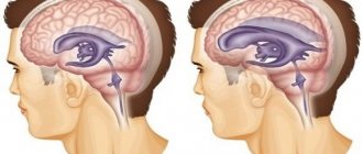
As for determining intracranial pressure (ICP), it is quite possible. To determine the ICP stimulus, magnetic resonance imaging is used. MRI can show the effects of prolonged intracranial pressure on the brain in the form of changes in the structure of certain areas of the brain. Typically, ICP is indicated by dilated fluid cavities in the brain and spreading of the brain matter around the ventricles of the brain, as well as internal hydrocephalus.
Using a tomograph, data is read from hydrogen nuclei in human cells that respond to the influence of a magnetic field. The received impulses are transformed into an accurate image with the detected pathology. The operating principle of the device is based on the phenomenon of nuclear magnetic resonance (NMR).
It will take about 30 minutes to identify the causes of intracranial pressure using the NMR phenomenon without contrast.
How to treat
Increased intracranial pressure begins to be treated after a comprehensive diagnosis. Therapy consists of eliminating the causes that caused the disorders.
The attending physician can prescribe suitable methods. Typically, the treatment of intracranial pressure in adults is carried out by a neurologist; other highly specialized specialists may also be involved.
Intracranial pressure can be relieved with medication if the condition is not life-threatening. In severe cases, surgery cannot be avoided.
Mild intracranial hypertension can also be eliminated using traditional medicine. But they can only be used under the supervision of a specialist, since only he knows how to reduce intracranial pressure. Therefore, if signs of venous insufficiency or moderate ICH appear, you should consult a specialist.
Cranial nerves
Magnetic resonance imaging of the skull also reveals pathology of the cranial nerves. Most often, an accurate diagnosis of neurovascular conflict is performed at the site where the nerve exits the trunk. Other pathologies leading to hemifacial spasm are also identified.
External symptoms for which the examination is performed are spasms of the facial muscles with tonic and clonic contractions.
To study cranial nerves, a specific pulse sequence is used and slice blocks are laid depending on which cranial nerve is being studied:
- to obtain a clear image of the olfactory nerve (bulb and tract), a block of slices is used in the axial plane, parallel to the course of the nerve itself;
- optic nerve - MRI makes it possible to examine the nerve itself, its chiasm, and tract;
- to visualize the oculomotor nerve, a block of slices is made, which is oriented in the coronal and sagittal plane;
- the trochlear nerve is visualized as a block of slices with one landmark - the inferior colliculus of the midbrain;
- the ternary nerve is examined by a block of sections in the region of the root of this nerve, with a guide to the location of its trunk in the lateral cisterns of the bridge, and is also examined in the sagittal, axial and coronal planes;
- The vestibulocochlear nerve is examined using a block of sections in the lower surface of the brain outside the olive of the medulla oblongata.
Preparing for an MRI
Usually the examination takes place without special preparation. In some cases, by decision of the doctor, the specialist may ask you to abstain from food or any drinks (coffee, tea) in the morning . Already in the laboratory, before the procedure itself, it is necessary to remove all gold or silver jewelry, otherwise they will interfere with the magnetic fields from doing their job.
You may also need to change to a special surgical shirt. The need for this is determined by how many metal parts there are on the clothing and how spacious it is.
Female representatives must inform their doctor about their pregnancy, even at the earliest stage. Until now, there has not been a single case where MRI has negatively affected the body of the mother or the unborn child, but doctors are not 100% confident in the complete safety of the study.
First, the radiologist must review the patient’s medical history, operations and find out whether the patient has artificially implanted implants.
Traditional methods
You can use traditional methods to eliminate the problem only if the problems arise due to excess weight, osteochondrosis, or stress.
Thanks to folk recipes, blood circulation improves and the functions of the nervous system are normalized. You can achieve similar results using:
- decoction of mulberry branches. They are dried, finely chopped and two tablespoons are placed in a liter of water and brought to a boil over low heat. Drink a glass for three weeks;
- decoction of poplar buds. A few spoons of kidneys are poured into half a liter of water, boiled for a quarter of an hour and consumed on an empty stomach for three weeks;
- mixtures of tinctures. Infusions of hawthorn, eucalyptus, peppermint, motherwort, and valerian are mixed in certain proportions and taken 25 drops three times a day, before meals. This remedy allows you to get rid of venous spasms, calms you down and makes you feel better.
These recipes should be used only after consulting a specialist.
The process of performing an MRI of the skull
The MRI equipment itself is a cylindrical machine with a tunnel in the middle, which is surrounded by a magnet. A table slides into this tunnel and the patient lies on it.
- To perform an MRI of the skull, the patient is placed on a table and secured to it with special belts and ties. For convenience and immobility, there are bolsters that secure the body.
- After fastening, the head is prepared for examination. Wired sensors that conduct and receive radiomagnetic waves are attached to it. It is these signals that provide information to the computer, which is then processed by the program and shows all sorts of pathologies and causes of intracranial pressure.
- Contrast-enhanced MRI uses contrast material that is injected through a catheter into a vein in the arm. In order to avoid clogging of the veins, which interferes with the passage of the substance, saline solution is poured in front of it.
- After preliminary preparation, the patient is placed on the table in the apparatus tunnel, and all medical staff must leave the room.
- The results of the study are given to the patient immediately after the procedure. If the examination is unsuccessful, the device may produce a blurred image, and a similar examination is carried out. When finished, the doctor removes the catheter.
The standard examination time is 45 minutes. At this time, several pictures are taken (one image - 5–8 minutes).
When scanning the head, magnetic resonance imaging has its own characteristics. In the process, blocks of sections of head tissue are made in layers. One level is 5 mm thick, and in one photo scan information is read from approximately 20 levels.
The accuracy of the survey results is determined by the magnetic field induction (measured in Tesla).
Remember! While the image is being taken, the patient must lie still and do everything the doctor says.
Spinal tap
The technique gained popularity due to the accuracy of the results. For diagnosis, a special long needle is inserted into the spinal cord to take a puncture, to which a pressure gauge is attached, as a result it is possible to obtain a specific pressure level. Normally, it should range from 80 to 170 mm of water column. The method can only be used in an inpatient setting in a neurosurgical or neurological department of a hospital.
Recognize pathology from the first days of life
Consequences of cranial MRI examination
Patients usually tolerate this procedure without pain or consequences. But there are some exceptions. During the examination, the patient may feel discomfort. It has to do with immobility. There are manifestations of attacks of claustrophobia even in trained people. In this case, the patient needs to take a sedative.
Additionally, the temperature of the head may increase, which is considered normal.
When using contrast material, you may feel a rush of blood and coolness, this is also considered normal. The process of inserting and removing the catheter may cause some discomfort and may cause irritation. Pain or nausea may occur. Due to the use of contrast agents after the examination, intracranial pressure or ocular itching may occur.
Prevention of ICP
To prevent intracranial pressure, the following rules should be followed:
- drink the amount of fluid recommended by your doctor;
- ensure intake of vitamins with food or complexes;
- walk more, ventilate the room;
- avoid heavy loads and stressful situations;
- engage in moderate physical activity;
- Night sleep should be at least 8 hours;
- avoid sudden changes in climate and time zones;
- do not fly by air;
- sleep on high pillows with your head elevated;
- get up immediately after waking up;
- If possible, massage the collar area.
Prevention and maintaining a healthy lifestyle is the best tool for avoiding complications with intracranial pressure.