Basal ganglia
, like the cerebellum, represent another auxiliary motor system, which usually does not function on its own, but in close connection with the cerebral cortex and the corticospinal motor control system. Indeed, the basal ganglia receive most of its input from the cerebral cortex, and almost all of its output goes back to the cortex.
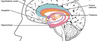
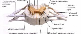
The figure shows the anatomical connections of the basal ganglia
with other brain structures. On each side of the brain, these ganglia consist of the caudate nucleus, putamen, globus pallidus, substantia nigra, and subthalamic nucleus. They are located mainly lateral to and around the thalamus, occupying most of the internal regions of both hemispheres of the cerebrum. It is also seen that almost all the motor and sensory nerve fibers connecting the cerebral cortex and the spinal cord pass through the space lying between the main structures of the basal ganglia, the caudate nucleus and the putamen. This space is called the internal capsule of the brain. Important for this discussion is the close connection between the basal ganglia and the corticospinal motor control system.
Neural circuit of the basal ganglia
. The anatomical connections between the basal ganglia and other brain elements that support motor control are complex. Shown on the left are the motor cortex, thalamus, and the brainstem and cerebellar circuits that work with them. On the right is the main outline of the basal ganglia system, showing the most important connections within the ganglia themselves and the extensive input and output pathways connecting other brain regions and the basal ganglia. In the following sections we will focus on two major circuits: the putamen circuit and the caudate circuit.
Physiology and function of the basal ganglia
One of the main functions of the basal ganglia
in motor control is their participation in the regulation of the execution of complex motor programs together with the corticospinal system, for example in the movement when writing letters. When the basal ganglia is severely damaged, the cortical motor control system can no longer support these movements. Instead, the person's handwriting becomes rough, as if he is learning to write for the first time.
To other complex motor acts
Activities that require the basal ganglia include cutting with scissors, hammering nails, throwing a basketball through a hoop, dribbling a football, throwing a baseball, shoveling while digging, most vocalizations, controlled eye movements, and virtually any of our fine movements. , in most cases performed unconsciously.
Nerve pathways of the putamen circuit
. The figure shows the main pathways through the basal ganglia involved in the execution of acquired forms of motor activity. These pathways primarily originate in the premotor cortex and somatosensory areas of the sensory cortex. They then pass into the putamen (mainly bypassing the caudate nucleus), from here to the internal part of the globus pallidus, then to the anterior ventral and ventrolateral nuclei of the thalamus and, finally, return to the primary motor cortex of the cerebrum and to areas of the premotor cortex and supplementary cortex, closely connected to the primary motor cortex. Thus, the main inputs to the putamen circuit come from brain regions adjacent to the primary motor cortex, but not from the primary cortex itself.
But the exits from this circuit
go mainly to the primary motor cortex or to closely related areas of the premotor and supplementary motor cortex. In close connection with this primary circuit of the putamen, auxiliary circuits function, coming from the putamen through the outer part of the globus pallidus, the subthalamus and the substantia nigra, ultimately returning to the motor cortex through the thalamus.
Movement disorders
when the shell contour is affected: athetosis, hemiballismus and chorea. How is the putamen circuit involved in ensuring the performance of complex motor acts? The answer is not clear. However, when part of the circuit is affected or blocked, some movements are significantly impaired. For example, lesions of the globus pallidus usually lead to spontaneous and often persistent wave-like movements of the hand, arm, neck, or face. Such movements are called athetosis.
Subthalamic nucleus lesion
often leads to sweeping movements of the entire limb. This condition is called hemiballismus. Multiple small lesions in the putamen lead to rapid twitching in the hands, face and other parts of the body, which is called chorea.
Lesions of the substantia nigra
lead to a widespread and extremely severe disease with characteristic rigidity, akinesia and tremor. This disease is known as Parkinson's disease and will be discussed in detail below.
Educational video lesson - basal ganglia, conducting pathways of the internal capsule of the brain
You can download this video and view it from another video hosting site on the page:
The human body consists of a large number of organs and structures, the main ones being the brain and heart. The heart is the engine of life, and the brain is the coordinator of all processes. In addition to knowledge about the main parts of the brain, you also need to know about the basal ganglia.
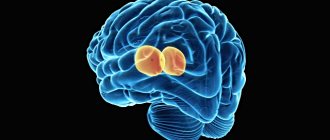
The basal ganglia are responsible for movement and coordination
The basal ganglia (ganglia) are accumulations of gray matter that form groups of nuclei. This part of the brain is responsible for movement and coordination.
Functions provided by ganglia
Motor activity occurs due to constant control of the pyramidal (corticospiral) tract. But it does not provide this completely. Some functions are taken over by the basal ganglia. Parkinson's disease or Wilson's disease is caused precisely by pathological disorders of subcortical accumulations of gray matter. The functions of the basal ganglia are considered vital, and their disorders are considered difficult to cure.
According to scientists, the main task of the nuclei is not the motor activity itself, but its control over the functioning, as well as the connection between muscle groups and the nervous system. The function of control over human movements is observed. This is characterized by the interaction of two systems, which includes the accumulation of subcortical substance. The striopallidal and limbic systems have their own functional characteristics. The first tends to control muscle contraction, which together forms coordination. The second is subject to the work and organization of vegetative functions. Their failure leads not only to human incoordination, but also to disruption of the mental activity of the brain.
Nuclear malfunctions lead to brain dysfunction
Structural features
The basal ganglia of the brain have a complex structure. According to their anatomical structure, they include:
- striatum (striatum);
- amygdaloidium (amygdala);
- fence.
Modern study of these clusters has created a new, convenient division of the nuclei into a cluster of substantia nigra and a nuclear tegmentum. But such a figurative structure does not give a complete picture of anatomical connections and neurotransmitters, so it is the anatomical structure that should be considered. Thus, the concept of the striatum is characterized by an accumulation of white and gray matter.
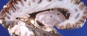
They are noticeable in a horizontal section of the cerebral hemispheres.
Basal ganglia is a complex term that includes concepts about the structure and functions of the striatum and amygdala. In addition, the striatum consists of the lenticular and caudate ganglion. Their location and connection has its own characteristics. The basal ganglia of the brain are separated by a neural capsule. The caudate ganglion is connected to the thalamus.
The caudate ganglion is connected to the thalamus
Features of the structure of the caudate ganglion
The second type of Golgi neurons is identical in structure to the caudate nucleus. Neurons play an important role in the formation of gray matter accumulations. This is noticeable by the similar features that unite them. The thinness of the axon and the shortness of the dendrites are identical. This nucleus provides its main functions with its own connections with individual areas and sections of the brain:
- thalamus;
- pale ball;
- cerebellum;
- substantia nigra;
- nuclei of the vestibules.
The versatility of the nuclei makes them one of the most important areas of the brain. The basal ganglia and their connections provide not only coordination of movements, but also autonomic functions. We must not forget that the ganglia are responsible for both integrative and cognitive abilities.
The caudate nucleus, with its connections with individual parts of the brain, forms a single closed neural network. And disruption of any of its sections can cause serious problems with a person’s neuromotor activity.
Neurons are critical to the gray matter of the brain
Features of the structure of the lenticular nucleus
The basal ganglia are interconnected by neural capsules. The lenticular nucleus is located outside the caudate and has an external connection with it. This ganglion has the shape of an angle with a capsule located in the middle. The inner surface of the nucleus is connected to the cerebral hemispheres, and the outer surface forms a connection with the head of the caudate ganglion.
The white matter is a septum that divides the lenticular nucleus into two main systems that differ in color. Those that have a dark shade are the shell. And those that are lighter belong to the structure of the globus pallidus. Modern scientists working in the field of neurosurgery consider the lenticular ganglion to be part of the striopallidal system. Its functions are associated with the vegetative effect of thermoregulation, as well as metabolic processes. The role of the nucleus significantly exceeds the hypothalamus in these functions.
The fence and the amygdala
The fence refers to a thin layer of gray matter. It has its own characteristics related to the structure and connections with the shell and the “island”:
- the fence is surrounded by a white substance;
- the fence is connected to the body and shell by internal and external neural connections;
- The putamen borders the amygdala.
Scientists are confident that the amygdala performs several functions. In addition to the main ones related to the limbic system, it is a component of the department responsible for the sense of smell.
The connection is confirmed by the nerve fibers that connect the olfactory lobe to the perforated substance. Therefore, the amygdala and its work are an integral part of the organization and control of mental work. The psychological state of a person also suffers.
The amygdala has a primarily olfactory function.
What problems does disruption of the ganglia cause?
Emerging pathological failures and disorders in the basal ganglia quickly lead to a deterioration in a person’s condition. Not only his well-being suffers, but also the quality of mental activity. If the functioning of this part of the brain is disrupted, a person may become disoriented, suffer from depression, etc. This is due to two types of pathologies - neoplasms and functional failure.
Any neoplasms in the subcortical part of the nuclei are dangerous. Their appearance and development leads to disability and even death. Therefore, at the slightest symptoms of pathology, you should consult a doctor for diagnosis and treatment. The formation of cysts or other neoplasms is caused by:
- degeneration of nerve cells;
- attack by infectious agents;
- injuries;
- hemorrhage.
Functional impairment is diagnosed less frequently. This is due to the nature of the occurrence of such pathology. It appears more often in infants during the maturation of the nervous system. In adults, failure is characterized by previous strokes or injuries.
Studies show that functional failure of the nuclei in more than 50% of cases is the main cause of the appearance of signs of Parkinson's disease in old age. Treatment of such a disease depends on the severity of the pathology itself and the timeliness of contacting specialists.
Symptoms of basal ganglia dysfunction
The physical condition of a person directly depends on the functioning of the basal ganglia. The causes of the development of pathologies of these structures can be: inflammatory diseases, infections, exacerbation of genetic abnormalities, injuries, metabolic disorders and pathologies of the development of the body.
Often the symptoms of the lesion remain unattended for a long time, due to the fact that the pathology develops gradually.
Characteristic symptoms of basal ganglia dysfunction include:
- Motor disturbances: tremors of the limbs, changes in muscle tone, loss of coordination of movements, the body adopting poses uncharacteristic for the given circumstances;
- Lethargy, apathy, lack of initiative, deterioration in health, changes in mood;
- Poverty of facial expressions, inability to express emotions;
- Speech disorders, changes in diction;
- Memory problems, confusion;
- Cardiac arrhythmia, respiratory failure, endocrinological disorders.
The appearance of various general cerebral abnormalities is explained by the functional purpose of the basal ganglia: the performance of the body depends on their condition and the quality of interaction with neighboring sections. However, this part of the brain remains poorly understood and not all principles of its functioning are fully understood.
Features of diagnosis and treatment
At the slightest sign of disturbance in the activity of the basal ganglia, you should contact a neurologist. This may be due to the following symptoms:
- violation of muscle motor activity;
- tremor;
- frequent muscle spasms;
- uncontrolled limb movements;
- memory problems.
Diagnosis of diseases is carried out on the basis of a general examination. If necessary, the patient may be referred for a brain scan.
This type of study can show dysfunctional areas not only in the basal ganglia, but also in other areas of the brain.
Treatment of dysfunctions of the basal ganglia is ineffective. Most often, therapy reduces symptoms. But in order for the result to be permanent, you should be treated for life. Any breaks can negatively affect the patient’s well-being.
The basal ganglia include the following anatomical structures: the striatum (striatum), consisting of the caudate nucleus and putamen; globus pallidus (pallidum), divided into internal and external sections; substantia nigra and Lewis's subthalamic nucleus.
BG functions:
1. Centers of complex unconditioned reflexes and instincts
2. Participation in the formation of conditioned reflexes
3. Coordination of muscle tone and voluntary movements. Control of amplitude, strength, direction of movements
4. Coordination of combined motor acts
5. Control of eye movements (saccades).
6. Programming complex targeted movements
7. Centers for inhibition of aggressive reactions
8. Higher mental functions (motivation, forecasting, cognitive activity). Complex forms of perception of external information (for example, comprehension of text)
9. Participation in sleep mechanisms
Afferent connections of the basal ganglia
. Most of the afferent signals coming to the basal ganglia enter the striatum. These signals come almost exclusively from three sources:
From all areas of the cerebral cortex;
From the intralamellar nuclei of the thalamus;
From the substantia nigra (via the dopaminergic pathway).
Efferent fibers from the striatum go to the globus pallidus and substantia nigra. From the latter, not only the dopaminergic path to the striatum begins, but also the paths going to the thalamus.
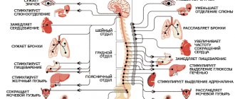
The most important of all efferent tracts of the basal ganglia originates from the internal part of the globus pallidus, ending in the thalamus, as well as in the roof of the midbrain. Through the stem formations with which the basal ganglia are connected, centrifugal impulses follow to the segmental motor apparatus and muscles along descending conductors.
From the red nuclei - along the rubrospinal tract;
From the Darkshevich nucleus - along the posterior longitudinal fasciculus to the nuclei of nerves 3, 4,6 and through it to the nucleus of the vestibular nerve;
From the nucleus of the vestibular nerve - along the vestibulospinal tract;
From the quadrigeminal region - along the tectospinal tract;
From the reticular formation - along the reticulospinal tract.
Thus, the basal ganglia play mainly the role of an intermediate link in the chain connecting the motor areas of the cortex with all its other areas.
In early phylogenesis, when the cerebral cortex was not yet developed, the striopallidal system was the main motor center that determined the behavior of the animal. Sensitive impulses flowing from the visual thalamus were processed here into motor ones, heading to the segmental apparatus and muscles. Due to the strio-pallidal apparatus, diffuse body movements of a rather complex nature were carried out: locomotion, swimming, etc.
At the same time, support for general muscle tone, “readiness” of the segmental apparatus for action, and redistribution of muscle tone during movements were ensured.
With the further evolution of the nervous system, the leading role in movements passes to the cerebral cortex with its motor analyzer and pyramidal system. Finally, a person experiences complex actions that are purposeful, voluntary in nature, with fine differentiation of individual movements.
Nevertheless, the striopallidal system has not lost its importance in humans. It only moves into a subordinate, subordinated position, ensuring the “tuning” of the motor apparatus, their “readiness for action” and the muscle tone necessary for the rapid implementation of movement.
Formation of the function of the basal ganglia in ontogenesis
. The basal ganglia develop more intensively than the visual thalamus. The nucleus pallidus myelinates earlier than the striatum and cerebral cortex. It has been established that myelination in the globus pallidus is almost completely completed by 8 months of fetal development. In the striatum, myelination begins in the fetus and ends only by 2 months of life. The caudate body doubles in size during the first 2 years of life, which is associated with the development of automatic motor acts in the child.
The motor activity of a newborn is largely associated with the pallidum, impulses from which cause uncoordinated movements of the head, torso and limbs.
In a newborn, the pallidum already has connections with the optic thalamus, subtuberculous region and substantia nigra. The connection between the pallidum and the striatus develops later; some of the striopallidal fibers become myelinated in the first month of life, and the other part only by 6 months and later.
It is believed that acts such as crying are motorically accomplished by the pallidum alone. The development of the striatum is associated with the appearance of facial movements, and then the ability to sit and stand. Since the striatum has an inhibitory effect on the pallidum, a gradual separation of movements is created. In order to sit, the child must be able to hold his head and back upright. This appears to him by the age of two months. Starts sitting at 6-8 months.
In the first months of life, the child has a negative support reaction: when trying to put him on his legs, he lifts them and pulls them towards his stomach. Then this reaction becomes positive: when you touch the support, the legs unbend. At 9 months the child can stand with support; at 10 months he can stand freely.
From 4-5 months of age, voluntary movements develop quite quickly, but for a long time they are accompanied by a variety of additional movements.
The appearance of voluntary (such as grasping) and expressive movements (smiling, laughter) is associated with the development of the striatal system and motor centers of the cerebral cortex. A child begins to laugh loudly at 8 months.
As all parts of the brain and the cerebral cortex grow and develop, the child’s movements become less generalized and more coordinated. Only by the end of the preschool period is a certain balance of cortical and subcortical motor mechanisms established.
The basal ganglia (basal nuclei)
is a striopallidal system consisting of three pairs of large nuclei immersed in the white matter of the telencephalon at the base of the cerebral hemispheres, and connecting the sensory and associative areas of the cortex with the motor cortex.
Structure
The phylogenetically ancient part of the basal ganglia is the globus pallidus, the later formation is the striatum, and the youngest part is the cervix.
The globus pallidus consists of outer and inner segments; striatum - from the caudate nucleus and putamen. The fence is located between the putamen and the insular cortex. Functionally, the basal ganglia also include the subthalamic nuclei and substantia nigra.
Functional connections of the basal ganglia
Exciting afferent impulses enter predominantly the striatum (caudate nucleus) mainly from three sources:
1) from all areas of the cortex directly and indirectly through the thalamus;
2) from nonspecific nuclei of the thalamus;
3) from the substantia nigra.
Among the efferent connections of the basal ganglia, three main outputs can be noted:
- from the striatum, inhibitory pathways go to the globus pallidus directly and with the participation of the subthalamic nucleus; from the globus pallidus the most important efferent path of the basal ganglia begins, going mainly to the ventral motor nuclei of the thalamus, from them the excitatory path goes to the motor cortex;
- part of the efferent fibers from the globus pallidus and striatum goes to the centers of the brain stem (reticular formation, red nucleus and then to the spinal cord), as well as through the inferior olive to the cerebellum;
- from the striatum, inhibitory pathways go to the substantia nigra and, after switching, to the nuclei of the thalamus.
Therefore, the basal ganglia are an intermediate link. They connect the associative and, in part, sensory cortex with the motor cortex. Therefore, in the structure of the basal ganglia there are several parallel functioning functional loops that connect them with the cerebral cortex.
Fig.1. Diagram of functional loops passing through the basal ganglia:
1 – skeletal-motor loop; 2 – oculomotor loop; 3 – complex loop; DC – motor cortex; PMC – premotor cortex; SSC – somatosensory cortex; PFC – prefrontal association cortex; P8 – field of the eighth frontal cortex; P7 – field of the seventh parietal cortex; FAC – frontal association cortex; VLN – ventrolateral nucleus; MDN – mediodorsal nucleus; PVN – anterior ventral nucleus; BS – globus pallidus; SN – black substance.
The skeletal-motor loop connects the premotor, motor, and somatosensory cortices to the putamen. The impulse from it goes to the globus pallidus and substantia nigra and then through the motor ventrolateral nucleus returns to the premotor area of the cortex. It is believed that this loop serves to regulate such movement parameters as amplitude, strength, direction.
The oculomotor loop connects the areas of the cortex that control gaze direction with the caudate nucleus. From there, the impulse goes to the globus pallidus and substantia nigra, from which it is projected, respectively, into the associative mediodorsal and anterior relay ventral nuclei of the thalamus, and from them returns to the frontal oculomotor field 8. This loop is involved in the regulation of saccadic eye movements (saccal).
It is also assumed that there are complex loops through which impulses from the frontal association zones of the cortex enter the caudate nucleus, globus pallidus and substantia nigra. Then, through the mediodorsal and ventral anterior nuclei of the thalamus, it returns to the associative frontal cortex. It is believed that these loops are involved in the implementation of higher psychophysiological functions of the brain: control of motivation, forecasting, cognitive activity.
Functions
Functions of the striatum
Influence of the striatum on the globus pallidus. The influence is carried out primarily by the inhibitory neurotransmitter GABA. However, some neurons of the globus pallidus give mixed responses, and some only EPSPs. That is, the striatum has a dual effect on the globus pallidus: inhibitory and excitatory, with a predominance of inhibitory action.
Influence of the striatum on the substantia nigra. There are bilateral connections between the substantia nigra and the striatum. Neurons of the striatum have an inhibitory effect on neurons of the substantia nigra. In turn, neurons of the substantia nigra have a modulating effect on the background activity of neurons in the striatum. In addition to influencing the striatum, the substantia nigra has an inhibitory effect on the neurons of the thalamus.
Influence of the striatum on the thalamus. Irritation of the striatum causes the appearance of high-amplitude rhythms in the thalamus, characteristic of the slow-wave sleep phase. Destruction of the striatum disrupts the sleep-wake cycle by reducing sleep duration.
Influence of the striatum on the motor cortex. The caudate nucleus of the striatum “inhibits” degrees of freedom of movement that are unnecessary under given conditions, thereby ensuring the formation of a clear motor-defensive reaction.
Stimulation of the striatum. Stimulation of the striatum in its various parts causes different reactions: turning the head and torso in the direction opposite to the stimulation; delay in food-producing activity; suppression of the sensation of pain.
Damage to the striatum. Damage to the caudate nucleus of the striatum leads to hyperkinesis (excessive movements) - chorea and athetosis. Functions of the globus pallidus
From the striatum, the globus pallidus receives predominantly inhibitory and partially excitatory influence. But it has a modulating effect on the motor cortex, cerebellum, red nucleus and reticular formation. The globus pallidus has an activating effect on the center of hunger and satiety. Destruction of the globus pallidus leads to adynamia, drowsiness, and emotional dullness.
Results of the activity of all basal ganglia:
- development, together with the cerebellum, of complex motor acts;
- control of movement parameters (force, amplitude, speed and direction);
- regulation of the sleep-wake cycle;
- participation in the mechanism of formation of conditioned reflexes, complex forms of perception (for example, comprehension of a text);
- participation in the act of inhibiting aggressive reactions.
Movement and thinking are the qualities that allow a person to live and develop fully.
Even minor disturbances in brain structures can lead to significant changes or complete loss of these abilities.
Responsible for these essential life processes are groups of nerve cells in the brain called the basal ganglia.
Basal ganglia of the brain
The gray matter on the surface of the brain forms the cortex. In addition, it is contained in the form of small accumulations in the thickness of the white matter, in the subcortical structures. In them it is represented by paired units called the basal ganglia or ganglia.
The basal ganglia of the brain are connected to the white matter and the cerebral cortex of the head. They are responsible for motor activity, the functioning of the ANS and the integration of processes of higher nervous activity.
With the development of pathology of these structures, their functionality suffers.
This is primarily reflected in muscle tone: the position of a person’s body changes when at rest or walking, the posture becomes unnatural, movements are chaotic and excessive.
What are the basal ganglia?
Gray matter in the form of separate clusters is located in the thickness of the base of the anterior part of the brain. There it forms the basal nuclei: paired structures, the parts of which are symmetrical with each other. Physiologically, they are connected with the white matter of the brain and the mediobasal cortex.
The basal ganglia coordinate the transmission of impulses from one hemisphere to the other, thereby facilitating the coordinated functioning of the organ. Communication with other parts of the brain is carried out using long processes - axons.
The basal ganglia of the brain include:
- Amygdala. Located in the thickness of the temporal lobes of the cerebral hemispheres. Belongs to the structures of the limbic system of the brain, which is responsible for the production of the mood hormone - dopamine. Thus, the amygdala provides control over the emotional component of a person’s state.
- Striped body. It is formed by the caudate and lenticular nuclei of the brain. In cross-section, this structure appears as alternating stripes of white and gray matter, which is why it got its name. With its help, muscle tone is regulated in the direction of weakening; the work of internal organs is controlled; behavioral reactions are realized and conditioned reflexes are formed.
- Fence. It is a thin layer of gray matter that is adjacent to the inner layer of the new cortex (neocortex) in the center of the brain. Also refers to the limbic system. Some scientists believe that the fence is involved in the formation of sexual feelings.
The subcortical nuclei of the brain are functionally combined into two systems. The first group represents its striopallidal part. These include the caudate nucleus, putamen and globus pallidus. And the second - extrapyramidal - in addition to the remaining basal ganglia, includes the medulla oblongata, cerebellum, substantia nigra and structures of the vestibular apparatus.
Functions of the basal ganglia
The main purpose of the basal ganglia is to maintain the performance of the body and the functioning of life support systems. Like any other nerve center of the brain, they carry out their activities through connections with neighboring structures.
For example, the striopallidal system has many contacts with the cortical regions and the brain stem. Their coordinated work is ensured by efferent and afferent pathways.
Among the main functions of the basal ganglia are:
- Control of the motor system: maintaining posture in space, ensuring standard actions, regulating muscle tone when performing conscious movements and reflex reactions, controlling fine motor skills;
- Vocabulary, speech patterns;
- Regulation of sleep-wake processes;
- Control over the autonomic nervous system: breathing, cardiac activity, maintaining optimal body temperature, metabolism, regulating the tone of the walls of blood vessels during changes in blood pressure;
- The production of specific active chemicals, with the help of which impulses are transmitted from one nerve cell to another.
The basal ganglia are also involved in the formation of behavioral reactions, conditioned and unconditioned reflexes.
Pathological states of nuclei
Pathologies of the basal ganglia are expressed by a number of diseases, since the vital activity of the body depends on their functioning. The degree of their manifestations may vary.
- Functional deficiency. The first signs of pathology appear at an early age. It is usually a consequence of genetic abnormalities and is inherited. In adults, the deviation can lead to the development of Parkinson's disease or paralysis.
- Neoplasms and cysts. Like any other structure of the brain, cells of the basal ganglia are capable of mutating into atypical ones and forming tumor-like neoplasms. Their localization may vary. The impetus for the development of a tumor is a metabolic disorder in cells, atrophy and necrosis of brain tissue. The appearance of neoplasms can occur both in utero and after the birth of a child, as he grows up. For example, some experts associate cerebral palsy with damage to the basal ganglia in the second half of pregnancy. Some types of pathologies can be triggered by a difficult course of labor, head injuries, and infectious diseases in the first year of a child’s life. An obvious manifestation of damage to the basal ganglia of the brain are neurological abnormalities in which excessive irritation (excitation) of formations occurs: hyperactivity, attention deficit disorder. There are also asymptomatic small cysts that may disappear over time.
- Calcification of the basal ganglia. A striking example of pathology is Idiopathic calcification of the basal ganglia or Fahr syndrome. It is characterized by the appearance of calcium accumulations (calcification) on the surface of the ganglia. The causes of the pathology are unknown, but there is an opinion that it can develop as a result of a chromosomal malfunction. The patient experiences degradation of motor functions, dementia, convulsions, headache, fatigue, dysarthria, and muscle spasms. Signs of parkinsonism may also appear - tremors, muscle rigidity, shuffling gait, “rolling” movements of the fingers. In the final stages, mental disorders develop.
- Corticobasal degeneration. Refers to progressive pathologies of the central nervous system. When it occurs, self-destruction of ganglion cells occurs due to disruption of the metabolic processes of the brain. The manifestation of pathology depends on the functioning (to one degree or another) of the part of the brain to which the affected area belongs. For example, the first symptom is often a feeling of numbness or awkwardness in a limb, a disorder of its sensitivity. Then other symptoms appear: various forms of muscular dystonia, myoclonus, postural tremor, etc.
Treatment of pathologies of the basal ganglia should be comprehensive. A psychotherapist, speech therapist and some other specialists, depending on the manifestations of the disease, must take part in this.
Diagnosis and prognosis of pathology
Identification of pathologies of the basal ganglia begins in the office of a neurologist. If other deviations are present, then the help of functional diagnostic specialists may be needed.
The final diagnosis is made based on the following studies:
- History;
- General neurological and physical examination;
- MRI or CT;
- Examination of the blood supply to the brain;
- Ultrasound;
- Electroencephalography.
The prognosis of the pathology depends on many external factors: age, gender, general condition of the patient, the degree of the disease, the time of its detection and the effectiveness of the proposed treatment. However, according to statistics, in 50% of cases it is unfavorable.
After therapy and rehabilitation, the remaining patients still have a chance to adapt and lead a normal life in society.
Consequences of basal ganglia pathologies
Manifestations of pathology, even with successful treatment, will accompany the patient throughout his life and can cause disability. The development of the disease is most often corrected by taking medications, physiotherapeutic procedures, physical exercise, and strengthening the nervous system.
As you know, the adaptive forces of the body are great. But at the same time, the sick person and his relatives need to be patient and follow all the specialists’ instructions: the effectiveness of rehabilitation measures and future adaptation in society depend on this.
Basal ganglia of the brain Link to main publication
Source: //GolovaiMozg.ru/stroenie/bazalnye-yadra-golovnogo-mozga
What you need to know about the basal ganglia
The large hemispheres of the human brain on the outside are a cortex formed by gray matter, and on the inside by a subcortex of white matter. The basal ganglia (ganglia, nodes), which are also called central or subcortical, are concentrations of gray matter in the white matter of the subcortex.
The basal ganglia are located at the base of the brain, which explains their name, outside the thalamus (optic thalamus). These are paired formations that are symmetrically represented in both hemispheres of the brain. With the help of nerve processes, they interact bilaterally with various areas of the central nervous system.
The main role of the subcortical nodes is to organize motor function and various aspects of higher nervous activity. Pathologies that arise in their structure affect the functioning of other parts of the central nervous system, causing problems with speech, coordination of movements, memory, and reflexes.
Brain nuclei and their functions
One of the most inexplicable things in the universe is the brain. Almost nothing is known about it with regard to its operating principles. From a physiological point of view, this organ has been well studied, but most people have more than a superficial understanding of its structure.
The majority of educated people know that the brain is two hemispheres, covered with a cortex and convolutions; it consists of several sections and somewhere there is gray and white matter. We will talk about all this in special topics, and today we will look at what the basal ganglia of the brain are, which few have heard of and know about.
Structure and location
The basal ganglia of the brain is a collection of gray matter in the white matter, located at the base of the brain and part of its anterior lobe. As we can see, the gray matter not only forms the hemispheres, but is also located in the form of separate clusters called ganglia. They have a close connection with the white matter and cortex of both hemispheres.
The structure of this region is based on a slice of the brain. It includes:
- amygdala;
- striatum (composed of the caudate nucleus, globus pallidus, putamen);
- fence;
- lenticular nucleus.
Between the lenticular nucleus and the thalamus there is a white substance called the internal capsule, and between the insula and the fence is the external capsule. Recently, a slightly different structure of the subcortical nuclei of the brain has been proposed:
- striatum;
- several nuclei of the midbrain and diencephalon (subthalamic, pedunculopontine and substantia nigra).
Together they are responsible for motor activity, motor coordination and motivation in human behavior. This is all that can be said for sure about the function of the subcortical nuclei. Otherwise, they, like the brain as a whole, are poorly understood. Absolutely nothing is known about the purpose of the fence.
Physiology
All subcortical nuclei are again conventionally combined into two systems. The first is called the striopallid system, which includes:
- pale globe;
- caudate nucleus of the brain;
- shell.
The last two structures consist of many layers, which is why they are grouped under the name striatum. Ballus pallidus is a brighter, lighter color and is not laminated.
The lenticular nucleus is formed by the globus pallidus (located inside) and the shell, which forms its outer layer. The amygdala and the amygdala are components of the limbic system of the brain.
Let's take a closer look at what these brain nuclei are.
Black substance
The component of the system that is most involved in the coordination of movements and motor skills, maintaining muscle tone and controlling postures. Participates in many autonomic functions, such as breathing, cardiac activity, and maintaining vascular tone.
Physically, the substance is a continuous strip, as was believed for decades, but anatomical sections have shown that it consists of two parts. One of them is a receiver that sends dopamine to the striatum, the second - a transmitter - serves as a transport artery for transmitting signals from the basal ganglia to other parts of the brain, of which there are more than a dozen.
Lenticular body
Its location is between the caudate nucleus and the thalamus, which, as stated, are separated by the external capsule. In front of the structure, it merges with the head of the caudate nucleus, which is why its frontal section has a wedge-shaped shape.
This nucleus consists of sections separated by a thin film of white matter:
- shell – darker outer part;
- pale ball.
The latter is very different in structure from the shell and consists of type I Golgi cells, which predominate in the human nervous system, and are larger in size than their type II. According to neurophysiologists, it is a more archaic brain structure than other components of the brain nucleus.
Other nodes
The fence is the thinnest layer of gray matter between the shell and the island, around which there is a white substance.
The basal ganglia are also represented by the amygdala, located under the shell in the temporal region of the head. It is believed, but not known for sure, that this part belongs to the olfactory system. It is also where the nerve fibers coming from the olfactory lobe end.
Features of the structure of the basal ganglia
The basal ganglia are located in the frontal and partially temporal lobes of the telencephalon. These are clusters of neuron bodies that form groups of gray matter. The white matter surrounding them is represented by processes of nerve cells and forms layers that separate the individual basal ganglia and other brain structural and functional elements.
The basal nodes include:
- striatum;
- fence;
- amygdala.
In anatomical sections, the striatum appears as alternating layers of gray and white matter. It consists of caudate and lenticular nuclei. The first is located anterior to the visual thalamus. As the caudate nucleus becomes thinner, it becomes the amygdala. The lenticular nucleus is located lateral to the thalamus optic and the caudate nucleus. It is connected to them by thin bridges of neurons.
The fence is a narrow strip of neurons. It is located between the lenticular nucleus and the insular cortex. It is separated from these structures by thin layers of white matter. The amygdala is shaped like the amygdala and is located in the temporal lobes of the telencephalon. It consists of several independent elements.
This classification is based on the structural features and location of the ganglia on an anatomical section of the brain. There is also a functional classification, according to which scientists classify only the striatum and some ganglia of the diencephalon and mesencephalon as the basal ganglia. These structures collectively provide human motor functions and individual aspects of behavior responsible for motivation.
Functions and significance of the basal (subcortical) nuclei
One of the most complex organs in the human body is the brain. This organ coordinates all processes in the body, provides vital functions, and regulates metabolic processes.
However, most readers have a rather superficial understanding of the structures of the brain. In addition to the hemispheres, cerebellum, cortex and medulla oblongata, it has many sections and structures.
One of these important formations is the basal ganglia or basal ganglia.
Gray matter forms the cerebral cortex; in addition, it is located in the form of separate ganglia in the subcortical structures, in the white matter. These formations in the white matter are paired and form the subcortical nuclei.
The basal ganglia are directly connected to the white matter and cerebral cortex. It is the subcortical nuclei that are responsible for human motor activity and coordinate its activities .
When a pathological process occurs, the function of the basal ganglia is significantly impaired.
This affects muscle tone, body position at rest and dynamics, the posture becomes forced, movements are chaotic and excessive.
What is the structure of the basal ganglia?
The basal ganglia include the striatum, which is divided into the lentiform and caudate nuclei, the amygdala, and the cervical nucleus. This classification is based on the anatomical structure and location of these structures on sections of the cerebral hemispheres.
In recent years, the term “basal ganglia” has come to mean the subthalamic nucleus, substantia nigra and pedunculopontine tegmental nucleus.
The name "striatum" arose from the alternating areas of white and gray matter in horizontal sections. The lenticular and caudate nuclei are connected to each other by thin bridges of gray matter.
The caudate nucleus is located slightly higher and more in the middle of the lentiform nucleus, they are separated by a capsule formed by neurons of the brain or white matter.
The anterior part of the caudate nucleus is slightly thickened; it and its tail form the lateral or outer wall of the anterior horn of the lateral ventricle of the brain. The posterior part of the caudate ganglion is thinned and adjacent to the bottom of the lateral ventricle and is located approximately in its middle.
The surface of the caudate nucleus, facing the middle, borders the thalamus. They are separated by a narrow strip of white matter in the brain.
Detailed structure
The caudate nucleus in its structure is formed by Golgi neurons of the second type. Their structure has a number of similar characteristics: they have a thin axon and significantly shortened dendrites. These cells are small. The main functions of the caudate nucleus are determined by their connections with other structures of the brain .
The caudate nucleus receives descending commands from the extrapyramidal system. This structure has a widespread network of neurons that interact with sensory areas through the globus pallidus and thalamus, forming closed pathways.
However, the caudate nucleus actively interacts with other structures of the brain, for example, with the substantia nigra, cerebellum, vestibular ganglia and other structures. Such diverse and stable connections make it possible to say that the basal ganglia of the hemispheres are multifunctional.
It participates in the vegetative functions of the whole organism, plays an important role in integrative and cognitive abilities, coordinates and stimulates human motor activity.
The lentiform body is located outward from the caudate nucleus and thalamus, they are separated by the external capsule. The middle surface of the lenticular ganglion has the shape of an angle, with its rounded part facing the median capsule.
It is located on parallel planes with the caudate nucleus and thalamus. The surface located inside has a hemispherical shape and is adjacent to the outside of the cerebral hemispheres. Anteriorly, the lentiform nucleus and the head of the caudate nucleus merge.
In cross sections, the shape of the lenticular nucleus is similar to a wedge, the wide part of which is directed outward.
The lenticular nucleus is divided by thin strips of white matter into 3 main structures: the darker part is the putamen, the lighter areas form a structure called the globus pallidus.
In its histological structure, the globus pallidus differs significantly from the shell and is presented in the form of Golgi cells of the first type; there are much more of them in the human body, and they are much larger than cells of the second type.
The globus pallidus is considered to be one of the most ancient formations of the higher nervous system; it developed much earlier than the putamen and caudate nucleus, constantly underwent changes and improved, but did not lose its importance as the basal ganglia.
At the present stage of development of neurology and neurosurgery, it is generally accepted that the lenticular nucleus is only a topographical landmark. While the structure at the junction of the body of the caudate and lentiform nuclei is organized into a striopallidal system.
The striopallidal system is the basis of the extrapyramidal system, and is also the main center for regulating the autonomic functions of the body in the field of thermoregulation and carbohydrate metabolism, significantly exceeding the hypothalamus in its importance.
Additional structures included in the basal ganglia
The fence is a thin layer of gray matter located between the island and the shell, and surrounded on all sides by white matter, which in turn forms 2 capsules: between the fence and the shell is the outer capsule, and the outer capsule separates them from the island.
The basal ganglia of the telencephalon are also represented by the amygdala. This accumulation of gray matter is located in the temporal lobe under the putamen. It is believed that it belongs, like part of the temporal lobe, to the olfactory centers and limbic system of the brain. The amygdala terminates in nerve fibers coming from the anterior perforated substance and the olfactory lobe.
The limbic system, or, as it is sometimes called, the visceral brain, is a very complex structure in its organization, which includes sections of the telencephalon, midbrain and medulla oblongata.
Its functions are as multifaceted as its structure; it is responsible for vegetative processes in the body, cognitive activity, brightly colored emotional reactions and active psychological processes, and also maintains constant homeostasis of the body.
What are the subcortical nuclei responsible for in the body?
Despite the fact that these structures are negligible compared to the body as a whole, their functions can hardly be overestimated! The main functions of the basal ganglia are to ensure and maintain active, adequate motor skills and human movements . Their coordinated functioning is the key to normal human well-being and full-fledged nervous activity.
The basal ganglia of the brain form two systems:
- Striopallidal (part of the extrapyramidal);
- Limbic.
The striopalidal system is responsible for the coordination of movements, correct and timely contraction of muscle fibers. When pathology occurs in this part of the nervous system, the first symptoms appear when a person moves and walks in the form of weakened muscle strength or discoordinated movements.
When their structures are damaged, the entire nervous system suffers, the most noticeable disorders being the hypothalamus and pituitary gland.
Types of pathology
When the subcortical nuclei are damaged, the symptoms appear gradually, but the patient’s general well-being suffers, he is weakened, disorganized, loses self-confidence, and often becomes despondent and depressed.
There are 2 main types of pathology:
- Cysts and neoplasms of the subcortical ganglia - these lesions can occur as a result of degeneration of nerve cells, infectious agents, trauma, ischemic damage and hemorrhage. This pathology is well diagnosed with CT and MRI studies and requires timely and adequate treatment, otherwise the patient faces disability or death.
- Functional failure of the basal ganglia is more often observed in children and is caused by underdevelopment of the nervous system as a whole. The main theory of development is genetic. In adults, it usually occurs as a result of injuries and strokes. Patients also need treatment and observation by a neurologist. In old age, it is this pathology that leads to the development of parkinsonism in 57% of cases.
Signs of damage to the basal ganglia
The main symptoms indicating disorders in the subcortical ganglia include:
- tremor;
- spontaneous movements of the limbs;
- muscle weakness or spasms;
- involuntary repetitive movements;
- impairment of memory and comprehension of what is happening.
Symptoms appear gradually. They can grow rapidly or, conversely, very slowly. However, even their one-time appearance, for example, twitching, cannot be ignored.
Diagnostics
The main methods for making a diagnosis are an examination by a neurologist, based on the results of which clinical tests and studies are prescribed. The most informative is magnetic resonance imaging of the brain, in which pronounced foci of dysfunction are clearly visible.
Forecast
The prognosis for the patient depends on the degree of damage to the basal ganglia, the timeliness of seeking medical help and the adequacy of treatment. As a rule, such patients receive lifelong maintenance therapy.
Rate this article:
Total: 198
4.48 198
Source: //mozgius.ru/stroenie/bazalnye-yadra.html
Anatomy and physiology of the basal ganglia
Although all basal ganglia are collections of gray matter, they have their own complex structural features. To understand what role this or that basal center plays in the functioning of the body, it is necessary to take a closer look at its structure and location.
Caudate nucleus
This subcortical node is located in the frontal lobes of the brain hemispheres. It is divided into several sections: a thickened large head, a tapering body and a thin long tail. The caudate nucleus is highly elongated and curved. The ganglion consists mostly of microneurons (up to 20 microns) with short thin processes. About 5% of the total cell mass of the subcortical ganglion consists of larger nerve cells (up to 50 microns) with highly branching dendrites.
This ganglion interacts with areas of the cortex, thalamus and nodes of the diencephalon and midbrain. It acts as a link between these brain structures, constantly transmitting neural impulses from the cerebral cortex to its other parts and back. It is multifunctional, but its role is especially significant in maintaining the activity of the nervous system, which regulates the activity of internal organs.
Lenticular nucleus
This basal node is shaped like a lentil seed. It is also located in the frontal regions of the cerebral hemispheres. When the brain is cut in the frontal plane, this structure is a triangle, the apex of which is directed inward. White matter divides this ganglion into a putamen and two layers of the globus pallidus. The shell is dark and located externally in relation to the light layers of the globus pallidus. The neuronal composition of the putamen is similar to the caudate nucleus, but the globus pallidus is represented mainly by large cells with small inclusions of microneurons.
Evolutionarily, the globus pallidus is recognized as the most ancient formation among other basal ganglia. The putamen, globus pallidus and caudate nucleus constitute the striopallidal system, which is part of the extrapyramidal system. The main function of this system is the regulation of voluntary movements. Anatomically, it is connected with many cortical fields of the cerebral hemispheres.
Fence
The slightly curved, thinned plate of gray matter that separates the putamen and the insula of the telencephalon is called the fence. The white matter around it forms two capsules: the outer and the “outermost”. These capsules separate the fence from neighboring gray matter structures. The fence is adjacent to the inner layer of the neocortex.
The thickness of the fence varies from fractions of a millimeter to several millimeters. Along its entire length it consists of neurons of various shapes. The fence is connected via neural pathways to the centers of the cerebral cortex, hippocampus, amygdala and partially striatal bodies. Some scientists consider the fence to be a continuation of the cerebral cortex or include it as part of the limbic system.
Amygdala
This ganglion is a group of gray matter cells concentrated under the shell. The amygdala consists of several formations: the nuclei of the cortex, the median and central nuclei, the basolateral complex, and interstitial cells. It is connected by nerve transmission to the hypothalamus, thalamus, sensory organs, cranial nerve nuclei, olfactory center and many other formations. Sometimes the amygdala is classified as part of the limbic system, which is responsible for the activity of internal organs, emotions, smell, sleep and wakefulness, learning, etc.
Functions of the basal ganglia
The main purpose of the basal ganglia is to maintain the performance of the body and the functioning of life support systems. Like any other nerve center of the brain, they carry out their activities through connections with neighboring structures.
For example, the striopallidal system has many contacts with the cortical regions and the brain stem. Their coordinated work is ensured by efferent and afferent pathways.
Among the main functions of the basal ganglia are:
- Control of the motor system: maintaining posture in space, ensuring standard actions, regulating muscle tone when performing conscious movements and reflex reactions, controlling fine motor skills;
- Vocabulary, speech patterns;
- Regulation of sleep-wake processes;
- Control over the autonomic nervous system: breathing, cardiac activity, maintaining optimal body temperature, metabolism, regulating the tone of the walls of blood vessels during changes in blood pressure;
- The production of specific active chemicals, with the help of which impulses are transmitted from one nerve cell to another.
The basal ganglia are also involved in the formation of behavioral reactions, conditioned and unconditioned reflexes.
The importance of the subcortical nodes for the body
The functions of the basal ganglia are determined by their interaction with other areas of the central nervous system. They form neural loops connecting the thalamus and the most important areas of the cerebral cortex: motor, somatosensory and frontal. In addition, the subcortical nodes are connected to each other and to some areas of the brain stem.
The caudate nucleus and putamen perform the following functions:
- control of direction, strength and amplitude of movements;
- analytical activity, learning, thinking, memory, communication;
- control of eye, mouth, and face movements;
- maintaining the functioning of internal organs;
- conditioned reflex activity;
- perception of sensory signals;
- control of muscle tone.
Specific functions of the shell include respiratory movements, saliva production and other aspects of feeding behavior, ensuring trophism of the skin and internal organs.
Functions of the globus pallidus:
- development of an orienting reaction;
- control of arm and leg movements;
- eating behavior;
- facial expressions;
- display of emotions;
- providing auxiliary movements and coordination abilities.
The functions of the fence and amygdala include:
- speech;
- eating behavior;
- emotional and long-term memory;
- development of behavioral reactions (fear, aggression, anxiety, etc.);
- ensuring social integration.
Thus, the size and condition of individual basal ganglia affects emotional behavior, voluntary and involuntary movements of a person, as well as higher nervous activity.