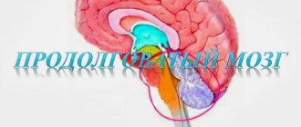
Location
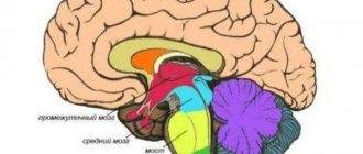
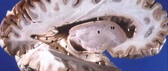
The thalamus is part of the cerebral hemispheres of the forebrain. It is located lateral to the lateral ventricles - cavities of the brain that are part of the cerebrospinal fluid (CSF) circulation system. Below it is the hypothalamus, from which the visual hillocks are separated by a groove.
Above and slightly outside the thalamus are the basal ganglia. These formations are necessary for precise, coordinated movements. These structures are separated from each other by the internal capsule - a bundle of white matter of the forebrain, through which pathways pass from the periphery to the center.
The right and left parts of the thalamus are connected to each other by interthalamic gray matter. It is present in 70% of people.
Classification of thalamic nuclei
In total, there are about 120 nuclei in the visual thalamus of the brain. Depending on their location, they are divided into three groups:
- medial;
- lateral;
- front
In the lateral group of nuclei, the medial and lateral geniculate bodies, as well as the cushion, are distinguished.
There is also a classification depending on the function performed by the kernels:
- specific;
- associative;
- nonspecific.
Comparative anatomy
Comparative anatomical and embryological studies have revealed a very complex nature of the development of the Thalamus and its individual complexes, determined both by the general patterns of evolution of the chordate brain and by environmental factors in the formation of sensory systems in various representatives of this type.
The formations of the ventral T., especially the most ancient ventral part of the lateral geniculate body, are differentiated already in selachians, that is, much earlier than the structures of the dorsal T., the division of which into separate formations is revealed only in reptiles. In birds and reptiles, the most developed structure of the dorsal thorax is the round nucleus, which has no direct homologs in the dorsal thorax of mammals. The structures of the dorsal T. are most difficult to differentiate in mammals. The most ancient formations of the dorsal T.—the posterior and pretectal nuclei, well developed in lower mammals, are reduced in higher ones. A similar process occurs with the intralaminar (intralamellar, T.) nuclei, as well as with the ventral part of the nucleus of the lateral geniculate body, which belongs to the ventral T.
The increasing complexity of the differentiation of dorsal T. structures is associated with an even greater complication of the structure of the new cortex (neocortex) of the mammalian brain, where the patterns of divergent (multidirectional) development are manifested in the relative progression of the newest and relative regression of the phylogenetically ancient zones of the cerebral cortex. This process is explained by the fact that with the evolutionary complexity of the brain in cortical-subcortical relationships, cortical-thalamic connections become decisive, and in them, in turn, associative connections become predominant (compared to projection, relay connections). Thus, in monkeys, projection zones occupy an even larger area of the cerebral cortex compared to associative zones; for example, only the occipital projection zone is approx. 20% of the area of the entire cortex. In humans, the relationships are reversed—associative zones are dominant, and the occipital projection zone makes up only 12% of the area of the neocortex. Therefore, in the thalamic complexes of the human brain, associative nuclei are the most developed.
Specific kernels
The specific nuclei of the thalamus have a number of distinctive features. All formations of this group receive sensory information from second neurons (nerve cells) of sensitive pathways. The second neuron, in turn, can be located in the spinal cord or in one of the structures of the brain stem: medulla oblongata, pons, midbrain.
Each of the signals coming from below is processed in the thalamus and then goes to the corresponding area of the cortex. Which area the nerve impulse goes to depends on what information it carries. Thus, information about sounds goes to the auditory cortex, about objects seen - to the visual cortex, and so on.
In addition to impulses from the second neurons of the pathways, specific nuclei are responsible for the perception of information coming from the cortex, reticular formation, and brain stem nuclei.
The nuclei, which are located in the anterior part of the thalamus, ensure the conduction of impulses from the limbic cortex of the brain through the hippocampus and hypothalamus. After processing the information, it again enters the limbic cortex. Thus, the nerve impulse circulates in a certain circle.
Physiology
The study of the structure and function of thalamic formations using modern complex methods has revealed the extremely complex nature of the relationship of T. both with the higher located parts of the forebrain and with the lower structures of the brain stem, cerebellum and spinal cord. The close integration of somatic, visceral, motivational influences, carried out at the level of thalamic complexes, determines the leading role of these formations of the diencephalon in the formation of not only cortico-subcortical relations, but also in the formation and development of integral behavioral acts of animals and humans.
The study of T.'s functions is carried out by its electrical stimulation or destruction in combination with analysis of the dynamics of various behavioral reactions, registration of EEG, evoked potentials or electrical activity of single neurons in response to various peripheral stimuli in experiments on animals and in treatment. purposes - in humans.
It has been established that in the relay nuclei of T. impulses of tactile, kinesthetic, temperature, pain (localized pain), taste and visceral sensitivity are switched (see). Each nucleus receives impulses from the opposite side of the body; only the face area has bilateral representation in the posterior ventral nucleus of T. The parts of the body that perform the most intense sensory-motor functions (face, tongue, distal limbs) have a more extensive representation. The medial section of the posterior ventral nucleus, whose somatotopic organization has been well studied, receives afferentation from the rostral parts of the body and impulses from taste receptors, and the lateral section receives impulses from the receptors of the trunk and limbs. Projections of tactile and visceral sensitivity have a wide overlap in T., which can cause the phenomenon of irradiation of visceral pain (see) on the surface of the body. Tactile and deep (proprioceptive) sensitivity also have common projections in T. Finely differentiated afferent impulses from thermo- and nociceptors pass predominantly along the spinothalamic pathway to the posterior ventral nucleus, while the more coarse forms of these types of sensitivity (diffuse pain, differentiation of large temperature ranges) are associated primarily with the nonspecific intralaminar nuclei of T. (parafascicular complex), as well as , possibly with back nuclei. Electrical stimulation of the posterior ventral nucleus in humans during stereotactic operations (see Stereotactic neurosurgery, Stereotactic method) causes paresthesia and, less commonly, a violation of the body diagram (see Body diagram).
Non-sensory relay formations include the anterior group of nuclei, which transmits impulses from the mammillary bodies to the limbic region of the cortex, as well as two nuclei of the ventral group of T. - the anterior ventral and lateral ventral, which transmit the bulk of excitations from the cerebellum and globus pallidus to the motor parts of the cerebral cortex, which determines their role in the regulation of involuntary motor activity and muscle tone. These connections of the lateral ventral nucleus justify the expediency of its destruction in patients with parkinsonism and various hyperkinesis.
The group of associative nuclei of T. has as its main source of afferentation relay and partly nonspecific structures of T. and the hypothalamus. Recently, it has been shown that some sensory projections (somatosensory, auditory, visual, visceral) may end in these nuclei. The interaction of various afferent impulses on the neurons of the associative nuclei of T., their complex intrathalamic and interthalamic connections explain the important role of this group of formations in the integrative activity of the brain. There are thalamoparietal and thalamofrontal associative systems. The first is primarily related to the formation of complexes of afferent influences necessary for assessing the signal significance of complex types of stimuli underlying the gnostic function, especially visual and auditory perception, as well as sensory support for the control of voluntary movements. The medial dorsal nucleus belongs to the thalamofrontal associative system, which is primarily involved in affective-emotional and programming activities. Stimulation of the medial dorsal nucleus of T. in patients causes a variety of positive and negative emotional reactions; destruction of this nucleus in laying down. purposes, as well as in experiments on animals, it reduces feelings of fear, anxiety and tension, but at the same time mild symptoms of the so-called appear. frontal syndrome - a decrease in initiative and intellectual abilities against the background of a persistent decrease in emotional reactivity. With the spread of patol. process on the medial sections of T. observe the development of dementia (see).
A group of nonspecific nuclei of T. receives afferentation primarily from the reticular formations of the brain stem, but there is evidence of the possibility of fibers ending in them that run as part of specific ascending systems. Although the nuclei of this group are projected onto the cortex more diffusely than the first two groups, some of them are characterized by more or less local connections with the cortex and striatum (striopallidum). This group includes the intraplate (intralaminar) nuclei (parafascicular nucleus, lateral central and paracentral nuclei), the reticular nucleus of the thalamus, the midline nuclei of T. The discovery of non-specific nuclei of T. is associated with the work of Dempsey and Morison (EW Dempsey, RS Morison, 1943 ), who showed that when these nuclei are stimulated, a widespread entrainment reaction occurs in the cerebral cortex. Later, S. P. Narikashvili, E. S. Moniava, V. I. Guselnikov and A. Ya. Supin (1968) and others discovered that in the genesis of this reaction, as well as the amplification reaction, the leading role is played by the interaction of non-specific T. nuclei with its specific formations.
Data on the functional significance of the nuclei of this group are contradictory: on the one hand, it has been established that the destruction of the complex parafascicular nucleus - central median nucleus (median center) affects positive and negative learning in rats; on the other hand, the effect of damage to this complex largely depends on the previous emotional background; The important role of afferentation of this complex in the activity of neurons of the caudate nucleus is also shown (see Basal ganglia). The idea of the nonspecificity of this group of nuclei is relative. Based on data on their structure, connections and function, we can talk about the existence in T. of complex complexes formed by relay and nonspecific nuclei. The study of the functions of the midline nuclei is complicated by their close proximity to the associative and relay nuclei of the T. Destruction of the medial parts of the T. may be accompanied by disturbances of conditioned reflexes (see), memory (see), sleep (see).
In the group of nonspecific nuclei, a special place is occupied by the reticular nucleus of the T. The latter adjoins externally the various relay nuclei of the dorsal T. and partially shares a common projection with them on the cortex. It is assumed that this nucleus is an intermediary both in the activity of other T. nuclei and in the thalamo-cortical interaction of its specific and nonspecific formations. Nowadays, the important role of T. is obvious in the implementation of complex functions of perception and processing of various signals that go further to the cerebral cortex and basal ganglia, in maintaining the level of wakefulness and emotional state, in the normal provision of simple and more complex forms of behavior and memory. Complete bilateral removal of T. in dogs and cats reveals a sharp deficit in adaptive reactions, disturbances in sensitivity and alternations in the wakefulness-sleep cycle. Surgical isolation of the cortex and basal nuclei from the brain stem (the so-called thalamic animal) leads to massive retrograde degeneration of the neurons of the brain and is accompanied by sudden and irreversible changes in adaptive behavior, up to the impossibility of independent nutrition.
Recently, in connection with the accumulation of data on interhemispheric asymmetry, indications have appeared of the functional disparity of the right and left T. in humans. Thus, the use of verbalized material in short-term memory tests is apparently carried out with the participation of predominantly the left T., non-verbalized material - with the participation of the right T.; The most significant syndrome of left-sided lesions of the thalamus is lack of content of speech.
In the 70s a number of new principles of thalamocortical interaction were formulated that are relevant to understanding the integration of neural processes at the level of the forebrain: the principles of “nuclear” and “scattered” types of thalamocortical projections, the interlayering of these different projections among themselves, their different distribution in the projection and associative areas of the neocortex, mono- and oligosynaptic transmission of excitations from T. to the cortex, as well as topographic correspondence (mainly for relay and associative structures) and inconsistency (mainly for nonspecific T. structures) of thalamocortical and corticothalamic influences. In neurophysiol. Research has revealed the dynamic integration of specific and non-specific influences at the diencephalic and cortical levels; it has been established that the rhythmic activity of the cerebral cortex is determined by the activity of the thalamic nuclei, and that the formation of the a-rhythm in the cortex occurs with the participation of both thalamic and cortical structures. It was found that neurophysiol. the basis of the rhythmic activity of the brain are the mechanisms of recurrent inhibition (see) and excitation (see), playing out at the thalamic and cortical levels. New ways of relationships between associative and projection thalamocortical systems have been identified and studied, and a position on complexly functioning thalamocorticothalamic associative systems has been formulated. The problem of cortical regulation of the activity of thalamic nuclei has received new coverage: it has been shown that the regulation of relay thalamic nuclei is carried out by Ch. arr. direct corticothalamic pathways, and nonspecific nuclei - mainly with the participation of the reticular formation of the brain stem. It has been established that the reticular systems of the brain are able to differentiate afferent signals and direct them to the corresponding brain structures under the influence of corticofugal modulation.
Associative kernels
The association nuclei are located closer to the posteromedial part of the thalamus, as well as in the cushion area. The peculiarity of these structures is that they do not participate in the perception of information that comes from the underlying formations of the central nervous system. These nuclei are necessary to receive already processed signals in other nuclei of the thalamus or in overlying brain structures.
The essence of the “associativity” of these nuclei is that any signals are suitable for them, and the neurons are able to adequately perceive them. Signals from these structures enter the correspondingly named areas of the cortex—associative zones. They are located in the temporal, frontal and parietal parts of the cortex. Thanks to the receipt of these signals, a person is able to:
- recognize objects;
- connect speech with movements and objects seen;
- be aware of the position of your body in space;
- perceive space as three-dimensional and so on.
Neurons
They exhibit high sensitivity to pharmacological agents and hormones. Drugs such as Reserpine, Aminazine, Serpazil and others can reduce the activity of the formation. Upstream and downstream signals interact in neurons. The pulses are in constant circulation in the circuits. Due to this, activity is maintained. It, in turn, is necessary to maintain the tone of the nervous system. In the event of destruction of the formation, especially its upper sections, deep sleep occurs, although afferent signals continue to enter the cortex through other pathways.
Nonspecific nuclei
This group of nuclei is called nonspecific because it receives information from almost all structures of the central nervous system:
- reticular formation;
- nuclei of the extrapyramidal system;
- other nuclei of the optic thalamus;
- brain stem structures;
- formations of the limbic system.
The impulse from nonspecific nuclei also goes to all areas of the cerebral cortex. Such selectivity, as in the case of associative and specific nuclei, is absent here.
Since it is this group of nuclei that has the largest number of connections, it is believed that thanks to it, the harmonious, coordinated work of all parts of the brain is ensured.
Separately about the visual thalamus
Previously, it was believed that the thalamus processed only visual impulses, and then the organ received the name - visual thalamus.
Now this name is considered obsolete, since the organ processes almost the entire range of afferent systems (except for smell). The system that provides visual perception is one of the most interesting. The main external organ of vision is the eye, a receptor that has a retina and is equipped with special cells (cones, rods) that transform the light beam and electrical signal. The electrical signal, in turn, passing through the nerve cells enters the lateral center of the thalamus, which sends the processed signal to the central part of the cerebral cortex. Here the final analysis of the signal occurs, thanks to which what is seen is formed, that is, the picture.
Metathalamus
A separate group of nuclei of the visual thalamus is distinguished called the metathalamus. This structure consists of the medial and lateral geniculate bodies.
The medial geniculate body receives hearing information. From the underlying parts of the brain, information arrives through the upper humps of the midbrain, and from above the structure receives impulses from the auditory cortex.
The lateral geniculate body belongs to the visual system. Sensitive information to the nuclei of this group comes from the retina through the optic nerves and optic tract. The information processed in the thalamus then goes to the occipital cortex, where the primary center of vision is located.
Functions of the thalamus
How is sensitive information coming from the periphery processed and then transmitted to the forebrain cortex? This is the main role of the visual thalamus.
Thanks to this function, in case of damage to the cortex, it is possible to restore sensitivity through the thalamus. Thus, reparation of pain, temperature, as well as rough touch is possible.
Another important function of the thalamus is the coordination of movement and sensitivity, that is, sensory and motor information. This is due to the fact that not only sensory impulses enter the thalamus. It also receives impulses from the cerebellum, ganglia of the extrapyramidal system, and cerebral cortex. And these structures, as is known, take part in the implementation of movements.
The visual thalamus is also involved in maintaining conscious activity, regulating sleep and wakefulness. This function is carried out due to the presence of connections with the locus coeruleus of the brain stem and the hypothalamus.
Hypothalamus brain
The structure of the hypothalamus is very complex, so this article will only discuss its functions. They consist in human behavioral responses, as well as in the influence on vegetation. In addition, the hypothalamus actively takes part in the regeneration of reserves.
The hypothalamus also has many nuclei, which are divided into posterior, middle and anterior. The nuclei of the posterior category regulate the sympathetic reactions of the body - increased blood pressure, rapid pulse, dilation of the pupil of the eye. On the contrary, nuclei of the middle category reduce sympathetic manifestations.
The hypothalamus is responsible for:
- thermoregulation;
- feeling of fullness and hunger;
- fear;
- sexual desire and so on.
All these processes occur as a result of activation or inhibition of various nuclei.
For example, if a person’s blood vessels dilate and he becomes cold, it means that the anterior group of nuclei has been irritated, and if the posterior nuclei are damaged, this can provoke lethargic sleep.
The hypothalamus is responsible for the regulation of movements; if excitation occurs in this area, a person can make chaotic movements. If disturbances occur in the so-called gray mound, which is also part of the hypothalamus, then the person begins to suffer from metabolic disorders.
Symptoms of the lesion
Since almost all signals from other structures of the nervous system pass through the thalamus, damage to the thalamus can manifest itself in a variety of symptoms. Extensive damage to the thalamus can be diagnosed by the following clinical signs:
- disturbance of sensitivity, primarily deep;
- burning, sharp pain that first appears when touched, and then spontaneously;
- motility disorders, among which there is the so-called thalamic hand, manifested by excessive flexion of the fingers in the metacarpophalangeal joints and extension in the interphalangeal joints;
- visual disturbances - hemianopsia (loss of visual fields on the side opposite to the lesion).
Thus, the thalamus is an important structure of the brain that ensures the integration of all processes in the body.
Pathologies of the hypothalamus
All diseases of the hypothalamus are associated with dysfunction of this structure, or more precisely with the characteristics of hormonal synthesis. Diseases can occur due to excess production of hormones, due to decreased secretion of hormones, but illnesses can also appear due to the normal production of hormones from the hypothalamus. There is a very close connection between the hypothalamus and the pituitary gland - they have a common blood circulation, a similar anatomical structure and identical functions. Therefore, diseases are often combined into one group, which is called pathologies of the hypothalamic-pituitary system.
Often the cause of pathological symptoms is the occurrence of a pituitary adenoma or the hypothalamus itself. In this case, the hypothalamus begins to produce a large amount of hormones, as a result of which the corresponding symptoms appear.
A typical lesion of the hypothalamus is prolactinoma, a tumor that is hormonally active because it produces prolactin.
Another dangerous disease is hypothalamic-pituitary syndrome; this disease is associated with impaired functionality of both the pituitary gland and the hypothalamus, which leads to the development of a characteristic clinical picture.
Due to the fact that there are many diseases affecting the hypothalamic-pituitary system, below are general symptoms that can be used to suspect pathologies of this part of the brain:
- Problems with saturation of the body. The situation can develop in two directions - either a person completely loses his appetite, or does not feel full no matter how much he eats.
- Problems with thermoregulation. This manifests itself in an increase in temperature, while no inflammatory processes are observed in the body. In addition, an increase in temperature is accompanied by chills, increased sweating, increased thirst, obesity and uncontrollable hunger.
- Epilepsy on a hypothalamic basis - interruptions in heart function, high blood pressure, pain in the epigastric region. During an attack, a person loses consciousness.
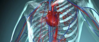
- Changes in the functioning of the vegetative-vascular system. They manifest themselves in the functioning of the digestion (belching, abdominal pain, bowel movements), in the functioning of the respiratory system (tachypnea, difficulty breathing, suffocation) and in the functioning of the heart and blood vessels (irregularities in the heart rhythm, high or low blood pressure, chest pain).
Neurologists, endocrinologists and gynecologists treat diseases of the hypothalamus.