Functions of the lobes of the brain
The brain is a powerful control center that sends commands throughout the body and controls the progress of their implementation. It is thanks to him that we perceive the world and are able to interact with it. The kind of brain a modern person has, his intelligence, thinking, are the result of millions of years of continuous evolution of mankind, his structure is unique.
The brain is characterized by division into zones, each of which specializes in performing its own specific functions. It is important to have information about what functions each zone performs.
Then you can easily understand why specific symptoms appear in such common diseases as Parkinson's disease, Alzheimer's disease, stroke, etc.
Violations can be regulated with medication, as well as with the help of special exercises and physical procedures.
The brain is structurally divided into:
- rear;
- average;
- front.
Each of them has their own role.
In an embryo, the head develops faster than other parts of the body. In a one-month-old embryo, all three parts of the brain can be easily seen. During this period they look like “brain bubbles”. The brain of a newborn is the most developed system in his body.
Scientists attribute the hindbrain and midbrain to more ancient structures. It is this part that is entrusted with the most important functions - maintaining breathing and blood circulation. The boundaries of their functions are clearly separated.
Each gyrus does its job. The more pronounced the groove became during development, the more functions it could perform.
But the anterior section provides everything that connects us with the external environment (speech, hearing, memory, ability to think, emotions).
There is an opinion that a woman's brain is smaller than a man's brain. Data from modern hardware studies, in particular on a tomograph, have not confirmed this. This definition can easily be called erroneous. The brain of different people may differ in size and weight, but this does not depend on gender.
Knowing the structure of the brain, you can understand why certain diseases appear and what their symptoms depend on.
Structurally, the brain consists of two hemispheres: right and left. Outwardly, they are very similar and are interconnected by a huge number of nerve fibers. Each person has one side that is dominant, right-handers have the left side, and left-handers have the right side.
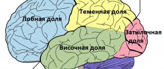
There are also four lobes of the brain. You can clearly see how the functions of the shares are differentiated.
What are shares?
The cerebral cortex has four lobes:
- occipital;
- parietal;
- temporal;
- frontal
Each share has a pair. All of them are responsible for maintaining the vital functions of the body and contact with the outside world. If injury, inflammation, or disease of the brain occurs, the function of the affected area may be completely or partially lost.
Frontal
These lobes have a frontal location, they occupy the forehead area. Let's figure out what the frontal lobe is responsible for. The frontal lobes of the brain are responsible for sending commands to all organs and systems. They can be figuratively called a “command post.” It would take a long time to list all their functions.
These centers are responsible for all actions and provide the most important human qualities (initiative, independence, critical self-esteem, etc.). When they are defeated, a person becomes carefree, changeable, his aspirations have no meaning, he is prone to inappropriate jokes.
Such symptoms may indicate atrophy of the frontal lobes, leading to passivity, which is easily mistaken for laziness.
Each lobe has a dominant and auxiliary part. For right-handed people, the left area will be dominant and vice versa. If you separate them, it is easier to understand which functions are assigned to a specific area.
It is the frontal lobes that control human behavior. This part of the brain sends commands that prevent a specific antisocial action from being performed. It is easy to notice how this area is affected in dementia patients. The internal limiter is turned off, and the person can tirelessly use obscene language, indulge in obscenities, etc.
The frontal lobes of the brain are also responsible for planning, organizing voluntary actions, and mastering the necessary skills.
Thanks to them, those actions that seem very difficult at first become automatic over time.
But when these areas are damaged, the person performs the actions as if anew each time, and automaticity is not developed. Such patients forget how to go to the store, how to cook, etc.
When the frontal lobes are damaged, perseveration can occur, in which patients literally become fixated on performing the same action. A person may repeat the same word, phrase, or constantly move objects around aimlessly.
The frontal lobes have a main, dominant, most often left, lobe. Thanks to her work, speech, attention, and abstract thinking are organized.
It is the frontal lobes that are responsible for maintaining the human body in an upright position. Patients with their lesions are distinguished by a hunched posture and a mincing gait.
Temporal
They are responsible for hearing, turning sounds into images. They provide speech perception and communication in general. The dominant temporal lobe of the brain allows you to fill the words you hear with meaning and select the necessary lexemes in order to express your thoughts. The non-dominant helps to recognize intonation and determine the expression of a human face.
The anterior and middle temporal regions are responsible for the sense of smell. If it is lost in old age, it may signal incipient Alzheimer's disease.
The hippocampus is responsible for long-term memory. It is he who stores all our memories.
If both temporal lobes are affected, a person cannot assimilate visual images, becomes serene, and his sexuality goes through the roof.
Parietal
In order to understand the functions of the parietal lobes, it is important to understand that the dominant and non-dominant side will do different jobs.
The dominant parietal lobe of the brain helps to understand the structure of the whole through its parts, their structure, order. Thanks to her, we know how to put individual parts into a whole. The ability to read is very indicative of this. To read a word, you need to put the letters together, and you need to create a phrase from the words. Manipulations with numbers are also carried out.
The parietal lobe helps to link individual movements into a complete action. When this function is disrupted, apraxia is observed. Patients cannot perform basic actions, for example, they are not able to get dressed. This happens with Alzheimer's disease. A person simply forgets how to make the necessary movements.
The dominant area helps you feel your body, distinguish between the right and left sides, and relate parts and the whole. This regulation is involved in spatial orientation.
The non-dominant side (in right-handed people it is the right side) combines information that comes from the occipital lobes and allows you to perceive the world around you in three-dimensional mode. If the non-dominant parietal lobe is disrupted, visual agnosia may occur, in which a person is unable to recognize objects, landscapes, or even faces.
The parietal lobes are involved in the perception of pain, cold, and heat. Their functioning also ensures orientation in space.
Occipital
The occipital lobes process visual information. It is with these lobes of the brain that we actually “see.” They read signals that come from the eyes. The occipital lobe is responsible for processing information about shape, color, and movement. The parietal lobe then turns this information into a three-dimensional image.
If a person stops recognizing familiar objects or loved ones, this may indicate a dysfunction in the occipital or temporal lobe of the brain. In a number of diseases, the brain loses the ability to process received signals.
How the hemispheres of the brain are connected
The hemispheres are connected by the corpus callosum. This is a large plexus of nerve fibers through which the signal is transmitted between the hemispheres. Adhesions are also involved in the joining process. There is a posterior, anterior, and superior commissure (fornix commissure). This organization helps to divide the functions of the brain between its individual lobes. This feature has been developed over millions of years of continuous evolution.
Conclusion
So, each department has its own functional load. If a separate lobe suffers due to injury or disease, another zone may take over some of its functions. Psychiatry has accumulated a lot of evidence of such redistribution.
It is important to remember that the brain cannot function fully without nutrients. The diet should have a variety of foods from which the nerve cells will receive the necessary substances. It is also important to improve blood supply to the brain. It is promoted by sports, walks in the fresh air, and a moderate amount of spices in the diet.
If you want to maintain full brain function until old age, you should develop your intellectual abilities. Scientists note an interesting pattern - people with intellectual work are less susceptible to Alzheimer's and Parkinson's diseases.
The secret, in their opinion, lies in the fact that with increased brain activity in the hemispheres, new connections are constantly created between neurons. This ensures constant tissue development.
If a disease affects one part of the brain, its functions are easily taken over by the neighboring zone.
Temporal
They are responsible for hearing, turning sounds into images. They provide speech perception and communication in general. The dominant temporal lobe of the brain allows you to fill the words you hear with meaning and select the necessary lexemes in order to express your thoughts. The non-dominant helps to recognize intonation and determine the expression of a human face.
The anterior and middle temporal regions are responsible for the sense of smell. If it is lost in old age, this may signal a nascent one.
The hippocampus is responsible for long-term memory. It is he who stores all our memories.
If both temporal lobes are affected, a person cannot assimilate visual images, becomes serene, and his sexuality goes through the roof.
The work of the frontal lobes of the brain. Frontal lobe: functions, structure and damage
The frontal lobes occupy about 28% of the total area of cortical structures. Their mass is approximately half the weight of the entire brain - about 450 g.
The frontal lobes are structures located in the frontal plane of the brain that are responsible for human mental activity, which determines their most important role in the formation and use of the mind.
The functions of the frontal lobes, located in the brain, include the ability to reflect, analyze, abstract, and generalize.
Structural features
The frontal lobes are a part of the brain that is responsible for communication skills (the ability to talk, think, remember).
The anatomical structure of the frontal lobe suggests a clear limitation from the temporal regions by the lateral grooves.
On the frontal side, the frontal part is separated from the parietal area by the central sulcus. The lower edge of the section is limited by the Sylvian fissure.
The main gyri located in the frontal lobe include the vertical precentral and superior, middle, inferior, running horizontally. The gyri are separated by grooves. During wakefulness, neurons and neurotransmitters in this area are more active than during sleep.
The area of the prefrontal cortex is functionally connected and actively interacts with parts of the limbic nervous system - brain structures located on both sides of the thalamus. The limbic system is involved in regulating the sense of smell, the functioning of internal organs, the expression of emotions, sleep and wakefulness, and memory.
The hypothalamus, part of the limbic system, controls the autonomic nervous system with the help of hormones, due to which a person feels thirst and hunger, wakes up and falls asleep in accordance with nature’s biological clock, and experiences sexual attraction to members of the opposite sex. The hippocampus is involved in the process of forming the base of long-term memory. This structure is responsible for the perception, analysis and storage of spatial information.
The frontal lobe is considered the cortical region of the limbic system. In the frontal part of the brain there are association zones where information received from outside is processed and compared with data stored in memory. The anatomy of this brain structure involves division into sections:
- Motor.
- Premotor.
- Dorsolateral prefrontal.
- Medial prefrontal.
- Orbitofrontal. It is the intersection of neural pathways through which information is transmitted to the associative zones of the cortex - a place of close interaction between the prefrontal and limbic systems.
The prefrontal region regulates complex behaviors and patterns, ensuring the interconnection of motivational, emotional and thought processes. The department participates in assessing the environment, taking into account the circumstances, formulating the order of planned actions, analyzing the consequences of planned actions, and developing typical behavior patterns for specific situations.
Main functions
The frontal lobes, located in the frontal plane of the brain, perform many important functions, including forecasting, planning, regulation and control of human psycho-emotional activity. The frontal lobes, consisting of the right and left parts, are responsible for the coordinated work of all departments in the brain, which indicates their leading role. Main functions:
- Decision making, long-term planning, implementation of targeted actions.
- Performing voluntary movements with all parts of the body. Impulses of voluntary motor activity move along the pyramidal path. The extrapyramidal system helps to perform voluntary movements, uses learned, typical motor patterns, maintains and redistributes muscle tone when performing movements.
- Motor mechanisms of speech function. The innervation of organs involved in the reproduction of sounds (tongue, facial muscles, lips) ensures the functioning of the speech apparatus. Broca's center coordinates the transmission of nerve impulses to the speech organs from the motor analyzer, which carries out the perception and analysis of information, the synthesis of certain stimuli.
- Individual personality characteristics. Character, type of temperament, worldview, behavior, initiative, critical self-esteem, features of communication with other members of society.
The left and right frontal lobes, located in the brain, are partially responsible for the vertical position of the body, which makes it possible to suspect damage to the brain matter in this area if a person develops a mincing, clumsy gait or begins to stoop heavily, stands and walks with a half-bent back.
The cortical part of the frontal lobes is responsible for voluntary eye movements in any direction, including in the opposite direction from the course along which the body is moving.
Tissue damage in the left lobe is accompanied by disturbances in verbal behavior (verbal reactions such as speaking, understanding and remembering words, responding to phrases), in the right lobe - non-verbal disorders.
brain
The human brain is an organ weighing 1.3-1.4 kg, located inside the cranium. The human brain is made up of more than one hundred billion neuron cells that form the gray matter, or cortex, the vast outer layer of the brain.
Neuronal processes (kind of like wires) are the axons that make up the white matter of the brain. Axons connect neurons to each other through dendrites.
The adult brain consumes about 20% of all the energy that the body needs, while the child's brain consumes about 50%.
- How does the human brain process information?
- Functions of the right and left hemispheres of the brain
- Emotions
- The structure of the human brain, the trinity of the brain
- white and gray matter
- prefrontal cortex
- hippocampus
- islet of Reil
- Broca's area
- Brain reward system
- Brain differences between men and women
- Human brain aging
- Sources
How does the human brain process information?
Today it is considered proven that the human brain can simultaneously process on average about 7 bits of information[2]. These can be individual sounds or visual signals, shades of emotions or thoughts distinguished by consciousness.
The minimum time required to distinguish one signal from another is 1/18 of a second. Thus, the perceptual limit is 126 bits per second. Conventionally, we can calculate that over the course of a 70-year life, a person processes 185 billion bits of information, including every thought, memory, and action.
Information is recorded in the brain through the formation of neural networks (a kind of routes).
Motor area
Allows you to ensure the normal functioning of the facial muscles, which is necessary for pronouncing complex phrases and words. To put it differently, thanks to the speech motor area, speech is formed in a person as a whole. If he is right-handed, then in the left hemisphere the speech motor zone takes up much more space than in the right, and if he is left-handed, everything is exactly the opposite.
If the zone is destroyed or severely damaged, the ability to speak is automatically lost. In this case, a person will be able to sing and scream without words. Also, if damaged, the ability to read to oneself and formulate one’s thoughts is lost. Such damage does not affect the function of understanding the speech of other people.
The left and right hemispheres contain the motor cortex, which is necessary to ensure the activity of the striated muscles. In the left hemisphere, the activity of the right side of the body, coordination of precise movements, and orientation on the ground are controlled. The internal organs send their impulses to this zone.
If the motor cortex is damaged, the following problems will occur:
- disturbances in the functioning of the cardiovascular system and respiratory organs;
- paresis of limbs;
- ataxia.
Frontal lobes of the brain - functions and symptoms of damage
Scientists consider the frontal cortex as a set of formations that, from an early age, exhibit pronounced individuality in their anatomical structure. Among these formations there are those that are new, “ human ” fields that develop at a later age. These include field 46.
Field 46 is a “human field”, because it is an evolutionary neoplasm that differentiates late. Field 46 is the last to mature and reaches 630% of its original size. Because this field is inhibitory, you can notice that children do not control their movements and grab everything that is not lying well. This behavior is typical of monkeys.
General
It is impossible to specifically develop the frontal lobes of the brain in children.
There is a misconception in society that physical activity promotes increased blood circulation in the brain, thereby developing all areas of the brain.
Physical activity fills the motor centers of the brain, while the rest of the brain ' rests ', because When performing different tasks, the brain uses specific centers, rather than the entire brain.
Based on the above, in order to determine exercises for the development of the frontal lobes, we need to find out what functions the frontal lobes are responsible for, with which we can develop the frontal lobes.
The frontal lobe, like the others, consists of white and gray matter.
Location
The frontal lobe is located in the anterior parts of the hemispheres. The frontal lobe is separated from the parietal lobe by the central sulcus, and from the temporal lobe by the lateral sulcus. Anatomically it consists of four convolutions - vertical and three horizontal. The convolutions are separated by grooves. The frontal lobe makes up one third of the mass of the cortex.
Assigned functions
Evolutionarily, it so happened that the active development of the frontal lobes is not associated with mental and intellectual activity. The frontal lobes arose in humans through evolution. The more a person could share food within his community, the more likely the community could survive.
In women, the frontal lobes arose for a specific purpose: sharing food. The men got this area as a gift.
Without those assigned tasks that lie on the shoulders of women, men began to use the frontal lobes in a variety of ways (thinking, building, etc.) to demonstrate Dominance.
Essentially, the frontal lobes are inhibitory centers . Also, many people ask what the left or right frontal lobe of the brain is responsible for. The question is not posed correctly, because... in the left and right frontal lobes there are corresponding fields that are responsible for specific functions. Roughly speaking, the frontal lobes are responsible for:
Fields and subfields are responsible for specific functions that are generalized under the frontal lobes. Because The polymorphism of the brain is enormous; the combination of the sizes of different fields makes up a person’s individuality.
Why do they say that over time a person changes. Throughout life, neurons die, and the remaining ones form new connections.
This introduces an imbalance in the quantitative ratio of connections between different fields that are responsible for different functions.
Not only do different people have different margin sizes, but some people may not have these margins at all. Polymorphism was identified by Soviet researchers S.A. Sarkisov, I.N. Filimonov, Yu.G. Shevchenko. They showed that the individual ways in which the cerebral cortex is structured within one ethnic group are so great that no common features can be seen.
- Field 8 - located in the posterior parts of the middle and superior frontal gyri. Has a center for voluntary eye movements
- Area 9 - dorsolateral prefrontal cortex
- Area 10 - Anterior prefrontal cortex
- Area 11 - olfactory area
- Area 12 - control of the basal ganglia
- Field 32 - Receptor area of emotional experiences
- Area 44 - Broca's Center (processing information about the location of the body relative to other bodies)
- Field 45 - music and motor center
- Field 46 - motor analyzer of head and eye rotation
- Field 47 - nuclear zone of singing, speech motor component Subfield 47.1
- Subfield 47.2
- Subfield 47.3
- Subfield 47.4
- Subfield 47.5
Symptoms of the lesion
Symptoms of the lesion are revealed in such a way that the selected functions are no longer adequately performed. The main thing is not to confuse some symptoms with laziness or imposed thoughts on this matter, although this is part of frontal lobe diseases.
- Uncontrollable grasping reflexes (Schuster reflex)
- Uncontrolled grasping reflexes when the skin of the hand is irritated at the base of the fingers (Yanishevsky-Bekhterev Reflex)
- Extension of the toes due to irritation of the skin of the foot (Hermann's sign)
- Maintaining an awkward arm position (Barre's sign)
- Constantly rubbing your nose (Duff's sign)
- Speech Impairment
- Loss of motivation
- Inability to concentrate
- Memory impairment
The following injuries and illnesses may cause these symptoms:
- Alzheimer's disease
- Frontotemporal dementia
- Traumatic brain injuries
- Strokes
- Oncological diseases
With such diseases and symptoms, a person may not be recognizable. A person may lose motivation, and his sense of defining personal boundaries becomes blurred. Impulsive behavior associated with the satisfaction of biological needs is possible. Because disruption of the frontal lobes (inhibitory) opens the boundaries to biological behavior controlled by the limbic system.
Answers to popular questions
- Where is the speech center in the brain? Located in Broca's center, namely in the posterior part of the inferior frontal gyrus
- Memory can be different (auditory, visual, gustatory, etc.). Depending on which center processes certain sensors, information from this sensor is stored in those centers
Didn't find a suitable answer? Find a doctor and ask him a question!
Occipital
The occipital lobes process visual information. It is with these lobes of the brain that we actually “see.” They read signals that come from the eyes. The occipital lobe is responsible for processing information about shape, color, and movement. The parietal lobe then turns this information into a three-dimensional image.
If a person stops recognizing familiar objects or loved ones, this may indicate a dysfunction in the occipital or temporal lobe of the brain. In a number of diseases, the brain loses the ability to process received signals.
Frontal lobe of the brain: structure, functions, symptoms of damage
If the brain is the control point of the human body, then the frontal lobes of the brain are a kind of “center of power.” Most scientists and physiologists in the world clearly recognize the “palm” of this part of the brain.
They are responsible for many important functions. Any damage to this area leads to serious and often irreversible consequences. It is these areas that are believed to control mental and emotional manifestations.
What are the frontal lobes responsible for?
To understand what the frontal zone is responsible for, it is necessary to identify the correspondence of their individual areas to the controlled parts of the body.
The central anterior gyrus is divided into three parts, each of which is responsible for its own area of the body:
- The lower third is associated with facial motor skills.
- The middle part controls the functions of the hands.
- The top third is all about footwork.
- The posterior parts of the superior gyrus of the frontal lobe control the patient's body.
This same area is part of the human extrapyramidal system. This is an ancient part of the brain that is responsible for muscle tone and voluntary control of movements, for the ability to fix and maintain a certain body position.
Nearby is the oculomotor center, which controls eye movements and helps to freely navigate and move in space.
The main functions of the frontal lobes are perception of the surrounding reality, control of speech and memory, manifestation of emotions, will, and motivational actions. From a physiological point of view, this area controls urination, coordination of movements, speech, handwriting, controls behavior, regulates motivation, thinking, cognitive functions, and socialization.
Symptoms indicating LD damage
Since the frontal part of the brain is responsible for numerous activities, manifestations of deviations can affect both physiological and behavioral functions of a person.
Symptoms are related to the location of the lesion in the frontal lobe. All of them can be divided into manifestations of behavioral disorders from the psyche and disorders of motor and physical functions.
Mental symptoms:
- fast fatiguability;
- worsening mood;
- sudden mood swings from euphoria to the deepest depression, transitions from a good-natured state to pronounced aggression;
- fussiness, loss of control over one’s actions. It is difficult for the patient to concentrate and complete the simplest task;
- distortion of memories;
- disturbances of memory, attention, smell. The patient may not smell or may be haunted by phantom odors. Such signs are especially characteristic of a tumor process in the frontal lobes;
- speech disorders;
- violation of critical perception of one’s own behavior, lack of understanding of the pathology of one’s actions.
Other disorders:
- coordination disorders, movement disorders, balance;
- convulsions, seizures;
- reflexive grasping actions of an obsessive type;
- epileptic seizures.
Signs of pathology depend on which area of the LD is affected and how severely.
Treatment methods for LD injuries
Since there are many reasons for the development of frontal lobe syndrome, treatment is directly related to the elimination of the original disease or disorder. These causes may be the following diseases or conditions:
- Neoplasms.
- Damages of cerebral vessels.
- Alzheimer's disease.
- Pick's pathology.
- Gilles de la Tourette's syndrome.
- Frontotemporal dementia.
- Traumatic brain injury, including that received at birth, when the child’s head passed through the birth canal. Previously, such injuries often occurred when obstetric forceps were applied to the head.
- Some other diseases.
With concussions and other skull injuries, the depth of damage to the frontal lobe is usually small, so the main symptoms often appear at the very beginning. With rest and appropriate treatment, they usually gradually disappear. In a tumor process with deep “germination” of the tumor, signs of ill health intensify over time.
Treatment of vascular disorders in the frontal lobes includes a whole range of medications that are selected individually for a particular patient. No two cases are the same, so there is no single treatment regimen. But the actions taken are approximately similar: the walls of blood vessels are strengthened, the blood is thinned, and cerebral circulation is improved.
In cases with tumors, whenever possible, surgery is used to remove the tumor; if this is not possible, then palliative treatment is used to maintain the vital functions of the body.
Specific diseases such as Alzheimer's disease do not yet have effective treatment and drugs that can cope with the disease, however, timely therapy can prolong a person's life as much as possible.
What could be the consequences of LD damage?
If the frontal lobe of the brain, the functions of which actually determine a person’s personality, is affected, then after an illness or serious injury the worst thing that can happen is a complete change in the behavior and the very essence of the patient’s character.
In a number of cases, it is noted that a person became the complete opposite of himself. Sometimes damage to the parts of the brain responsible for controlling behavior, the concept of good and evil, and a sense of responsibility for one’s actions led to the emergence of antisocial personalities and even serial maniacs.
Even if extreme manifestations are excluded, LD lesions lead to extremely serious consequences. If the sensory organs are damaged, the patient may suffer from disorders of vision, hearing, touch, smell, and ceases to orientate normally in space.
In other situations, the patient is deprived of the opportunity to normally assess the situation, be aware of the world around him, learn, and remember. Such a person sometimes cannot take care of himself, so he needs constant supervision and help.
If there are problems with motor functions, it is difficult for the patient to move, navigate in space and take care of himself.
The severity of manifestations can only be reduced by promptly seeking medical help and taking emergency measures to prevent further development of damage to the frontal lobe.
Functions of the frontal lobes of the human brain
The frontal lobes occupy about 28% of the total area of cortical structures. Their mass is approximately half the weight of the entire brain - about 450 g.
The frontal lobes are structures located in the frontal plane of the brain that are responsible for human mental activity, which determines their most important role in the formation and use of the mind.
The functions of the frontal lobes, located in the brain, include the ability to reflect, analyze, abstract, and generalize.
Symptoms of tissue damage in the frontal lobe of the brain
Among the most severe lesions of the tissues of the frontal lobes are ischemic, atrophic changes in the tissue structure, stroke with a focus of ischemia or hemorrhage in this area. Impaired blood supply to this part of the brain negatively affects cognitive abilities. The main reasons for the deterioration and cessation of blood flow in this area:
- Exacerbation of arterial hypertension.
- Atherosclerotic damage to vascular walls.
- Developmental anomalies and acquired defects of elements of the circulatory system (vascular malformations, aneurysms).
- Disorder in the hemostasis system (increased thrombosis, blood clotting disorder).
Frontal lobe syndrome occurs due to damage to the tissues of the prefrontal region, which is responsible for multi-stage thought processes, behavior and cognitive abilities. General cerebral symptoms of pathology:
- Acute pain in the frontal plane of the head.
- Nausea, often leading to bouts of vomiting.
- Dizziness, confusion.
- Sometimes increased body temperature.
Frontal lobe syndrome is classified as an organic personality disorder in the international medical classification of diseases. It often develops against the background of other pathologies - Pick's disease and Alzheimer's disease, brain tumors, trauma to the head, damage to the vascular system in this area. Specific signs of the disease:
- Activation of rudimentary reflexes (sucking, grasping, searching). Occurs when a large area of tissue is affected.
- Loss of self-control, inability to plan and manage actions.
- Loss of awareness of one's own personality.
- Motor and speech dysfunction.
- Impossibility of abstract thinking and planning.
- Perseveration. Unconscious repeated repetition of one word, phrase, or action.
- Impaired concentration and memory functions.
Similar disorders are often observed in patients with Alzheimer's disease, who experience pathological structural changes in tissues - the formation of protein structures around neurons that interfere with and disrupt the relationships between nerve cells. Alzheimer's disease often causes dementia.
Consequences of damage to the frontal lobe
When these brain structures are damaged, a person becomes carefree, he is characterized by changeable moods, meaningless, illogical actions and aspirations.
Atrophy of the brain matter in this area is accompanied by a loss of motivation, which leads to passivity, indifference, apathy, and lack of interest in the world around us.
The condition is often mistaken for laziness, not realizing that the cause of the change in behavior is due to the death of nerve cells in the forehead area.
The consequences of a frontal stroke develop in two types - abulic and disinhibited. In the first case, symptoms such as indifference, apathy, lack of initiative, loss of creative thinking and curiosity predominate. In the second, impulsive behavior is observed, actions lose their meaning and logic, and a person is not able to foresee the result of his actions.
The frontal lobes are structures that control an individual’s mental activity and behavior. When these parts of the brain are damaged, the individual loses the ability to weigh himself as a person, objectively perceive events and react to the situation, plan and take purposeful actions.