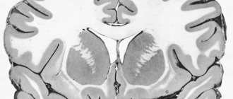
Glia are a structure of the nervous system formed by specialized cells of various shapes that fill the spaces between neurons or capillaries, accounting for 10% of the brain volume.
The size of glial cells is 3-4 times smaller than nerve cells; their number in the central nervous system of mammals reaches 140 billion. With age, the number of neurons in the brain decreases, and the number of glial cells increases.
Functions of neuroglia
Astroglia - represented by multi-processed cells. Their sizes range from 7 to 25 microns. Most of the processes end on the walls of blood vessels. The nuclei contain DNA, the protoplasm has the Golgi apparatus, centrisome, and mitochondria. Astroglia serves as a support for neurons, ensures reparative processes of nerve trunks, insulates nerve fiber, and participates in the metabolism of neurons.
Oligodendroglia are cells that have a single process. The number of oligodendroglia increases in the cortex from the upper to the lower layers. In the subcortical structures, in the brain stem, there are more oligodendroglia than in the cortex. It is involved in the myelination of axons and the metabolism of neurons.
Microglia are the smallest glial cells and belong to the wandering cells. They are formed from the structures of the membranes of the brain, penetrate into the white and then into the gray matter of the brain. Microglial cells are capable of phagocytosis.
Types of Glial Cells
Previous10Next
When Rudolf Vikhrov first introduced the term “glia”, he described those elements that were later called astroglia
. The name “glia” was later transferred to other cells, later discovered, that did not have the specialization of neurons, but were located in the nervous tissue. Much credit for the classification of glia belongs to the Spanish neurohistological school, especially Ramon y Cajal and Rio del Gortega. Morphological differences in individual types of glial cells correspond to their different purposes and different specializations.
4.2.1.
Ependyma From a phylogenetic and ontogenetic point of view, the ependymal cell is the earliest glial element. In some lower animals, the entire glial population of the nervous system consists entirely of these cells. In higher vertebrates, they form only the lining of the cavities of the central nervous system, which in its structure resembles columnar epithelium and is called ependyma.
The surface of the ependymal cell facing the cavity is covered with short finger-like processes of the cell membrane - microvilli
and
eyelashes
.
The coordinated movement of cilia (up to 6 times per second) is one of the mechanisms that sets in motion the fluid medium of the cavities and ventricles of the central nervous system, which is otherwise called cerebrospinal fluid
. On the lateral surfaces of ependymal cells there are tight gap junctions that strengthen the layer of cells, creating a continuous structure like an epithelial one. On the basal side of the ependymal cell, a process is often formed that contacts other glial cells. This type of glial cells performs three functions, which are carried out more or less sequentially in individual development.
The first function is proliferative - the cambial reserve of cells of the germinal neural tube
The second function is support. As the wall of the neural tube thickens, the ependymal cells lining it give off long processes that reach its outer surface and for some time contribute to the formation of the outer membrane surrounding the neural tube.
The third function is the formation of a continuous epithelial lining of the ventricles of the brain. This lining is also preserved in the central canal of the spinal cord. In some areas of the ventricles, ependyma participates in the formation of the choroid plexuses of the brain.
Astrocytic glia
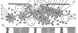
The distribution of astrocytes in the central nervous system is uniform, but they are less common in the matter than in the gray substance. The nuclei of astrocytes are larger than the nuclei of the ependyma, they are lighter, and often have a complex shape (polymorphic). Astrocytes are characterized by a large number of processes that radiate away from the cell body. Astrocytes have a lot of fibrillar structures, hence their main function is support.
In general, all astrocytes are divided into two large categories - fibrillar and protoplasmic (Fig. 12). Fibrillar astrocytes contain a large number of fibril bundles; these cells are located mainly in the white matter. For a long time, it was believed that only fibrillar astrocytes contain fibrils, but with the help of an electron microscope such bundles were also identified in protoplasmic astrocytes. The latter have branching cytoplasmic processes extending from the cell body like thickets of bushes. Their processes are shorter and the branches are more numerous than those of fibrillar astrocytes.
Astrocytes permeate the entire central nervous system, contacting each other and with the receptor part of neuronal synapses. Astrocytes make up a quarter of the glial cell population. In their configuration, astrocytes are completely dependent on the system of neurons that they surround. Neuronal axons are often enveloped in astrocyte cytoplasm. Astrocytes are also located between the capillaries of the bloodstream and the bodies of neurons, and transport substances from the blood to neurons and back. In addition, astroglia connects cerebrospinal fluid with the bloodstream.
Since degenerating astrocytes are sometimes found, it is possible that degeneration is in equilibrium with the formation of new astrocytes, suggesting that the population of these cells may be slowly being renewed. Thus, the main functions of astrocytic glia:
musculoskeletal
trophic
insulating
Oligodendroglia
Oligodendrocytes have the same origin as astrocytes. They are smaller in size than astrocytes and have fewer processes. Oligodendrocytes are found both in the gray matter (where they represent most of the perineuronal elements) and in the white matter (where they are responsible for the formation of myelin,
these oligodendrocytes have long processes).
One type of oligodendroglia is Schwann
cells.
These cells are located in the peripheral nervous system and are able to spirally wrap their cytoplasm around the axons of neurons and form myelin sheaths
.
Those oligodendrocytes that are located in the gray matter of the central nervous system are located so tightly adjacent to the bodies of neurons that they are called satellite cells
. Main functions of oligodendrocytes:
insulating - Schwann cells completely limit all neuronal parts of the peripheral nervous system from connective tissue
synthetic - Schwann cells take on the task of creating the myelin protein and ensuring its structural and functional integrity. Myelination in embryonic ontogenesis occurs quite late and continues for weeks and months after birth. The main function is that myelin allows you to increase the speed of nerve impulses.
metabolism of the adjacent neuron is a common function of all oligodendrocytes (both satellites and Schwann cells)
Microglia
Microglial cells originate from another germ layer, the mesoderm, and play an important role in pathological conditions. They grow into the brain in the later stages of embryogenesis, accumulating in those places where the pia mater is directly adjacent to the white matter. From here they spread throughout the tissue of the central nervous system.
Some authors, for example Arthur Ham and David Cormack, believe that “... it would be simpler if microglia were not classified as neuroglia at all, since they do not develop from the epithelium of the neural tube (like other cells of brain tissue) and do not perform a supporting function
».
Microglia (as the name suggests) are the smallest glial element in the CNS. In quantitative terms, microglial cells are smaller than astrocytes and oligodendrocytes. These cells do not have a predominant localization in nervous tissue, but there are more of them in the gray matter than in the white matter. They are often found in the form of satellites around nerve cells and blood vessels. The cytoplasm of these cells surrounds the nucleus only in a narrow strip. Numerous processes extend from the cell body, which are richly branched.
Microglia are characterized by increased mobility and the ability to accept and process metabolites (waste products of other cells). Microglial elements are activated during disease processes accompanied by the breakdown of nervous tissue. The main function of microglia is the ability, like other tissue macrophages, to phagocytose (digest, utilize) dying cells of the central nervous system. However, as studies have shown, microglia retain their functional activity under normal conditions. There is a constant exchange between nervous tissue, microglia and blood.
Structure of nerves
The processes of neurons covered with membranes are called nerve fibers. As we have already mentioned, depending on the degree of surrounded by oligodendrocytes, they are non-myelinated (myelinated) and myelinated (myelinated). Bundles of nerve fibers surrounded by connective tissue membranes are called nerves
or
nerve trunks
. Some nerves contain single nerve cells and small ganglia or nodes (clusters of neurons). Nerves are divided into:
cranial
– 12 pairs
spinal
– 31 pairs
All nerves and their branches (together with the terminal devices - receptors and effectors) make up the peripheral nervous system. Through nerves and their branches, the central nervous system communicates with organs, organ systems are united and the integrity of the body is ensured.
Nerves are quite durable structures, unlike the tissues of the brain or spinal cord. This is explained by the fact that the connective tissue sheath of the nerves is a tubular structure of three orders (Fig. 13):
epineurium
- outer sheath of the nerve trunk. It is represented by loose, unformed connective tissue, rich in collagen fibers. Contains fibroblasts, fat cells, and blood and lymphatic vessels
perineurium
- thin layers of connective tissue that divide the nerve into separate nerve bundles. Also contains blood and lymphatic vessels.
endoneurium
– divides the nerve bundle into individual nerve fibers.
In nerves of small diameter, the epineurium is absent. Small nerves are composed of a perineurium containing endoneurial tubes surrounding individual nerve fibers.
The same membranes - epi-, peri- and endoneurium - are also present in the cerebrospinal ganglia.
Nerves, depending on the composition of their fibers, are divided into:
sensitive - contain centripetal fibers
motor - centrifugal fibers
mixed - both types of fibers
Most peripheral nerves are of the mixed type. Toward the distal end of the nerve, afferent and efferent fibers are sorted, and individual nerve bundles become predominantly sensory or motor. The number and diameter of nerve fibers in the bundle vary. The distal portions of some nerves have more fibers than the proximal portions. The increase in the number of fibers in a nerve is associated with their branching.
Section 2
Previous10Next
Date added: 2017-11-21; views: 4220; ORDER A WORK WRITING
Similar articles:
Nervous tissue
Nervous tissue consists of two types of cells: nerve (neurons) and glial. Glial cells are closely adjacent to the neuron, performing supporting, nutritional, secretory and protective functions.
Neuron is the basic structural and functional unit of nervous tissue. Its main feature is the ability to generate nerve impulses and transmit excitation to other neurons or muscle and glandular cells of working organs. Neurons can consist of a body and processes. Nerve cells are designed to conduct nerve impulses. Having received information on one part of the surface, the neuron very quickly transmits it to another part of its surface. Since the processes of a neuron are very long, information is transmitted over long distances. Most neurons have two types of processes: short, thick, branching near the body - dendrites, and long (up to 1.5 m), thin and branching only at the very end - axons. Axons form nerve fibers.
A nerve impulse is an electrical wave traveling at high speed along a nerve fiber.
Depending on the functions performed and structural features, all nerve cells are divided into three types: sensory, motor (executive) and intercalary. Motor fibers running as part of nerves transmit signals to muscles and glands, sensory fibers transmit information about the state of organs to the central nervous system.
Now we can combine all the information received into a table.
Connective tissue
Connective tissue Consists of cells, intercellular substance and connective tissue fibers. It consists of bones, cartilage, tendons, ligaments, blood, fat, it is present in all organs (loose connective tissue) in the form of the so-called stroma (framework) of organs.
In contrast to epithelial tissue, in all types of connective tissue (except adipose tissue), the intercellular substance predominates over the cells in volume, i.e., the intercellular substance is very well expressed. The chemical composition and physical properties of the intercellular substance are very diverse in different types of connective tissue. For example, blood - the cells in it “float” and move freely, since the intercellular substance is well developed.
In general, connective tissue makes up what is called the internal environment of the body. It is very diverse and is represented by various types - from dense and loose forms to blood and lymph, the cells of which are in the liquid. The fundamental differences in the types of connective tissue are determined by the ratios of cellular components and the nature of the intercellular substance.
Dense fibrous connective tissue (muscle tendons, joint ligaments) is dominated by fibrous structures and experiences significant mechanical stress.
Loose fibrous connective tissue is extremely common in the body. It is very rich, on the contrary, in cellular forms of different types. Some of them are involved in the formation of tissue fibers (fibroblasts), others, which is especially important, provide primarily protective and regulatory processes, including through immune mechanisms (macrophages, lymphocytes, tissue basophils, plasma cells).