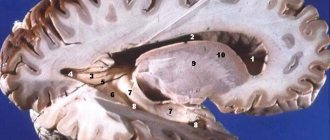
The human brain consists of white and gray matter. The first is everything that is filled between the gray matter on the cortex and on the surface there is a uniform layer of gray matter with nerve cells, the thickness of which is up to four and a half millimeters.
Let's study in more detail what gray and white matter is in the brain.
Space limitations prevented the inclusion of electro- or magnetoencephalographic data, but it is certain that, given the dynamic spatial and temporal dimensions of these data, it would be necessary to speak of “networks.” Many problems need to be solved such as.
Relationship between age and white matter status
The fact that intelligence may be associated with increased gray matter volume and decreased brain glucose consumption under certain conditions. Know if experience and training can increase gray matter. Gender differences noted in some studies.
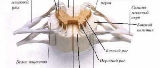
Gray and white matter of the spinal cord
The heterogeneous substance of this organ is gray and white. The first is formed by a huge number of neurons, which are concentrated in nuclei and come in three types:
Lack of research related to intelligence measurement, imaging and genetic studies. Limits inherent in identifying the neural basis for general intelligence. Select several conversation options. A map that connects the brain with mind maps.
The paratheto-frontal integration theory of intelligence: A major contribution to the theory of intelligence. The sleeping brain, states of consciousness and the human mind. On the neural basis of crystallized intelligence. Integrative action in the frontoparietal network: caring for the distracted brain.
About correlation images. Intelligence and reasoning are two different things. Intelligence, hormones, gender, brain size and biochemistry: demonstrate causation before integration. A global approach is needed. Can parietal frontal integration theory of intelligence be extended to explain individual differences in performance and daily living skills?
- radicular cells;
- tufted neurons;
- internal cells.
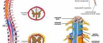
The white matter of the spinal cord surrounds the gray matter. It includes nerve processes that make up three fiber systems:
- intercalary and afferent neurons connecting different parts of the spinal cord;
- sensory afferents, which are long centripetal;
- motor afferent or long centrifugal.
Anatomy of gray matter
According to anatomical features, gray matter is an integral part of the white component. It is located on a cross section, which visually resembles the wings of a butterfly. In the middle there is a central channel, it is filled with a special liquid - liquor.
The ventricles of the brain are located here and have close interaction with each other. The fullness of cerebrospinal fluid depends on certain patterns. This is due to the complex plexuses of the cerebrospinal fluid. Thanks to its research, one can learn about a person’s condition and diagnose many diseases. These include:
- infections;
- tumors;
- inflammation;
- parasites.
In the gray matter of the spinal cord there are several columns interconnected by adhesions. In the middle of it you can see a hole, which represents the central channel. The back and front plates are the main components. When analyzing the cross-section, you will notice that the interconnected pillars resemble the wings of a butterfly, as mentioned above.
Here the protrusions are easily visible, gradually diverging to the sides. In literature they are called horns. Their composition is unique, due to the presence of paired wide and narrow parts. The plates are based on neurons, consisting of a centralized core and auxiliary components. The roots of the spinal cord are also located here.
https://www.youtube.com/watch?v=ytpolicyandsafetyru
Gray matter is characterized by the presence of columns. The dorsal horn includes neurons, they are located in a certain sequence. They are often called axons; due to their unique structure, they tend to the anterior commissure. This makes it easy to get to the other side of the brain. Interneurons are branched dendrites.
All of them together form the main component. It is based on three projections that extend along the entire length of the brain. This design is represented by pillars. There are three types of protrusions, respectively, they are located in the front, rear and side parts.
hindbrain
This brain consists of the pons and the cerebellum. Let's look at the gray and white matter in them. The bridge is a large white ridge on the back side of the base. On the one hand, its border with the cerebral peduncles is pronounced, and on the other, with the medulla oblongata. If you make a cross section, the white matter of the brain and the gray nucleus will be visible very clearly. Transverse fibers divide the bridge into ventral and dorsal sections. In the ventral part, the white matter of the pathways is mainly present, and the gray matter forms its nuclei here.
The dorsal part is represented by nuclei: switching, sensory systems and cranial nerves.
The cerebellum is located under the occipital lobes. It includes the hemispheres and the middle part called the “worm”. Gray matter makes up the cerebellar cortex and nuclei, which are tent-shaped, spherical, corky and dentate. The white matter of the brain in this part is located under the cerebellar cortex. It penetrates into all gyri as white plates and consists of different fibers that either connect the lobules and gyri, or are directed to the internal nuclei, or connect sections of the brain.
Roots
When considering the functions of the spinal cord and its structure, one cannot fail to mention the so-called spinal cord roots.
In short, the roots of the spinal cord are bundles of nerve fibers that enter any segment of the organ and form the spinal nerves.
The roots form the sensitive part of the spinal nerve. The root consists of motor nerve fibers, which are processes of the anterior horns of the gray matter.
This is interesting! How we are made: human structure - internal organs in detailed description and location diagram
Midbrain
It starts from the mesencephalon. On the one hand, it corresponds to the surface of the brain stem between the superior medullary velum, and on the other, to the area between the mammillary bodies and the anterior part of the pons.
It includes the cerebral aqueduct, on one side of which the boundary is provided by the roof, and on the other by the covering of the cerebral peduncles. In the ventral region, the posterior perforated substance and the peduncles of the cerebrum are distinguished, and in the dorsal region, the roof plate and the handles of the inferior and superior colliculi are distinguished.
If we look at the white and gray matter of the brain in the cerebral aqueduct, we will see that the white surrounds the central gray matter, which consists of small cells and has a thickness of 2 to 5 millimeters. It consists of the trochlear, trigeminal and oculomotor nerves, together with the accessory nucleus of the latter and the intermediate nucleus.
INTRODUCTION
The brain and spinal cord together form the central nervous system - system nervorum centrale.
The nervous system regulates the activities of various organs, organ systems and the entire body. The nervous system communicates between different organs and organ systems, being the coordinator of the coordinated activity of all organs and organ systems, determining the integrity of the body. The nervous system communicates the body with the external environment. The brain is the material basis of thinking and related speech. The brain - encephalon - is the part of the central nervous system enclosed in the cranial cavity. The spinal cord - medulla spinalis - is the part that is located in the spinal canal. The lower part of the brain - the medulla oblongata - borders the spinal cord1.
https://www.youtube.com/watch?v=ytcreatorsru
Gray matter and white matter of the spinal cord and brain are the components of the spinal cord and brain, their substrate.
The gray matter of the spinal cord and brain consists mainly of clusters of nerve cell bodies and the nearest branches of their processes. The white matter of the spinal cord and brain consists mainly of clusters of nerve fibers, processes of nerve cells that have a myelin sheath (hence the white color of the fibers and substance).
Nerve fibers form the pathways of the spinal cord and brain and connect various parts of the central nervous system and various nuclei (nerve centers). The nerve fibers of the white matter of the cerebral hemispheres are divided into three groups: associative, commissural and projection. They make up the pathways of the same name in the brain and spinal cord.
Associative nerve fibers emerge from the cerebral cortex, that is, they are extracortical. They are located within one hemisphere and connect its various nerve centers.
Commissural nerve fibers pass through the commissures of the brain: corpus callosum, anterior commissure.
Projection nerve fibers provide bilateral connections between the cerebral hemispheres and all underlying parts of the nervous system and constitute the internal capsule and its corona radiata.
In our abstract we will look at the anatomical structure of the brain and spinal cord, study what the white and gray matter of the spinal cord and brain are and determine the significance of the connective tissue membranes of the spinal cord and brain.
The spinal cord is a cylindrical cord covered with membranes, freely located in the cavity of the spinal canal. At the top it becomes medulla oblongata; below the spinal cord reaches the region of the 1st or upper edge of the 2nd lumbar vertebra.
The diameter of the spinal cord is not the same everywhere; in two places two fusiform thickenings are found: in the cervical region - cervical thickening - intumescentia cervicalis (from the 4th cervical to the 2nd thoracic vertebra); in the lowest part of the thoracic region there is a lumbar thickening - intumescentia lumbalis - (from the 12th thoracic to the 2nd sacral vertebra).
The spinal cord is connected to the brain via the brain stem and, emerging from the foramen magnum, extends below approximately 45 cm to the 1st lumbar vertebra. In its middle part, the spinal cord is about 1.8 cm wide, no more than the width of a finger. The spinal cord is protected by the bones of the spine, and 31 pairs of spinal nerves emerge from the spaces between the vertebrae. The curvature of the spinal cord follows the curvature of the spinal column (Fig. 1).
Functions of the spinal cord – through the spinal nerves, the spinal cord transmits information from the brain to various organs. It is also involved in many reflex acts. These are ultra-fast automatic reactions, which are mainly of a defensive nature (blinking, sneezing, withdrawing the hand, and others).
Rice. 1. Brain and spinal cord
Structure of the Spinal Cord – The spinal cord is a central column of gray matter, which consists of interneurons, sensory neuron terminals, and motor neuron cell bodies. Gray matter is surrounded by white matter, which is made up of bundles of nerve fibers called tracts that run through the spinal cord.
The structure of nerves. Nerves are thin, cream-colored wire-like threads that form the peripheral nervous system. The cranial and spinal nerves have the same structure. A nerve consists of bundles of neurons, or rather, long nerve fibers (axons). Each bundle is surrounded by connective tissue called perineurium.
The spinal cord has two functions: reflex and conduction. As a reflex center, the spinal cord is capable of performing complex motor and autonomic reflexes. It is connected by afferent – sensitive – pathways to receptors, and by efferent pathways – to skeletal muscles and all internal organs.
The spinal cord connects the periphery with the brain through long ascending and descending tracts. Afferent impulses along the spinal cord pathways are carried to the brain, carrying information about changes in the external and internal environment of the body. Along descending pathways, impulses from the brain are transmitted to effector neurons of the spinal cord and cause or regulate their activity.
Reflex function. The nerve centers of the spinal cord are segmental, or working, centers. Their neurons are directly connected to receptors and working organs. In addition to the spinal cord, such centers are present in the medulla oblongata and midbrain. Suprasegmental centers, for example, the diencephalon and cerebral cortex, do not have a direct connection with the periphery.
In addition to the motor centers of skeletal muscles, the spinal cord contains a number of sympathetic and parasympathetic autonomic centers. In the lateral horns of the thoracic and upper segments of the lumbar spinal cord there are spinal centers of the sympathetic nervous system that innervate the heart, blood vessels, sweat glands, digestive tract, skeletal muscles, i.e. all organs and tissues of the body. This is where the neurons directly connected to the peripheral sympathetic ganglia lie4.
In the upper thoracic segment, there is a sympathetic center for pupil dilation, in the five upper thoracic segments there are sympathetic cardiac centers. The sacral part of the spinal cord contains parasympathetic centers that innervate the pelvic organs (reflex centers for urination, defecation, erection, ejaculation).
https://www.youtube.com/watch?v=ytcopyrightru
The spinal cord has a segmental structure. A segment is a segment that gives rise to two pairs of roots. If the back roots of a frog are cut on one side and the front roots on the other, then the legs on the side where the back roots are cut will lose sensitivity, and on the opposite side, where the front roots are cut, they will be paralyzed. Consequently, the dorsal roots of the spinal cord are sensitive, and the anterior ones are motor.
In experiments with transection of individual roots, it was found that each segment of the spinal cord innervates three transverse segments, or metameres, of the body: its own, one above and one below. Consequently, each metamer of the body receives sensory fibers from three roots and, in order to desensitize an area of the body, it is necessary to cut three roots (safety factor). Skeletal muscles also receive motor innervation from three adjacent segments of the spinal cord.
Each spinal reflex has its own receptive field and its own localization (location), its own level. For example, the center of the knee reflex is located in the II – IV lumbar segment; Achilles - in the V lumbar and I - II sacral segments; plantar - in the I - II sacral, the center of the abdominal muscles in the VIII - XII thoracic segments.
Conducting function of the spinal cord. The spinal cord performs a conductive function due to the ascending and descending tracts passing through the white matter of the spinal cord. These pathways connect individual segments of the spinal cord with each other and also with the brain5.
Spinal shock. Transection or injury of the spinal cord causes a phenomenon called spinal shock. Spinal shock is expressed in a sharp drop in excitability and inhibition of the activity of all reflex centers of the spinal cord located below the site of transection. During spinal shock, the stimuli that normally trigger reflexes are rendered ineffective.
A paw prick does not cause a flexion reflex. At the same time, the activity of centers located above the transection is maintained. A monkey whose spinal cord has been transected in the area of the upper thoracic segments, after anesthesia has passed, takes a banana with its front paws, peels it, brings it to its mouth and eats it.
Diencephalon
It is located between the corpus callosum and the fornix, and on the sides it fuses with the Dorsal region consists of the visual tuberosities, on the upper part of which there is the epitubercle, and in the ventral part there is the inferior tuberosity region.
The gray matter here consists of nuclei that are associated with centers of sensitivity. White matter is represented by conducting pathways in different directions, guaranteeing the connection of formations with the cerebral cortex and nuclei. The diencephalon also includes the pituitary gland and pineal gland.
Finite brain
It is represented by two hemispheres, which are separated by a gap running along them. It is connected in depth by the corpus callosum and commissures.
The cavity is represented by the lateral ventricles located in one and the second hemisphere. These hemispheres consist of:
- a cloak of neocortex or six-layer cortex, distinguished by nerve cells;
- striatum from the basal ganglia - ancient, old and new;
- partitions.
But sometimes there is another classification:
- olfactory brain;
- subcortex;
- gray matter of the cortex.
Without touching on the gray matter, let's focus immediately on the white matter.
On the characteristics of the white matter of the hemispheres
The white matter of the brain occupies all the space between the gray and basal ganglia. There is a huge number of nerve fibers here. The white matter contains the following areas:
- central substance of the internal capsule, corpus callosum and long fibers;
- radiant crown of radiating fibers;
- semi-oval center in outer parts;
- a substance found in the convolutions between the furrows.
Nerve fibers are:
- commissural;
- associative;
- projection.
The white matter includes nerve fibers that are connected by the convolutions of one and the other cerebral cortex and other formations.
Nerve fibers
Mostly commissural fibers are found in the corpus callosum. They are located in the cerebral commissures, which connect the cortex on different hemispheres and symmetrical points.
Association fibers group areas on one hemisphere. In this case, short ones connect neighboring convolutions, and long ones connect those located at a far distance from each other.
Projection fibers connect the cortex with those formations located below, and then with the periphery.
If the internal capsule is viewed in section from the front, the lenticular nucleus and the posterior limb will be visible. Projection fibers are divided into:
- fibers located from the thalamus to the cortex and in the opposite direction, they excite the cortex and are centrifugal;
- fibers directed to the motor nuclei of the nerves;
- fibers that conduct impulses to the muscles of the whole body;
- fibers directed from the cortex to the pontine nuclei, providing a regulatory and inhibitory effect on the work of the cerebellum.
Those projection fibers that are located closest to the cortex create the corona radiata. Then their main part passes into the internal capsule, where the white matter is located between the caudate and lenticular nuclei, as well as the thalamus.
There is an extremely complex pattern on the surface, with alternating grooves and ridges between them. They are called convolutions. Deep grooves divide the hemispheres into large areas called lobes. In general, the grooves of the brain are deeply individual; they can vary greatly from person to person.
The hemispheres have five lobes:
- frontal;
- parietal;
- temporal;
- occipital;
- island.
The central sulcus originates at the top of the hemisphere and moves down and forward to the frontal lobe. The area posterior to the central sulcus is the parietal lobe, which ends in the parieto-occipital sulcus.
The frontal lobe is divided into four convolutions, vertical and horizontal. The lateral surface is represented by three convolutions, which are delimited from each other.
The furrows of the occipital lobe are variable. But everyone, as a rule, has a transverse one, which is connected to the end of the interparietal groove.
On the parietal lobe there is a groove that runs horizontally parallel to the central one and merges with another groove. Depending on their location, this lobe is divided into three convolutions.
The island has a triangular shape. It is covered with short convolutions.
Forebrain tissues
The spinal cord is fundamentally different in structure from the brain. In it, light and dark substance is concentrated in nuclei, which are of the following types:
- Internal;
- Beam;
- Radicular.
Unlike the brain tissues of the head, in the back the substantia alba is located outside the substantia grisea. Among other features, the components of the white matter of the spinal cord can be distinguished:
- Intercalary and afferent neurons, which serve to connect different parts of the spinal cord;
- Afferent neurons (sensitive);
- Motor neurons.
Directly above the medulla oblongata is the pons, and to the right is the cerebellum. The first section is presented in the form of a light-colored roller. It is associated with the cerebral peduncles and myelencephalon.
Transverse fibers divide the bridge into the following parts:
- Ventral (gastric). In this area, the substantia alba is represented predominantly by conductive fibers, and the substantia grisea has its nuclei here;
- Dorsal (dorsal). It consists of the following elements: Switching cores;
- Network formation;
- Sensory systems;
- Nerve pathways.
The cerebellum is located just below the occipital part of the brain. It consists of 2 hemispheres and a middle part. Gray matter is presented in the form of nuclei (dentate, cork-shaped, spherical, tent-shaped) and cortex. The white substance is under the dark shell. It is located in all convolutions and mainly consists of fibers that perform the following purposes:
- Connect the cerebral lobes and gyri;
- They follow the nuclei localized inside;
- Link departments.
The anterior section is also called the terminal section. It consists of two hemispheres separated by a depression. It runs along the entire section and connects below with the corpus callosum. The cavity of the terminal brain tissue contains the lateral ventricles, and the hemispheres themselves consist of the following components:
- Isocortex;
- Striatum;
- Partitions.
It plays the role of conducting pathways, which are divided into 3 groups:
- Associative. This type of fiber serves to connect different parts of the cortex in the region of the 1st hemisphere. There are short and long associative paths. The first type is presented as an arc-shaped accumulation of substance. It connects parts of the cortex of adjacent gyri. Long paths connect the lobes of the hemispheres;
- Commissural. They are localized in brain adhesions and are responsible for connecting formations in both hemispheres. The basis of the commissural fibers is the corpus callosum. Parts of this formation connect the gray matter of certain lobes with each other;
- Projection. The fibers of this group form the capsule and corona radiata. The first formation is a plate of white matter. It is surrounded by the lenticular and caudate nuclei and the hypothalamus. The capsule itself contains 2 legs and a knee. Fibers localized closer to the cortex form the corona radiata. The role of these pathways is to connect the cortex with the formations below.
On the surface of the brain (cortex) you can see a rather interesting and complex pattern. From an anatomical point of view, the alternation of grooves and ridges is clearly visible. The latter are located between them and are called convolutions.
The size of the grooves and medullary lobes is most often individual and differences may be observed in each person. However, there are certain standards that experts focus on:
- Central groove. It begins on the superior surface of the hemispheres and separates the parietal and frontal lobes. On the sides of it remain the temporal parts;
- Frontal lobe. It includes 4 convolutions and this area borders the parietal and temporal parts;
- Temporal. It consists of 3 convolutions separated from each other. Border this area with all other shares;
- Occipital lobe. In many people it differs in the structure of the grooves, but in most cases the transverse depression is associated with the interparietal one. This lobe borders the temporal and parietal;
- Parietal. It includes three convolutions and borders this area with all the others.
Brain lesions
Thanks to the achievements of modern science, it has become possible to conduct high-tech brain diagnostics. Thus, if there is a pathological focus in the white matter, it can be detected at an early stage and therapy can be prescribed in a timely manner.
Among the diseases that are caused by damage to this substance are its disorders in the hemispheres, pathologies of the capsule, corpus callosum and syndromes of a mixed nature. For example, if the hind leg is damaged, one half of the human body can be paralyzed. This problem may develop with sensory disturbances or visual field defects. Malfunctions of the corpus callosum lead to mental disorders. In this case, the person ceases to recognize surrounding objects, phenomena, etc., or does not perform purposeful actions. If the lesion is bilateral, swallowing and speech disorders may occur.
The importance of both gray and white matter in the brain cannot be overstated. Therefore, the earlier the presence of pathology is detected, the greater the chance that treatment will be successful.
Abstracts

