| White matter | |
| |
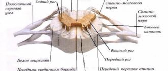
In the spinal cord, gray matter is located around the central canal, surrounded by white matter (cross section) | |
| Latin name | substantia alba |
| System | Central nervous |
White matter
(lat.
substantia alba
) - a component of the central nervous system of vertebrates and humans, consisting mainly of bundles of axons covered with myelin. Contrasted with the gray matter of the brain, which consists of the cell bodies of neurons. The color differentiation of the white and gray matter of nervous tissue is due to the white color of myelin.
In the spinal cord, the white matter is located outside the gray matter. Macroscopically, the white matter of the spinal cord is divided into anterior cords (funiculus anterior), lateral cords (funiculus lateralis) and posterior cords (funiculus posterior).
In the brain, white matter, on the contrary, is located inside and surrounded by gray matter. However, the white matter also contains areas of gray matter - clusters of nerve cells. They are called nuclei.
White matter in the spinal cord
White matter is present in the human body not only in the brain, but also in the spinal cord. However, in this part of the human nervous system, the white matter is located around the gray matter, outside it. Here it is intended to provide communication with certain areas of the brain (for example, the motor center), as well as the interconnection of parts of the spinal cord.
Excerpt characterizing White Matter
But Rostov snatched his hand away and with such malice, as if Denisov were his greatest enemy, directly and firmly fixed his eyes on him. - Do you understand what you are saying? - he said in a trembling voice, - there was no one in the room except me. Therefore, if not this, then... He could not finish and ran out of the room. “Oh, what’s wrong with you and with everyone,” were the last words that Rostov heard. Rostov came to Telyanin’s apartment. “The master is not at home, they have left for headquarters,” Telyanin’s orderly told him. - Or what happened? - added the orderly, surprised at the upset face of the cadet. - There is nothing. “We missed it a little,” said the orderly. The headquarters was located three miles from Salzenek. Rostov, without going home, took a horse and rode to headquarters. In the village occupied by the headquarters there was a tavern frequented by officers. Rostov arrived at the tavern; at the porch he saw Telyanin's horse. In the second room of the tavern the lieutenant was sitting with a plate of sausages and a bottle of wine. “Oh, and you’ve stopped by, young man,” he said, smiling and raising his eyebrows high. “Yes,” said Rostov, as if it took a lot of effort to pronounce this word, and sat down at the next table. Both were silent; There were two Germans and one Russian officer sitting in the room. Everyone was silent, and the sounds of knives on plates and the lieutenant’s slurping could be heard. When Telyanin finished breakfast, he took a double wallet out of his pocket, pulled apart the rings with his small white fingers curved upward, took out a gold one and, raising his eyebrows, gave the money to the servant. “Please hurry,” he said. The gold one was new. Rostov stood up and approached Telyanin. “Let me see your wallet,” he said in a quiet, barely audible voice. With darting eyes, but still raised eyebrows, Telyanin handed over the wallet. “Yes, a nice wallet... Yes... yes...” he said and suddenly turned pale. “Look, young man,” he added.
White matter of the brain
"Substantia alba" or white matter is the fluid that occupies the cavity between the basal ganglia and the "substantia grisea". White matter consists of many nerve fibers, which are conductors that diverge in different directions. Its main functions include not only its conduction of nerve impulses, but also creates a safe environment for the functioning of the nuclei and other parts of the cerebrum (translated from Latin as “brain”). White matter is fully formed in humans in the first six years of their life.
In medical science, it is customary to divide nerve fibers into three groups:
- Associative fibers, which, in turn, also come in different types - short and long, they are all concentrated in one hemisphere, but perform different functions. The short ones connect neighboring convolutions, and the long ones, accordingly, maintain the connection of more distant areas. The paths of associative fibers are as follows: the superior oblong fasciculus of the frontal lobe to the temporal, parietal and occipital cortex; hook-shaped bun and belt; inferior longitudinal fasciculus from the frontal lobe to the occipital cortex.
- Commissural fibers are responsible for the function of connecting the two hemispheres, as well as for the compatibility of their functions in brain activity. This group of fibers is represented by the anterior commissure, the commissure of the fornix and the corpus callosum.
- Projection fibers connect the cortex with other centers of the central nervous system, up to the spinal cord. There are several such types of fibers: some are responsible for motor impulses sent to the muscles of the human body, others lead to the nuclei of the cranial nerves, others lead from the thalamus to the cortex and back, and the last from the cortex to the nuclei of the bridge.
Links
-
Afferent nerve/Sensory neuron GSA GVA SSA SVA Nerve fibers (Muscle spindles (Ia), Neurotendon spindle (Ib), II or Aβ fibers, III or Aδ fibers, IV or C fibers) Efferent nerve/Motor neuron GSE GVE SVE Upper motor neuron Lower motor neuron (α motor neurons, γ motor neurons) Synapse Chemical synapse Neuromuscular synapse Ephaps (Electrical synapse) Neuropil Synaptic vesicle Touch receptor Meissner's corpuscle Merkel's corpuscle Pacini's corpuscle Ruffini's corpuscle Neuromuscular spindle Free nerve ending Olfactory neuron Photoreceptor cells Hair cells Taste bud Neuroglia Astrocytes (Radial glia) Oligodendrocytes Ependymal cells (Tanycytes) Microglia Myelin ( White matter ) CNS: Oligodendrocytes PNS: Schwann cells (Neurolemma · Node of Ranvier/Internodal segment · Myelin notch)
Connective tissue Epineurium Perineurium Endoneurium Nerve fiber bundles Meninges: dura mater, arachnoid membrane, pia mater
Functions of white matter of the brain
The white matter of the cerebral hemispheres “Substantia alba” is generally responsible for coordinating all human life activities, since it is this part that provides communication to all parts of the nerve chain. White matter:
- connects together the work of both hemispheres;
- plays an important role in transmitting data from the cerebral cortex to parts of the nervous system;
- ensures contact of the visual thalamus with the cerebrum cortex;
- connects the convolutions in both parts of the hemispheres.
Functioning of axons
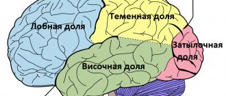
Frontal lobes
These lobes of the brain are more developed than others and have greater mass. The work of the white matter of the frontal lobes contributes to the formation of voluntary movements, regulates complex forms of behavior, mechanisms for reproducing speech and writing, and thinking processes. The white matter pathways of the brain contribute to absolutely all motor processes.
Temporal lobes
The following centers are located here: 1) understanding of oral speech, 2) perception of sound signals, 3) vestibular analyzer, 4) center of vision, 5) center of smell and taste, 6) center of music. The functioning of the temporal lobes is asymmetrical. If a person is left-handed, then the right hemisphere will have greater functionality;
if you are right-handed, then the left hemisphere will be more active (dominant). The functioning of the white matter of this hemisphere makes it possible to understand speech and learn based on the information heard. By combining olfactory, auditory and visual information, draw conclusions, creating images of a harmonious emotional background and long-term memory.
Parietal lobes
The centers located here give a person general sensitivity: pain, tactile and temperature. There are also centers that carry out complex coordinated movements, brought to the point of automatism, and actions of a purposeful nature, acquired through training and continuous practice throughout life.
These are eating, walking, dressing, writing habits, certain work activities and other actions that are unique to humans. The left dominant side provides the ability to write and read; is responsible for actions leading to the desired result; is responsible for feeling the position of your body as a whole and its individual parts;
Occipital lobes
Here, the pathways of the white matter of the brain are aimed at the perception of visual information, followed by its processing and memorization. Objects in the surrounding world are perceived by the eyes as a set of stimuli that reflect light differently onto the retina. The light signal is converted into information about the color and shape of the visible object, its movements.
Damage to the “substantia alba”
Deformation of the white matter threatens with a host of unpleasant consequences, among which are disorders of the hemispheres, problems with the corpus callosum and internal capsule, as well as other mixed syndromes.
Against the background of changes in the condition of this department, the following diseases may develop:
- Hemiplegia – paralysis of one part of the body;
- “Three hemi syndrome” - loss of sensation in half of the face, torso or limb - hemianesthesia; destruction of sensory perception - hemiataxia; visual field defect - hemianopsia;
- Mental illnesses – lack of recognition of objects and phenomena, untargeted actions, pseudobulbar syndrome;
- Disorders of the speech apparatus and impaired swallowing reflex.
Physical exercise
According to recent studies by scientists from the United States, physical activity can have a positive effect on the structure of white matter, and therefore on the health of the entire brain as a whole. First, exercise helps increase blood flow to myelin fibers. Secondly, exercise makes your brain matter denser, which allows it to quickly transmit signals from one part of the brain to another. In addition, it has been scientifically proven that physical activity is beneficial for both children and older people to maintain brain health.
Functions[ | ]
White matter is the tissue through which messages pass between different areas of gray matter in the central nervous system. White matter is white because of the fatty substance (myelin) that surrounds nerve fibers (axons). This myelin is found in almost all long nerve fibers and acts as electrical insulation. This is important because it allows messages to be passed from place to place quickly.
Unlike gray matter, which peaks in development in your twenties, white matter continues to develop and peaks in middle age.
Relationship between age and white matter status
Neuroscientists from the USA conducted an experiment: the scientific research group included people aged 7 to 85 years. Using diffusion tomography, more than a hundred participants were examined in the brain and in particular the volume of the “substantia alba”.
The conclusions are as follows: the largest number of high-quality connections was observed among subjects aged 30 to 50 years. The peak of thinking activity and the highest degree of learning develops to the maximum in the middle of life, and then declines.
Internal structure of the cerebral hemispheres
The surface of the hemispheres is covered with a thin layer of gray matter - the cerebral cortex, along which the nuclei - the basal ganglia of the cerebral hemispheres - are located in the thickness of the white matter.
The cerebral cortex has a thickness of 1.3 to 5 mm, the thickest cortex is located in the upper parts of the precentral and postcentral gyri. The cortex contains about 13 billion neurons, each of which forms synapses with 8–10 thousand others. In the cortex, pyramid-shaped cells predominate; a long axon and several basal dendrites extend from the base of the “pyramid,” and an apical branching dendrite extends from the apex. Small pyramidal cells are interneurons, large ones are effector neurons. Thus, Betz giant pyramidal cells of the precentral gyrus generate impulses of voluntary movements directed to skeletal muscles through the motor nuclei of the brain and spinal cord (pyramidal tracts). About 96% of the surface of the human hemispheres is covered with a new cortex (neocortex), which has a six-layer structure: the outer layer is molecular (small multipolar associative neurons, processes of cells of the underlying layers), the second layer is the outer granular layer (small pyramidal neurons, the dendrites of which are directed into the molecular layer , axons - also into the molecular layer or into the white matter), the third - middle pyramidal (the thickest, pyramidal cells of small and medium sizes, axons of small cells do not leave the cortex, axons of larger ones form commissural fibers in the white matter), fourth - internal granular (small pyramidal and stellate neurons), the fifth is a layer of large pyramidal cells (best developed in the precentral gyrus, where it is represented by Betz’s giant pyramidal cells), the sixth layer is polymorphic (neurons of different shapes and sizes, the axons of which go into the white matter, dendrites - into the molecular layer). The old cortex (archicortex) makes up just over 2%, is located in the limbic lobe, dentate gyrus and hippocampus, and is divided into 3 layers. The ancient cortex (paleocortex) occupies less than 1% of the surface, is located in the structures of the olfactory brain and in the transparent septum, and has no layers. There is also an intermediate type of bark.
Rice. 22. Layers of new crust
- – molecular; 2- external granular; 3- outer pyramidal;
4 – – internal granular; 5 – internal pyramidal; 6- polymorphic
Localization of functions in the cerebral cortex. The sensory zones of the cortex provide analysis of stimuli from the external and internal environment of the body and are the morphological substrate of sensations and perception. Primary (projection) zones receive information directly from subcortical sensory centers and analyze individual signs of the stimulus. The main afferentation comes through the nuclei of the thalamus to the 3rd and 4th layers of the cortex. Localization of projection zones: somatovisceral sensitivity - postcentral gyrus and superior parietal lobule, visual - “along the banks” of the calcarine sulcus, auditory - the middle part of the superior temporal gyrus (small transverse gyri of Heschl), vestibular - middle temporal and postcentral gyri, taste sensitivity - lower sections postcentral gyrus, parahippocampal gyrus, uncinus, hippocampus, olfactory – uncus, hippocampus, olfactory olfactory tracts, olfactory triangles, anterior perforated substance, septum pellucidum. Secondary sensory zones provide holistic perception of the stimulus and are located near the primary ones. In the associative sensory zones, which occupy a much larger area than the primary and secondary ones, intersensory integration and the formation of a holistic sensory picture of the world occur. Motor zones of the cortex - precentral gyrus (primary motor cortex, which directly mobilizes motor neurons), inferior parietal lobule, supramarginal gyrus (coordination of purposeful complex movements), extensive cortical fields anterior to the precentral gyrus (associative and premotor zones, where the strategy and plan of movements are formed ). Speech centers - the posterior sections of the inferior frontal gyrus (Broca's center, or speech motor center, regulates the work of the muscles involved in the speech act), the posterior sections of the middle frontal gyrus (the center for regulating movements associated with writing), the posterior sections of the superior temporal gyrus (Wernicke's center, or sensory speech center, ensures the perception and understanding of oral speech), angular gyrus (the center for comparing the visual image of a word with its acoustic analogue). Emotional zones - the limbic cortex and olfactory brain. Higher mental functions (consciousness, thinking, memory, emotions) are the result of the integrative activity of the entire cerebral cortex, primarily the frontal cortex.
The basal ganglia of the cerebral hemispheres functionally belong to the striopallidal and limbic systems. Nuclei of the striopallidal system: the caudate nucleus, the putamen (the striatum, corpus striatum, on sections it looks like alternating dark and light stripes) and the globus pallidus, globus pallidum, light in color and uniform in structure. The putamen and globus pallidus are anatomically united into the lenticular nucleus, separated from the caudate nucleus and thalamus by the internal capsule of white matter. The caudate nucleus is located lateral and above the thalamus, separated from it by the knee of the internal capsule, has a head (located in the frontal lobe, forms the lateral wall of the anterior horn of the lateral ventricle), a body (deep in the parietal lobe, forms the lateral wall of the central part of the lateral ventricle), and a tail ( extends into the anteromedial part of the temporal lobe, forms the superior wall of the inferior horn of the lateral ventricle). The putamen is the largest nucleus of the striopallidal system and lies below and lateral to the caudate nucleus. The globus pallidus is located medial to the putamen and consists of medial and lateral plates. The striopallidal system is part of the extrapyramidal system that controls movement and muscle tone. Nuclei of the limbic system: septal nuclei (in the septum pellucidum), septum (lateral to the putamen, separated from it by the outer capsule of the white matter and from the insular cortex by the outermost capsule), amygdala (in the white matter of the temporal lobe anterior to the inferior horn of the lateral ventricle).
The limbic system of the brain , in addition to the listed nuclei, includes the limbic lobe of the cortex, the olfactory brain, the hippocampus, as well as structures of the diencephalon - the anterior nuclei of the thalamus, the hypothalamus, and some reticular nuclei. All structures of the limbic system are connected to each other and form a vicious circle, which creates conditions for long-term circulation of excitation. The limbic system is the morphological substrate of emotions and motivations, the center of regulation of autonomic functions
Rice. 23. Brain structures related to the limbic system
|
| Rice. 24. Basal ganglia of the cerebral hemispheres | |
Horizontal section of the brain1 – cerebral cortex; 2 – genu corpus callosum;
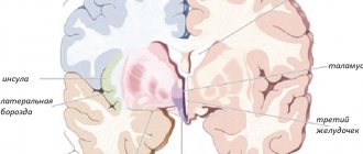
8 – shell; 9 – globus pallidus; 10 – third ventricle; 11 – posterior horn of the lateral ventricle; 12 – thalamus; 13 – islet cortex; 14 – head of the caudate nucleus; 15 – cavity of transparent partitions | Frontal incision at the mastoid levelny bodies
9 - globus pallidus; 10 – shell; 11 – vault; 12 – caudate nucleus; 13 – corpus callosum |
Rice. 25. “Undivided” brain
| Rice. 26 Layout of associative fibers of the left hemisphere (lateral surface) 1 - superior longitudinal fasciculus 2 - arcuate fibers 3 - uncinate fasciculus | Rice. 27. Layout of bundles of associative fibers of the right hemisphere (medial surface)1 – encircling bundle2 – superior longitudinal bundle 3 – arcuate fibers 4 – lower longitudinal beam |
| Rice. 28. Projection corticospinal fibers, frontal section 1 – vault; 2 – tail of the caudate nucleus; 3 – internal capsule; 4 – shell; 5 – globus pallidus; 6 – lower horn of the lateral ventricle; 7 – choroid plexus of the lateral ventricle; 8 – red core; 9- corticospinal tract; 10 – olive core; 11 – medulla oblongata; 12-bridge; 13- gray matter; 14 – thalamus; 15 – spinal fibers; 16 – corpus callosum | Rice. 29. Commissural fibers of the cerebrum, frontal section (diagram)
|
The white matter of the cerebral hemispheres is represented by three types of fibers: projection (connect the cortex with underlying structures, form capsules between the basal ganglia), associative (connect areas of the cortex within one hemisphere), commissural (connect the cortex of the right and left hemispheres). The largest commissure is the corpus callosum, which connects the neocortex of the hemispheres. The anterior part is the knee, bends downwards and backwards, passes into the beak, and then into the terminal plate. The middle part is the trunk, the rear thickened part is the roller. The anterior commissure connects the old and ancient cerebral cortex and is located posterior to the lamina terminalis of the corpus callosum in front of the columns of the fornix. The arch commissure connects the right and left parts of the arch. The cerebral vault is the conductive system of limbic structures. Consists of two arcuate cords located under the corpus callosum above the roof of the third ventricle. The middle part of the vault - the body anteriorly moves away from the corpus callosum, bends forward and downward and passes into the columns of the vault, ending in the mastoid bodies. Posteriorly, the body of the arch continues into the legs of the arch, connected to each other by a commissure of the arch. The crura of the fornix diverge, run along the hippocampus and end in the area of the uncinates of the temporal lobes.
Autonomic (autonomic) nervous system
The autonomic nervous system (ANS) coordinates and regulates the activity of internal organs, metabolism, and functional activity of tissues. Its main function is to maintain homeostasis (constancy of the internal environment of the body). The ANS has central and peripheral sections.
The central ANS consists of segmental and suprasegmental vegetative centers. Segmental centers (sympathetic and parasmpathetic) are located in the lateral horns of the spinal cord and in the brain stem (autonomic nuclei of the cranial nerves). There lie effector autonomic neurons connected (via the autonomic ganglia) with the working organs. The suprasegmental centers are the reticular formation, the diencephalon, the basal ganglia of the cerebral hemispheres, the cerebellum, and the cerebral cortex (mainly the limbic cortex). These structures do not have a direct connection with the working organs and influence their functions through segmental centers. The suprasegmental centers are not clearly divided into sympathetic and parasympathetic, but have ergotropic and trophotropic zones that partially overlap. Activation of ergotropic zones causes predominantly sympathetic effects in the periphery, activation of trophotropic zones – predominantly parasympathetic. At the suprasegmental level, the centers of the autonomic and somatic nervous systems are united and function cooperatively, at the level of segmental centers and in the peripheral part they are separated morphologically and functionally.
The peripheral ANS consists of autonomic nerves and autonomic ganglia (sympathetic and parasympathetic). According to their location, autonomic ganglia are paravertebral (located near the spine), prevertebral (remote from the spine), extramural (near innervated organs), intramural (in the walls of internal organs). In the ganglia, efferent pathways switch from segmental centers to working organs. Thus, the arc of the autonomic reflex differs from the arc of the somatic reflex in that the efferent pathway consists of two neurons: the bodies of the first effector autonomic neurons are located in the segmental autonomic centers, the bodies of the second effector neurons are in the autonomic ganglia. Preganglionic fibers have a myelin sheath, postganglionic fibers are unmyelinated.
The sympathetic division of the ANS has centers in the lateral horns of the C VIII – LII segments of the spinal cord. Ganglia are paravertebral ganglia located in the form of a chain to the right and left of the spine (right and left sympathetic trunks) and prevertebral ganglia of the abdominal cavity and pelvis. Postganglionic fibers form autonomic nerve plexuses that innervate internal organs. The plexus contains both sympathetic and parasympathetic fibers. Stimulation of sympathetic nerves causes increased heart rate and contractions, narrowing of the arteries of the skin, mucous membranes (arteries of the brain, skeletal muscles, heart can either narrow or expand), an increase in total blood pressure, weakening of motility and secretion of the gastrointestinal tract, urinary retention , dilation of the respiratory tract, dilation of the pupil, increased sweating.
The parasympathetic division of the ANS has centers in the autonomic nuclei of the III, VII, IX, X pairs of cranial nerves and in the lateral horns of the S II–V segments of the spinal cord (innervation of the pelvic organs). Preganglionic parasympathetic fibers are involved in the formation of autonomic plexuses. Ganglia - extra- and intramural (ciliary node - in the orbit, pterygopalatine, submandibular, auricular - near the bones of the skull, organ nodes - near or inside the organs of the neck, chest, abdomen). Stimulation of the parasympathetic nerves causes a slowdown and weakening of heart contractions, dilation of the arteries of the external genitalia, increased motility and secretion of the digestive tract, contraction of the walls of the bladder, narrowing of the airways and increased bronchial secretion, increased secretion of the lacrimal glands, and constriction of the pupil.
The metasympathetic nervous system is the most autonomous part of the autonomic nervous system, has no centers in the spinal cord or brain, is represented by intramural ganglia and short fibers, and carries out local regulation of individual muscle groups and glands of internal organs.
Rice. 30. Scheme of the arc of the autonomic reflex
1 – posterior root; 2 – spinal node; 3 – lateral horn; 4 – preganglionic autonomic fibers (as part of the anterior root);
5 – spinal nerve; 6.8 – connecting branches; 7 – node of the sympathetic trunk; 9 – postganglionic autonomic fibers (as part of the spinal nerve); 10 – postganglionic autonomic fibers (as part of the splanchnic nerve); 11 – node of the autonomic plexus; 12 – postganglionic fibers (as part of the visceral plexuses); 13- postganglionic fibers to the sweat glands of the skin, hair muscles and blood vessels
Rice. 31. Diagram of autonomic innervation of internal organs: a – parasympathetic part, b – sympathetic part
1 – upper cervical node;
2 – lateral columns of the spinal cord; 3 – upper cervical cardiac nerve; 4 – cardiac and pulmonary thoracic nerves; 5 – great splanchnic nerve; 6 – celiac plexus; 7 – inferior mesenteric plexus; 8 – hypogastric plexuses; 9 – small splanchnic nerve; 10 - lumbar splanchnic nerves; 11 – sacral splanchnic nerves; 12 – sacral parasympathetic nuclei; 13 – pelvic splanchnic nerves; 14 – pelvic nodes; 15 – parasympathetic nodes (as part of organ plexuses); 16 – vagus nerve; 17 – ear node; 18 – submandibular node; 19 – pterygopalatine node; 20 – ciliary node; 21 – posterior nucleus of the vagus nerve; 22 – lower salivary-secretory nucleus; 23 – superior salivary nucleus; 24 – accessory oculomotor nucleus Don’t know how to solve or complete a coursework or dissertation? Order a solution

