Main article
:
Color vision
Main article
:
Theory of three-component color vision (Mig version)
Fig.1.
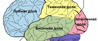
Human brain, rear view. Primary visual cortex V1
(Brodmann area 17) is indicated in red orange - field 18; yellow - field 19.[1] Fig.2. Human brain, left view. Above: lateral surface, below: medial surface. Brodmann area 17 (primary, or striate, visual cortex) is indicated in orange [2] Fig. 3. Dorsal (green) and ventral (lilac) visual pathways originating in the primary visual cortex.[3]
Visual cortex
(eng. visual cortex) is part of the cerebral cortex, responsible for processing visual information. It is mainly concentrated in the occipital lobe of each cerebral hemisphere[4].
The opposingly selected brightest signals of visible light rays S, M, L - GLC (not in color), focused object points on the exteroceptors (Mig version) of the cones of the retina (receptor level), are sent along the optic nerves here, to the visual cortex. Here a binocular (stereo) color optical image is formed (neural level). For the first time, subjectively we experience a color (the Mig version) that is personally ours. (When determining color using colorimetry, color is estimated by the data of the average observer of a large group of healthy people)
Concept of visual cortex
includes
the primary visual cortex
(also called
the striate cortex
or
visual area V1
) and the extrastriate visual cortex - areas
V2
,
V3
,
V4
, and
V5
.
(See about areas V2
,
V3
,
V4
, and
V5
in the article Visual cortex)
Primary visual cortex is anatomically equivalent to Brodmann area 17
, or BA17. The extrastriate visual cortex includes Brodmann areas 18 and [4].
The visual cortex is present in each of the cerebral hemispheres. Areas of the visual cortex of the left hemisphere receive signals from the right half of the visual field, and the right hemisphere receive signals from the left half.
In the future, the article will talk about the features of the visual cortex of primates (mainly humans).[5]
Introduction[edit]
Fig. 4, Scheme of color vision from the point of view of the three-component theory
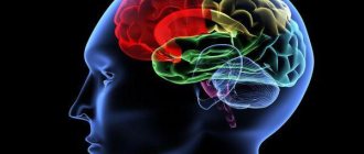
Visual parts of the brain
- perception of color and light, obtaining an optical image in the cerebral cortex - the second, final stage of the visual system for the formation of optical vision in the visual parts of the brain (see Fig. 3,4).
Even at the initial stage of visual perception of light and color in the visual system, within the retina, passing through the initial color mechanisms of the “opponent”.
Fig.3a. Optical paths after the meeting of signals from the right and left eyes into the layers of the geniculate body
Enemy mechanisms are known to resort to opposing color effects of red-green, blue-yellow, and black-white. (See Opponent Color Vision Theory (Mig version)). In this case, visual information is returned back through the optic nerve to the optic chiasm, where the two optic nerves meet and information from the temporal (contralateral) visual field chiasm to the opposite side of the brain. After the optic chiasm, the optic nerve fiber tracts are referred to as optic tracts, which enter the thalamus en:Thalamus through a synapse at the lateral lateral geniculate body (LCT). The LCT is a separate section of the brain made up of six layers: two magnocellular (large cell) colorless layers (M cells) and four parvocellular (small cell) color layers (P cells). Within the P-cell layers of the LCT there are two colored enemy types: red vs. green and blue vs. yellow (green/red)
.
After synapsis in the LCT, the visual tracts move back to the primary visual cortex
(PVC-V1), located behind the brain within the occipital lobe. Within layer V1 of the lateral geniculate body there is a distinct streak (striation). It is also referred to as "striate cortex", with other cortical visual areas referred to collectively as "extrastriate cortex". At this stage, color processing becomes much more complex.
Activating reticular system
The diffuse nervous system consists of nonspecific nuclei.
This system is located in the medial sections of the thalamus. It is the anterior part of the activating reticular system, which regulates the excitability of the cortex. A variety of sensory signals can activate this system. Sensory signals can be both visual and olfactory, somatosensory, vestibular, auditory. The activating reticular system is a channel that transmits signal data to the superficial layer of the cortex through nonspecific nuclei located in the thalamus. Excitation of the ARS is necessary for a person to be able to maintain a state of wakefulness. If disturbances occur in this system, comatose sleep-like states may occur.
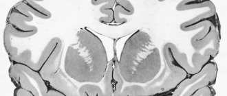
Primary visual cortex (VI)[edit]
Fig.4. Human brain. visual area V1
is indicated in red. Fig. 5. Photomicrograph shows the visual cortex (pink). The pia mater and arachnids including blood vessels are visible at the top of the image. Subcortical white matter (blue) - This is visible at the bottom of the image. OH-LFB spot..
Primary visual cortex
- the most studied visual area of the brain. Studies have shown that in mammals it occupies the posterior pole of the occipital lobe of each hemisphere (these lobes are responsible for processing visual stimuli). This is the simplest [6] and phylogenetically “older” of the cortical zones associated with vision. It is adapted for processing information about static and moving objects, especially for recognizing simple images.
An integral part of the functional architecture of the cerebral cortex - the primary visual cortex - almost completely corresponds to the anatomically defined striate cortex
.
The name of the latter goes back to the Latin “stripe, stripe” (lat. stria) and is largely due to the fact that here the strip of Gennari [en]
(outer strip of Baillarger), formed by the terminal sections of myelin-covered axons extending from the neurons of the lateral geniculate body and ending in
IV
of the gray matter.
The primary visual cortex is divided into six functionally distinct horizontal cytoarchitectonic layers
(see Fig. K), designated by Roman numerals from
I
to
VI
[4][7].
Layer IV
(
the internal granular layer
[7]), which receives the largest number of afferent fibers coming from the lateral geniculate body (LCT), is in turn divided into four sublayers, designated
IVA
,
IVB
,
IVCα
and
IVCβ
.
Nerve cells of the IVCα
mainly receive signals coming from neurons of the magnocellular (“large cell”, ventral) layers of the LCT [8] (“
magnocellular visual pathway
the IVCβ
sublayer - from neurons of the parvocellular (“small cell”, dorsal) layers of the LCT [8] ("
parvocellular visual pathway
").
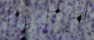
It is believed that the average number of neurons in the adult primary visual cortex is about 140 million in each hemisphere[9].
Function[edit]
Risk.
Band 6 is the primary visual cortex (also called striate cortex or visual area V1. Schematic diagram of P-cell neurons located within the parvocellular layers of the geniculate nucleus (LGN) of the thalamus. The primary visual cortex (V1) has very clear maps of spatial information in vision. For example In humans, the upper half of the calcarine region ("spur") of the fissure responds strongly to incoming visual signals. From the lower half of the visual field of the calcarine region, the flow goes to the upper half of the visual field. Conceptually, it is (retinotopic) or it is a representation of visual information from the retina, neurons , especially the visual flow of neurons. This is how mapping occurs - the transformation of the visual optical image from the retina to the V1 area.
The correspondence of this location in the V1 zone and in the subjective field of vision is correlated very accurately: even the blind spots of the retina are compared with the zone with data in V1. From an evolutionary point of view, this redirection occurs very simply in most animals that possess the V1 zone. In animals and humans with a fovea (the center of the macula - the macula) in the retina, most of the V1 area is mapped to a small central part of the visual field. A phenomenon known as cortical enlargement. Perhaps for the purpose of precise spatial coding, neurons in V1 have the smallest receptive field sizes of any visual cortex or microscopic areas.
The tuning properties of area V1 neurons (neuron responses) differ significantly over time. Early in the tuning time (40 ms onwards) individual V1 neurons have strong (tuning) characteristics of exposure to a small set of stimuli. That is, neuron responses may differ by small changes in the visual orientation of spatial frequencies and colors. Moreover, individual neurons in humans and animals of zone V1 of binocular vision in the ocular system, namely, adjust one of the two eyes. In area V1 and the primary sensory cortex in general, neurons with similar tuning properties tend to cluster together in cortical columns. David Hubel and Thorsten Wiesel proposed the classic “ice cube” model of cortical column organization to tune two properties: ocular dominance and orientation. However, this model cannot accommodate color, spatial frequency, and many other features that tune neurons [citation]. The precise organization of all these cortical columns in area V1 remains a hot topic in the present study.
The current consensus is that V1 neuronal responses appear to consist of tiled structures that represent selective spatiotemporal filters. The functioning of zone V1 in the spatial domain can be considered as an analogue of a set of spatial-local ones - the Fourier Transform complex or, more precisely, the Gabor transform. In theory, these filters together can handle neural processing of spatial frequency, orientation, motion, direction, speed (temporal frequency), and many other spatiotemporal characteristics. Neuronal experiments are required to substantiate these theories, but new questions are raised.
At later times (after 100 ms) V1 neurons are also sensitive to more global scene organization (Lamme & Roelfsema, 2000). These response parameters are likely due to repetitive processing (with high-level cortical areas influencing low-level cortical areas) and horizontal connections from pyramidal neurons (Hupe et al. 1998). While forward connections are primarily operational, feedback connections are primarily modulatory with their consequences (Angelucci et al., 2003; Hupe et al., 2001). Experience shows that feedback occurring at a higher level, in areas such as V4 OH or MT, from larger and more complex receptive fields, can change the shape of V1 responses, taking into account contextual or extra-classical receptive fields effect (Guo et al., 2007; Huang et al., 2007; Sillito et al., 2006).
The visual information conveyed to area V1 is not encoded in spatial (or optical) terms, but rather it is local contrast. For example, for an image consisting of half a black side and half a white side, the line break between black and white represents strong local contrasts and is encoded, and at the same time, in the form of several code neurons, luminance information (black or white per se) . As information for further retransmission to subsequent visual zones, all non-local frequencies and signal phases are also encoded in it. Crucially, at these early stages of cortical visual processing, the spatial location of visual information is well preserved against the backdrop of local encoding contrast.[10]
The principle of opposition in the selection of color signals[edit]
Main article
:
Color vision - differentiated perception of the main rays of focused object points (Mig version)
Main article
:
Trichromatism and the principle of opposition (Mig version)
Main article
:
The theory of opponent color vision (Mig version)
Scheme of trichromatism with opponent selection of color Schematic diagram of three-component color vision of humans, primates with opponent selection of the brightest main rays of light KZS with subsequent transmission of color signals S, M, L to the brain
The principle of opposition in the selection of the main light signals S, M, L - KZS
is a theory that defines the way in which the retina (MIG version) of the human eye allows the visual system to sense color (MIG version) at the neural level in the brain's cortex. This occurs at the level of perception of visible rays (receptor) by three types of RGB cones (S, M., L), based on opponent color selection (white-black, red-green, blue-yellow) with subsequent transmission of signals to the brain.[11 ][12]
Optical images in the brain and in photography[edit]
Optical imaging in the brain[edit]
Based on the above, it is clear that the optical image (or object points) on the focal surface - the retina (biological photosensor), as in photography, is perceived by cells consisting of a certain number of photosensors (pixels), for example, cones, sensitive to the main spectral rays, for example, to red, green, blue (RGB). Signals from photosensors or photoreceptors of cones (there are about 6 million of them) are transmitted to the brain through a strictly connected biological transmission system using synapses along neural channels, of which there are approximately 1.2 million. The question arises: how are there 6 million signals transduced by blue, green, red cones of each block or from 2 million? 1.2 million cells can be transmitted. channels? In this case, one should take into account the work of exteroreceptors (photosensors) of the ganglion layer of the retina ipRGC, synapsically connected by direct and feedback with cones, rods and the brain containing the photopigment melanopsin, which are capable of suppressing or enhancing phototransduction of biosignals of rods and cones.[citation needed].
At the initial stage of visual perception of light and color (within the retina), color perception begins at an early level in the visual system - already within the retina, passing through the initial color mechanisms of the “opponent” - the opposing selection of the brightest signals.
After synapsis in the LGN, the visual tracts move back to the primary visual cortex
(PVC-V1)en:Visual_cortex, located behind the brain within the occipital lobe en:Occipital_lobe. Within layer V1 of the lateral geniculate body there is a distinct streak (striation). It is also referred to as the "striate cortex" with other cortical visual areas referred to collectively as the "extrastriate cortex". At this stage, color processing becomes much more complex.
As a result, the biological ADC created by nature (at the level of the retina and brain) is a unique biological system for converting and obtaining optical images (color and gray) in the brain (including stereo). Achievements in the field of color and stereo photography are still far from the perfection of these visual biological systems created by nature, with which we visually enjoy the colorful world around us every day.
Optical image in photography[edit]
Main article
:
Bayer filter
Diencephalon
Between the cerebral hemispheres is the diencephalon, which is divided into two parts: the thalamus and hypothalamus. In addition to them, there is also the epithalamus, to which the pineal gland and pituitary gland are adjacent - endocrine glands. The thalamus is an “information funnel” that filters signals and passes them to the cerebral cortex: if all information flows passed into the cortex, it would not be able to function effectively. Blocking signals is carried out using inhibitory neurons. The structures of the thalamus correspond to different centers of the cerebral cortex: the anterior nuclei are responsible for transmitting information to the centers of emotions and memory, the ventral lateral ones are associated with motor control, the ventrobasal complex works with information about the sensitivity of the body, and above it are the auditory and visual centers. The medial nuclei of the thalamus are associated with the centers of sleep and wakefulness, as well as with taste and pain signals and vestibular sensitivity.
The hypothalamus is involved in neuroendocrine regulation and controls the activities of various internal organs. In addition, it contains the most important centers of biological needs: hunger and thirst, sexual and parental behavior, fear and aggression. Neurons of the hypothalamus evaluate the concentration of major hormones in the blood. The hypothalamus closely interacts with the pituitary gland, a gland that produces thyroid-stimulating hormone and thereby regulates the activity of the thyroid gland. Commands are given to the pituitary gland using releasing hormones produced by the hypothalamus. The hypothalamus also produces oxytocin and vasopressin, hormones that are respectively responsible for the contraction of the uterus during childbirth and the mammary glands when feeding a child and the body’s need for fluid. The pineal gland (or pineal gland) influences pubertal development and sexual behavior and produces melatonin, which is involved in synchronizing circadian rhythms.
About the work of the thalamus and hypothalamus
Notes
- Butler, Ann B. (2001). "Chordate Evolution and the Origin of Craniates: An Old Brain in a New Head". The Anatomical Record 261: 111–125.
- Amphibian brain
- Brain Facts and Figures. Retrieved June 8, 2012. Archived June 22, 2012.
- Heike Le Ker.
Neuronen-Nachschub: Neue Nervenzellen wachsen im menschlichen Gehirn nach (German).
Spiegel Online
(15. Februar 2007). Retrieved January 9, 2020. - Nerve cells of the human brain are still being restored, Swedish scientists say. NEWSru.com
(February 16, 2007). Retrieved January 9, 2020. - Evgenia Samokhin.
“Burner” of energy // Science and Life: magazine. - M., 2020. - No. 4. - P. 22-25. — ISSN 0028-1263.
Content
- 1 The brain as an organ of vertebrates
- 2 Brain tissue
- 3 Brain cells
- 4 Blood supply
- 5 Functions
- 6 Parts of the brain
- 7 Plasticity
- 8 Embryonic development
- 9 Research methods 9.1 Ablations
- 9.2 Transcranial magnetic stimulation
- 9.3 Electrophysiology
- 9.4 Electrical stimulation
- 9.5 Other techniques
Embryonic development
The brain of a four-week embryo
Embryonic development of the brain is one of the keys to understanding its structure and functions.
The brain develops from the rostral part of the neural tube. Most of the brain (95%) is a derivative of the pterygoid plate.
Embryogenesis of the brain goes through several stages.
- Stage of three brain vesicles - in humans, at the beginning of the fourth week of intrauterine development, the rostral end of the neural tube forms three vesicles: Prosencephalon (forebrain), Mesencephalon (midbrain), Rhombencephalon (diamond-shaped brain, or primary hindbrain).
- Stage of five brain vesicles - in humans, at the beginning of the ninth week of intrauterine development, the Prosencephalon is finally divided into Telencephalon (telecephalon) and Diencephalon (diencephalon), Mesencephalon is preserved, and Rhombencephalon is divided into Metencephalon (hindbrain) and Myelencephalon (medulla oblongata).
During the formation of the second stage (from the third to the seventh weeks of development), the human brain acquires three bends: midbrain, cervical and pavement. First, the midcerebral and pontine flexures are formed simultaneously and in one direction, then the cervical flexure is formed in the opposite direction. As a result, the linear brain “folds” in a zigzag manner.
During the development of the human brain, a certain similarity between phylogeny and ontogenesis can be noted. In the process of evolution of the animal world, the telencephalon was formed first, and then the midbrain. The forebrain is an evolutionarily newer brain formation. Also, during the intrauterine development of a child, the hindbrain is first formed as the most evolutionarily ancient part of the brain, and then the midbrain and then the forebrain. After birth, from infancy to adulthood, an organizational complication of neural connections in the brain occurs.
Motor zone
Let's talk about the motor zone separately. It should be noted that this zone of the cortex does not correlate in any way with the lobes discussed above. It is part of the cortex containing direct connections to motor neurons in the spinal cord. This name is given to neurons that directly control the activity of the muscles of the body.
The main motor area of the cerebral cortex is located in a gyrus called the precentral gyrus. This gyrus is a mirror image of the sensory area in many aspects. Between them there is contralateral innervation. To put it another way, the innervation is directed to the muscles that are located on the other side of the body. The exception is the facial area, which is characterized by bilateral control of the muscles located on the jaw and lower part of the face.
Slightly below the main motor zone is an additional zone. Scientists believe that it has independent functions that are associated with the process of outputting motor impulses. The supplementary motor area has also been studied by specialists. Experiments carried out on animals show that stimulation of this zone provokes the occurrence of motor reactions.
The peculiarity is that such reactions occur even if the main motor area has been isolated or completely destroyed. It is also involved in motor planning and speech motivation in the dominant hemisphere. Scientists believe that if the accessory motor is damaged, dynamic aphasia can occur. Brain reflexes suffer.