Coordinate movement and orientation in space
1) medulla oblongata and cerebellum
2) midbrain and forebrain
3) cerebellum and cerebral cortex
4) spinal cord and cerebral hemispheres of the forebrain
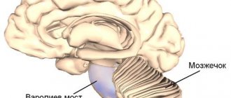
The cerebellum and cerebral cortex coordinate movement and orientation in space. The cerebellum is a part of the vertebrate brain responsible for coordinating movements, regulating balance and muscle tone. The cerebellum receives a copy of afferent information transmitted from the spinal cord to the cerebral cortex, as well as efferent information from the motor centers of the cerebral cortex to the spinal cord. The first signals the current state of the controlled variable (muscle tone, position of the body and limbs in space), and the second gives an idea of the required final state. Although the cerebellum is connected to the cerebral cortex, its activity is not controlled by consciousness.
Human motor skills are characterized by the accuracy of purposeful movements, which is ensured by the proportional work of many muscle groups, controlled voluntarily and automatically. This complex multifunctional system is implemented by a multineural coordinating apparatus that controls the balance of the body, stabilizes the center of gravity, regulates the tone and coordinated various activities of the muscles. The center of coordination of movements is the cerebellum; functionally, it contains the body of the cerebellum, consisting of two hemispheres, the vermis and three pairs of peduncles.
In the implementation of voluntary movement, the main role of the cerebellum is to coordinate the fast and slow components of the motor act. This becomes possible thanks to the bilateral connections of the cerebellum with the muscles and cerebral cortex. The cerebellum receives afferent impulses from all receptors irritated during movement (from proprioceptors, vestibular, visual, auditory, etc.). Receiving information about the state of the motor system, the cerebellum influences the red nucleus of the brain, which sends impulses to the γ-motoneurons of the spinal cord, which regulate muscle tone. In addition, part of the afferent impulses through the cerebellum enters the cerebral cortex of the motor zone (precentral and frontal gyri).
The main function of the cerebellum is carried out at a subconscious level. Efferent impulses from the cerebellar nuclei regulate proprioceptive stretch reflexes. During muscle contraction, the proprioceptor of the synergist muscles and antagonist muscles are excited. Normally, the transformation of voluntary movement into a complex reflex does not occur due to the inhibitory influence of cerebellar impulses. Therefore, when the cerebellum is damaged, disinhibition of segmental proprioceptive reflexes is manifested by movements of the limbs like ataxia.
The cerebellum has many afferent and efferent connections. Posterior spinocerebellar tract (Flexig's tract). The first neuron is located in the spinal ganglion; the axon in the dorsal root, through the dorsal horn, approaches the cells of Clark's column. The fibers of these second neurons are directed to the outer layers of the posterior part of the lateral funiculus on their side, ascend along the entire spinal cord and, at the level of the medulla oblongata, as part of the inferior cerebellar peduncle, enter the cerebellar vermis. This pathway is referred to as the posterior spinocerebellar tract. In the cortex of the cerebellar vermis there is a third neuron that contacts the piriform neurons of the cerebellar hemisphere cortex. The axons of the latter go to the dentate nucleus. The fibers of this fifth neuron are part of the superior cerebellar peduncle. The right and left superior cerebellar peduncles cross - Wernicke's cross - and end at the red nucleus cells of the opposite side. The axons of the cells of the red nucleus are immediately directed to the opposite side of the midbrain and form a ventral decussation in the tegmentum of the midbrain - the decussation of Forel, pass as part of the lateral cord of the spinal cord, and reach the cells of the anterior horns. The set of axons of the cells of the red nucleus is called the Monakov bundle.
Govers' anterior spinocerebellar tract. The first neuron is located in the spinal ganglion, the second neuron is a cell of the dorsal horn, but its axons move to the opposite side and go up the spinal cord, in the anterior part of the lateral cord, pass through the medulla oblongata, the pons of the brain, at the level of the superior medullary velum they pass to the opposite side side and as part of the superior cerebellar peduncle reach the cells of the cerebellar nuclei.
The cerebellum receives afferent proprioceptive impulses not only along the Flexig and Gowers pathways, they also arrive along the axons of the cells of the nuclei of the thin and cuneate fasciculi, some of which go through the lower cerebellar peduncles to its vermis.
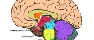
Structure
- Terminal, which includes both hemispheres
- Posterior, to which the cerebellum belongs
- Middle, located between the pons and the cerebellum
- Intermediate, above average
- Oblong, which is a direct continuation of the dorsal
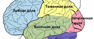
The concept of the telencephalon unites both hemispheres, while it is also usually divided into 4 lobes - frontal, temporal, parietal, occipital.
The coordinated work of all departments is aimed at the work of higher mental functions - perception, attention, memory, thinking. Our nervous system receives signals from the senses, and the brain processes them - hearing, vision, taste, smell, sense of balance. It also controls all vital processes - breathing, heartbeat, metabolism. Let's take a closer look at where this magic happens.
Functions of the cerebellum
The coordinated work of the main vital systems largely depends on the degree of damage to the “smallest” organ itself. If these parts of the brain are completely removed, the person simply will not be able to exist. With partial removal, this will lead to the main symptoms of its damage (tremor of the limbs, ataxia, etc.), but with proper therapeutic treatment, these symptoms go away.
However, if, when symptoms recede, the functionality of the frontal lobe of the brain is disrupted, then the symptoms return. Therefore, we can say that the cerebral cortex somewhat relieves pathological manifestations that are caused by damage to the cerebellum.
If we more accurately describe the symptoms when the part of the brain responsible for coordinating movements is damaged, then the manifestations can be expressed as follows:
- Intentional (intentional) tremor of the limbs, which occurs, for example, when trying to hit the nose with a finger
- Slowness of speech
- Lack of smoothness in limb movements
- Changed handwriting
- Gait disturbance and constant dizziness (ataxia)
- Loss of sensation
- Intestinal dysfunction
- Increased intensity of metabolic processes, for example, a sharp increase in blood sugar when eating sweets, while the sugar level remains for a long time
- Decreased appetite, tendency to anorexia
- Delayed healing of skin lesions
- Decreased vascular tone
In the case of complete removal of this part of the brain, the symptoms are expressed even more intensely. With ataxia, which manifests itself most intensely when the cerebellum is damaged or removed, the patient simply cannot get out of bed, an unsteady gait and eye twitching appear.
The cerebellum is directly involved in almost all systems of our life:
This “little brain” also influences the coherence of these systems through its implementation through other structures of the central nervous system; more precisely, it optimizes the communication between its various departments. However, it is worth noting that after damage to the cerebellum, functions are preserved, but some processes may be irreversible and this is clearly manifested in a person’s daily activities.
Intermediate department
This department processes all incoming information. Its main function is our ability to adapt, to adapt. The diencephalon consists of three parts:
- The thalamus receives signals from the nervous system and sends them to the appropriate organs.
- The hypothalamus is responsible for pleasure and the functioning of all internal organs. is the center of pleasure and also regulates the functioning of internal organs.
- The epithalamus produces melatonin, a hormone that regulates our sleep and wakefulness.
Average
This department is small in size and simple in structure, consisting of parts:
- roofs – visual and auditory centers are included;
- legs - includes pathways.
The midbrain is 2 cm long and is a narrow canal that circulates cerebrospinal fluid. The rate of renewal of cerebrospinal fluid is approximately 5 times a day.
The main functionality of the midbrain:
- Sensory. The contained subcortical centers are responsible for the auditory and visual departments.
- Motor. Along with the oblongata, it ensures the functioning of the reflex actions of the body, helps to navigate in space, and is also responsible for the reaction to surrounding stimuli: the volume of sound or the brightness of light. Responsible for controlling automatic actions: swallowing, chewing, walking, breathing.
- Ensures the functioning of the body's motor system, coordination and muscle tone.
- Conductor. Provides conscious work of body movements.
The midbrain provides control over the work of muscles, giving instructions for straightening or bending, i.e. allows a person to move.
Midbrain nuclei
The nuclei play a special role in the functioning of the body:
- The colliculus nuclei in the upper part belong to the visual centers of the brain. Signals are sent from the retina to the brain, and an orientation reflex occurs - turning the head towards the light. The pupils dilate, the lens changes curvature - this ensures clarity and clarity of vision.
- The colliculus nuclei in the lower part are auditory centers. They are responsible for reflex work - the head turns in the direction of the outgoing sound.
- When the sound is too loud and the light is too bright, the brain reacts to such stimuli - irritation, which pushes the human body to a sharp and quick reaction.
Oblong
Performs regulation of systems: respiratory, circulatory, digestive. Thanks to it, we have unconditioned reflexes, such as sneezing, as well as muscle tone. In addition, the production of various secretions is stimulated there - saliva, tears, gastrointestinal enzymes.
Science still has a lot to learn about the features of our most important organ. It is up to us to maintain his high performance through constant training. Train higher mental functions - attention, memory, thinking - on cognitive simulators so that the work of all departments is productive.
Cerebellar nuclei and signal transmission methods
All full-fledged operation of cerebellar signals is supported not without the help of nuclei. Therefore, damage to the nuclei has the same pathological manifestations as complete damage to the cerebellum.
The kernels are divided into the following:
- Tent cores. Located in the median plane of the cerebellum. Signals are received from cerebellar neurons, which carry information from various systems (auditory, vestibular, visual)
- Spherical and cork-shaped. The signal is received from the intermediate zone (vermis) and the nerve cells of the cerebellum
- Serrated. They are the largest nuclei in the cerebellum and are located on the side of the intermediate zone. Signal reception occurs from the lateral hemispheres and neurons
It is worth noting that the characteristic signaling is determined by the location of the nuclei themselves, that is, the nuclei that are located in the middle receive information from the central intermediate zone, the lateral ones - from the lateral section of the intermediate zone, etc.
There are 2 ways in which signals enter the cerebellum, which come through the following fibers:
- Bryophytes. These fibers come from the “pontine” nuclei, the spinal cord, then enter through granular cells, which activate Purkinje cells.
- Climbing. They enter the cerebellar cortex from the inferior olivary nucleus; subsequently, data from all parts of the brain are received there and transmitted to the cerebellum.
Symptoms of ataxia
Typical imbalances are often caused by cerebellar lesions. It is difficult for the patient to remain in one position. When walking, he spreads his legs wide and swings his arms chaotically. In the severe stage, he cannot sit, stand, or hold his head up without assistance.
The vestibular form develops when one of the elements of the vestibular apparatus is damaged. Patients with encephalitis suffer. Main symptoms:
- sense of rotation of objects;
- motion sickness;
- nausea;
- loss of orientation, dizziness.
The cortical form develops when the function of the fronto-pontocerebellar system is impaired. In addition to typical manifestations, the psyche changes, the grasping reflex weakens, and odors disappear. Causes of the condition: abscesses, problems with cerebral circulation. They also distinguish between kinetic, sensitive, and static ataxia with subjective symptoms.
Important! One of the dangerous symptoms is blockage of the auditory artery.
Thrombosis or embolism interferes with blood flow, causing severe pain, ataxia, noise, and loss of stability. With partial blockage, the patient suffers from high-frequency sounds.
The phenomenon is fraught with stroke and one-sided deafness. Attacks of Meniere's disease - dropsy of the endolymphatic space - are associated with impaired blood flow. Usually there is noise in anticipation of an attack. With chronic attacks, hearing decreases and the perception of individual sounds is difficult.
Vestibular neuritis occurs after infectious diseases. In addition to the classic symptoms, spontaneous rotatory nystagmus is observed with rotation of the eyes across and along the orbit. After treatment, the syndrome of incoordination disappears after 3 days.
After 60 years, vertebrobasilar insufficiency may develop. The disease is typical for people over 60. The causes of loss of balance are: cerebellar ischemia of the middle ear, problems with the vestibular nerve. Subjective signs: falls, nasal pronunciation of sounds, double vision.
Brain cells
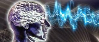
The main cells are called neurons. They process information, their number reaches 20 billion. There are 10 times more glial cells.
The body carefully protects the brain from external influences by placing it in the skull. Neurons are located in a semipermeable membrane and have processes: dendrites and one axon. The length of dendrites is small compared to the axon, which can reach several meters.
To transmit information, neurons send nerve impulses to an axon, which has many branches and is connected to other neurons. The impulse originates in the dendrites and is sent to the neuron. The nervous system is a complex web of neuron processes that are interconnected.
The structure of the brain and the chemical interaction of neurons have been studied superficially. At rest, a neuron has an electrical potential of 70 millivolts. Excitation of a neuron occurs through the flow of sodium and potassium across the membrane. Inhibition occurs as a result of the action of potassium and chlorides.
The job of a neuron is to interact between dendrites. If the excitatory effect prevails over the inhibitory effect, then a certain part of the neuron membrane is activated. Thanks to this, a nerve impulse arises that moves along the axon at a speed of 0.1 m/s to 100 m/s.
Thus, any planned movement is formed in the cortex of the frontal lobes of the cerebral hemispheres. Motor neurons give commands to parts of the body. Simple movement activates the functions of parts of the human brain. When talking or thinking, large parts of the gray matter are involved.
Violations and their causes in alphabetical order:
violation of movement coordination -
The term "coordination"
comes from the Latin coordinatio - mutual ordering. Coordination of movements refers to the processes of coordinating the activity of the muscles of the body, aimed at the successful completion of a motor task.
For the central nervous system, the control object is the musculoskeletal system. The uniqueness of the musculoskeletal system lies in the fact that it consists of a large number of links, movably connected at joints that allow rotation of one link relative to another. Joints can allow links to rotate about one, two or three axes, i.e., have one, two or three degrees of freedom. In order to reach any given point in three-dimensional space (within the length of the limb), it is enough to have a two-link limb with two degrees of freedom in the proximal joint (“shoulder”) and one degree of freedom in the distal (“elbow”). In fact, the limbs have a greater number of links and degrees of freedom. Therefore, if we wanted to solve a geometric problem about how the angles at the joints must change in order for the operating point of a limb to move from one given position in space to another, we would find that this problem has an infinite number of solutions.
In order for the kinematic chain to make the desired movement, it is necessary to exclude those degrees of freedom that are redundant for a given movement. This can be achieved in two ways: 1. excess degrees of freedom can be fixed by simultaneous activation of antagonistic muscle groups (coactivation); 2. it is possible to associate movements in different joints with certain relationships, thus reducing the number of independent variables that the central nervous system must “deal with.”
Such stable combinations of simultaneous movements in several joints aimed at achieving a common goal are called synergies.
What diseases cause loss of coordination of movement:
Impaired coordination of movements. Since many parts of the central nervous system are involved in the control of movements, disorders of motor coordination can be used for diagnostic purposes. They are manifested by disturbances in stability when standing and walking, asymmetry of movements of the right and left sides, disturbances in the accuracy of movements, a decrease in strength and a decrease in speed. Registration of spatial and temporal characteristics of movements with their quantitative representation makes it possible to assess the degree of movement disorders in various diseases, the progress of restoration of motor functions, and propose effective methods of motor rehabilitation.
One of the forms of impaired coordination of movements is ataxia. Ataxia (from the Greek disorder). Impaired coordination of movements is observed with damage to the frontal lobes of the brain, the cerebellum, and deep sensitivity pathways in the spinal cord and brain. It manifests itself as an imbalance when standing (static ataxia) or a disorder of motor coordination (dynamic ataxia).
Symptoms and course. Many parts of the nervous system are involved in movement. A person must feel his limbs, see them and surrounding objects, be in balance, all these sensations must be linked together - coordinated. With the disease, patients cannot stand with their eyes closed, cannot eat due to severe trembling in their hands, and often speak poorly - their speech is scanned. Ataxia can manifest itself as a significant deterioration in handwriting, which is characteristic of brain damage due to rheumatism. They cannot perform precise movements: thread a needle with normal vision, take a match from a box.
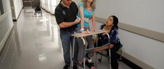
Recognition. If ataxia is suspected, ask the patient to stand with arms extended forward and eyes closed, legs closed, then touch the tip of the nose with a finger or, lying on his back, touch the heel of the knee of the opposite leg. If a person cannot complete these tasks, then it is necessary to consult a doctor.
Which doctors should you contact if movement coordination problems occur:
Have you noticed a lack of coordination of movement? Do you want to know more detailed information or do you need an inspection? You can make an appointment with a doctor
Euro lab
clinic is always at your service!
The best doctors will examine you, study external signs and help you identify the disease by symptoms, advise you and provide the necessary assistance. You can also call a doctor at home
.
Euro lab
clinic is open for you around the clock.
How to contact the clinic:
The telephone number of our clinic in Kyiv: (+38 (multi-channel). The clinic secretary will select a convenient day and time for you to visit the doctor. Our coordinates and directions are indicated. Look in more detail about all the clinic’s services on her.
(+38
If you have previously performed any tests, be sure to take their results to a consultation with your doctor.
If the studies have not been performed, we will do everything necessary in our clinic or with our colleagues in other clinics.
Have you lost coordination of movement? It is necessary to take a very careful approach to your overall health. People do not pay enough attention to the symptoms of diseases
and do not realize that these diseases can be life-threatening.
There are many diseases that at first do not manifest themselves in our body, but in the end it turns out that, unfortunately, it is too late to treat them. Each disease has its own specific signs, characteristic external manifestations - the so-called symptoms of the disease
.
Identifying symptoms is the first step in diagnosing diseases in general. be examined by a doctor
several times a year in order not only to prevent a terrible disease, but also to maintain a healthy spirit in the body and the organism as a whole.
If you want a doctor, use the online consultation section, perhaps you will find answers to your questions there and read tips on caring for yourself
.
If you are interested in reviews about clinics and doctors, try to find the information you need on. Also register on the Eurolab
medical to be constantly aware of the latest news and information updates on the site, which will be automatically sent to you by email.
The symptom chart is for educational purposes only. Do not self-medicate; For all questions regarding the definition of the disease and methods of its treatment, consult your doctor. EUROLAB is not responsible for the consequences caused by the use of information posted on the portal.
If you are interested in any other symptoms of diseases and types of disorders, or you have any other questions or suggestions, write to us, we will definitely try to help you.
If coordination is impaired, any movements and actions are difficult for a person to perform. This often happens when there is a disturbance in the central nervous system. The signal sent to the brain does not reach the target, or arrives in a distorted form.
When this disorder manifests itself, the patient’s movements are uncertain, uncoordinated, and unsteadiness is observed while walking. Often, when coordination is impaired, the patient experiences dizziness, accompanied by nausea and vomiting, and often there is congestion and tinnitus.
The reason for impaired coordination of movement can be the use of alcoholic beverages, drugs, age-related changes, and head injuries.
To identify the disorder, the patient is offered the simplest tests: closing his eyes, touching the tip of his nose with his finger, writing a simple sentence or drawing some figure in the air. If coordination is impaired, instead of a given figure, a person will draw a zigzag; when writing a sentence, the letters will be uneven and overlap one another.
Sometimes the patient makes uncontrolled movements called chorea or tremor. In some people suffering from this disorder, uncontrolled movements are observed at rest, in others when performing any actions. If the processes of cerebellar activity are disrupted, there is a possibility of developing ataxia. With ataxia, the patient is uncomfortable when his body is in an upright position, and in some cases there are difficulties when walking, others experience discomfort in a standing position (it is difficult to maintain balance).
Signs of impaired coordination of movements
People with such ailments move uncertainly, their movements show laxity, too much amplitude, and inconsistency. Having tried to outline an imaginary circle in the air, a person is faced with a problem - instead of a circle, he gets a broken line, a zigzag.
Another test for incoordination is to ask the patient to touch the tip of the nose, which also fails.
Looking at the patient’s handwriting, you will also be convinced that his muscle control is not all right, since letters and lines creep on top of each other, becoming uneven and sloppy.
Loss of coordination with dizziness
Often coordination disorders are accompanied by dizziness. This is a sign that the brain is involved in the pathological process, namely its vestibular center, which is responsible for the balance of the body in space. In addition to dizziness and incoordination, additional concerns may include:
- nausea, sometimes vomiting, sharply increasing when turning the head or any movements;
- pain and discomfort in the cervical spine;
- headaches, noise in the head;
- increased blood pressure.
Causes of poor coordination with dizziness:
- Stroke
in the veno-basilar region and other vascular pathology. - Cervical osteochondrosis
, in which there is a decrease in blood flow to the brain, and ischemia of its centers develops. - Meniere's disease
is a disease of the inner ear. - Hypertonic disease
. As pressure increases, the lumen of blood vessels decreases, which leads to disruption of the nutrition of the vestibular centers. - Tumors
, brain cysts.