The cerebral peduncle (CP) is one of two paired structures that make up the midbrain, which determines its functional purpose and importance.
Almost all chordates have a mesencephalon, which is responsible for performing vital functions that play an invaluable role in ensuring vitality, survival, pain regulation and the alternation of sleep and wakefulness.
It is also responsible for defense reflexes, protection, spatial-temporal orientation, and even for maintaining a constant body temperature, controlling movements and concentrating.
Characteristic
Human anatomy is a section that examines the parts of the brain. The human brain matter weighs about 1250 g. In men, the volume of the brain matter is 8-15% larger than in women. The mass of the brain matter is about 2% of the weight of the entire body. At the same time, the brain consumes 25% of all oxygen entering the body and 17% of glucose, which serves as an energy resource for living tissues.
Brain tissue is represented by gray and white matter, which have different functions and chemical composition. The proportion of water in gray is 84%, in white – 70%. Lipid (fat) fractions in white occupy 50% of the dry residue, in white – 30% of the dry residue. Brain tissue also contains proteins (8-9% of raw tissue weight) and minerals (1-2% of raw tissue weight).
The substance of the brain is covered with 3 membranes - soft, then arachnoid and hard, and the latter consists of 2 layers formed by connective tissue, however, when asked how many membranes there are, it is customary to answer that there are three. The arachnoid is represented by a thin membrane formed from connective tissue. The soft tissue is adjacent to the medulla; it is abundantly equipped with blood vessels and nerve endings.
The brain consists of neurons and glial cells that form the nervous tissue that forms the basis of the central nervous system and supports its functions. Nervous tissue is represented by a system of specialized structures that interact with each other. External stimuli (light, sound, tactile stimuli) are converted by receptors into nerve impulses, which enter parts of the brain through the neuronal system; in anatomy, afferent and efferent transmission pathways are distinguished.
In the first case, impulses arrive in the central parts of the brain system, in the second from control systems (associative centers) to the peripheral structures of the nervous system located in the muscles, glands, and executive organs. The brain tissue contains about 100 billion neurons, united in a complex neuronal network.
Neurons are surrounded by glial cells, which perform trophic (nutritional) and supportive functions. The number of glial cells exceeds the number of neurons. In the anatomical structure formed by nerve cells, there is an intercellular space, represented by a liquid containing salts, carbohydrates, fat and protein fractions, and nourishing brain tissue.
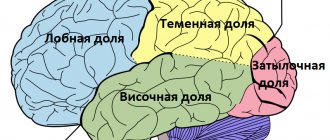
The brain is located inside the cranium, where in humans the cerebral part predominates over the visceral (facial) part. The human brain consists of 3 large segments - the hemispheres, the cerebellum and the brainstem, from which the spinal cord arises. The largest segment of the brain is the hemisphere (hemisphere), the second largest part is the cerebellum. The trunk ranks 3rd in size.
The hemispheres are separated from each other by a gap, which is clearly visible in the photo (top view). In the depths of the gap there is a commissure - the corpus callosum, which connects both hemispheres together. In a photograph of the brain showing the lower surface, segments are clearly visible - the entire part of the brainstem, the cerebellum, the lower side of the hemispheres, the cranial nerves extending from the medulla. The lateral view shows predominantly the cortex covering the cerebral hemispheres.
The structural scheme involves the division of large parts into separate segments, for example, the brain stem includes the medulla oblongata and middle sections, the pons, the cerebellum, which is responsible for human motor coordination, and the intermediate section are often included in the brain stem.
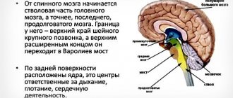
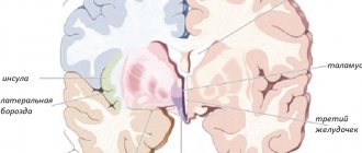
A vertical section of the human brain shows the corpus callosum, pons, cerebellum, medulla oblongata, pituitary gland, pineal gland. On the general plan of the structure, 5 main sections of the brain are distinguishable (taking into account the characteristics of embryonic development) - oblongata, posterior, middle, intermediate and anterior.
There is a theory that the brain is an organ of the central part of the nervous system, which works on the principle of a binary code in the process of generating or transmitting nerve impulses. Thanks to the neural networks through which nerve impulses are transmitted, all brain structures work harmoniously.
Roof of the midbrain
The roof of the midbrain, the dorsal part, is hidden under the corpus callosum (its posterior end). It is divided into 4 mounds located in pairs by means of two grooves (transverse and longitudinal), running crosswise. The two upper hillocks are the subcortical centers of vision, and the two lower ones are the hearing centers. Between the superior tubercles in a flat groove is the pineal gland.
The handle of the colliculus is directed laterally, upward and anteriorly, towards the diencephalon. Every mound goes into it. The handle of the superior colliculus runs under the thalamic cushion towards the lateral geniculate body. The manubrium of the inferior one disappears under the geniculate medial body. The geniculate bodies mentioned above no longer belong to the midbrain, but to the diencephalon.
Functions of departments
The main function assigned to the brain structures is to ensure interaction with the environment. Here, teams are formed that are responsible for processes in the body such as thermoregulation, metabolism, selective development of structural formations (systems) that ensure the body’s adaptive abilities, and neurohumoral regulation.
The functions of the brain are determined depending on the location of its parts. Moreover, individual sections of the brain matter simultaneously perform several tasks, in parallel closely interacting with all other sections. If any part of the brain is damaged, its compensatory functions can be performed by another brain structure.
Oblong
The human brain is designed in such a way that it includes separate areas characterized by functional differences, for example, such as the oblongata, which continues the dorsal. The control center for cardiac activity is located in the medulla oblongata. Within the heart center, nuclei are distinguished from which the vagus nerves depart. The functions of the vagus nerve include autonomic regulation of the heart and the condition of blood vessels.
In the vasomotor center, in the structure of the nuclei of the vagus nerve endings, pressor and depressor zones are distinguished, which interact with each other. The pressor zone is responsible for the narrowing of blood vessels, the depressor zone is responsible for the expansion of the vascular lumen. The pressor zone reacts to the chemical composition of the blood. Information is transmitted through chemoreceptors.
The depressor zone responds to information coming from baroreceptors (they perceive the pressure that is exerted on the vascular walls). The hypothalamus is a section of the diencephalon that is responsible for neuroendocrine function and determines the effects of the cardiovascular system, taking into account the external environment and the current needs of the body.
The structure of the hindbrain provides for the presence of the pons, where part of the respiratory center responsible for respiratory function is located. The area responsible for inhalation is located in the pons, the area regulating the process of exhalation is in the medulla oblongata. Both areas interact with each other. During the process of inhalation, the external intercostal muscles are involved, which, by contracting, contribute to the expansion of the lungs and the entry of air into them.
Exhalation occurs due to contraction of the internal intercostal muscles. Commands that cause muscle contraction come from the corresponding zones of inhalation and exhalation through the structures of the spinal cord. The vagus nerve, hypothalamus and cortical structures can influence the frequency of respiratory movements. The medulla oblongata contains the nuclei of 4 pairs of cranial nerves (vagus, hypoglossal, accessory, glossopharyngeal).
The functions of the glossopharyngeal nerve include the motor activity of the pharyngeal muscles, the implementation of reflex reactions (sneezing, swallowing, coughing, gagging, the formation of speech sounds). It is believed that in humans, the medulla oblongata located in the brain contains centers such as cough-sneezing and vomiting.
Rear
As can be seen in the figure, the brain is structured in such a way that a person has several important brain regions, including the posterior one, consisting of the cerebellum and the pons. The fourth ventricle in the ventricular system expands like the spinal canal, forming a cavity in which the posterior section is located. The pons Varoliev is formed by a dense plexus of conducting pathways.
The cerebellum is the brain's motor center and contains pathways that connect it to many other areas of the brain and body systems. Bundles consisting of nerve fibers form the cerebellar peduncles - 3 pairs. The lower legs connect the cerebellum with the medulla oblongata segment, the middle ones provide interaction with the pons and then with the cortex, the upper ones with areas of the midbrain.
The mass of the cerebellum is equal to 10% of the weight of the entire brain matter. However, more than 50% of all neurons of the nervous system are localized in this area. The functions of the cerebellum include regulating the tone of skeletal muscles, maintaining body posture, maintaining balance, performing purposeful, voluntary movements, and coordinating motor activity. The cerebellum is involved in the planning and execution of movements:
- Ballistic (performed under the influence of gravity).
- Sports (ball throw, javelin).
- Musical (playing instruments).
General neuronal systems formed by the cerebellum and cortical regions are involved in thinking processes aimed at organizing complex movements. In the area of the bottom of the 4th ventricle there are nuclei of nerve branches - facial, vestibulocochlear, abducens, segmental trigeminal.
Average
The structure of the brain involves the separation of the middle section, which, in short, reflects the stability and immutability of the human nervous system. This department has undergone virtually no evolutionary changes. A horizontal section of the brain reveals the structures that make up the middle section, which is responsible for alternating sleep-wake cycles, concentration, reflex reactions (defensive, associated with orientation in space), and human functions such as hearing and vision.
The middle section contains structures that receive information from the organs of vision, regulate pain sensitivity and body temperature. The red nucleus of the department controls postural movements (formation and maintenance of posture), together with the substantia nigra it participates in the regulation of the extrapyramidal system (muscle tone, posture). The structures of the middle section form the body's reactions to external stimuli - visual, auditory.
The visual center is located in the area of the superior colliculus, and the auditory center is located in the area of the inferior colliculus. The aqueduct of Sylvius passes through the structures of the middle section, connecting the 3rd and 4th ventricles. In the middle section there are nuclei of the cranial nerves - trochlear, oculomotor, as well as one trigeminal nucleus. The trochlear and oculomotor nerve branches regulate the movements made by the eyes.
Intermediate
The human brain is designed in such a way that it contains an intermediate section that is responsible for functions such as sensory perception. The main part of the human diencephalon is the thalamus - the cerebral visual, sensory tubercle. The thalamus receives information from all receptors concentrated in the sensory organs.
Information flows to the thalamus from the organs of vision and hearing, from receptors - taste, olfactory, tactile, proprioceptors (receptors located in muscles, skin, ligaments and joint capsules), interoreceptors (receptors located in internal organs, tissues, elements of the vascular system) , vestibuloreceptors (receptors located in the labyrinth of the inner ear perceive changes in speed and direction when the body moves in space).
Appendages (superior, inferior, posterior) and the optic chiasm extend from the thalamus. The superior epididymis contains the pineal gland, which produces melatonin. The pineal gland also secretes histamine, norepinephrine, and serotonin, which are involved in controlling circadian rhythms. Regulatory activities are focused on the level of illumination.
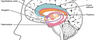
In the posterior appendage, secondary centers are concentrated - auditory and visual, represented by the medial (located closer to the median plane) and lateral (lying away from the median plane) geniculate bodies. The functions of the lower appendage, the hypothalamus, are associated with its location in the limbic system, a brief description of which requires mention of its important role in controlling the work of internal organs, the formation of emotions and memory.
The hypothalamus is the highest regulatory brain center of the autonomic (autonomous) part of the nervous system. In this part of the human brain, the chemical composition of blood and cerebrospinal fluid (cerebrospinal fluid) is analyzed. The pituitary gland, which is part of the hypothalamus, is a gland that secretes hormones that regulate the activity of other glands of the endocrine system. Chemoreceptors (receptors that perceive chemical stimuli) transmit information about the concentration of substances contained in body fluids to the hypothalamus.
Based on the information obtained, the hypothalamus weakens or stimulates metabolic processes through a direct influence on the autonomic centers using nerve impulses or through the production of biological peptide substances - statins (inhibit the secretory activity of the anterior pituitary gland) and liberins (stimulate the secretory activity of the anterior pituitary gland). The functions of the hypothalamus include the control of processes based on biological motivation:
- Eating behavior (taste preferences, diet, diet features).
- Sexual behavior (behavioral reactions aimed at realizing sexual needs).
- Aggressive-defensive reactions.
The location of the hypothalamus within the limbic system of the brain suggests its participation in the interaction between somatic and autonomic functions. We are talking about motor reactions based on information received from the senses and the control of somatic functions taking into account the current needs of the body.
For example, if defensive behavior is activated due to biological needs, effective functioning of the skeletal muscles and sensory organs is necessary. A person needs to hear and see clearly and move dynamically. To increase the efficiency of muscle work, important parameters are the speed of transmission of nerve impulses, the provision of nerve structures and muscle tissue with nutrients and oxygen.
In conclusion, it can be argued that the hypothalamus provides internal resources for the implementation of external behavior. The nuclei of the thalamus are divided into groups - switching, integrative, modulating. The first group is an intermediate link in the afferent pathways through which information flows from organs to nerve centers. Afferent pathways extend from the head, limbs, and parts of the body.
The signals enter the analyzing zones of the cortical layer, where the received data is processed. Integrative nuclei provide communication between all structural divisions of the thalamus among themselves and with the associative zones of the cortical layer. An example is combining visual stimuli and verbal information into a single image. The functions of the thalamus support gnostic (cognitive) activity.
Modulating nuclei are the most ancient structure of the thalamus. This is the basis of the reticular formation, which stretches along the brain stem, consists of reticular nuclei and a branched neuronal network with numerous axons and dendrites. The reticular formation activates the cortical areas of the brain, which leads to concentration and timely processing of information coming through ascending pathways. The reticular formation controls the reflex functions of the spinal cord.
Structure
– legs, where the conductive pathways mainly pass;
– subcortical centers of vision and hearing.
Its size is only two centimeters. The midbrain consists of the peduncles and quadrigeminalis. It is responsible for the following functions: visual, auditory, spatial orientation. The peduncles of the midbrain are red (due to the many blood vessels). Connecting with the cerebellum and vestibular apparatus, they provide balance to the body. The quadrigeminals connect the temporal lobe of the brain with bundles of auditory conductors.
What is the midbrain cavity?
Many terms can be found in such a section as the anatomy of the midbrain. Its structure and functions require strict scientific accuracy when describing it. We have omitted complex Latin names and left only basic terms. This is enough for the first acquaintance.
Let's say a few words about the midbrain cavity. It is a narrow channel and is called a water pipe. This canal is lined with ependyma, it is narrow, its length is 1.5-2 cm. The cerebral aqueduct connects the fourth ventricle to the third. The peduncle tire limits it ventrally, and dorsally – the roof of the midbrain.
Brain stems
We continue to describe the human midbrain, functions and structure. The next thing we'll focus on is his legs. What is it? This is the ventral part, which contains all the pathways leading to the forebrain. Note that the legs are two semi-cylindrical thick white strands, diverging at an angle from the edge of the bridge and plunging into the hemispheres.
They are divided into the base of the stalk (ventral part) and the tire. The substantia nigra serves as the boundary between them. It owes its color to melanin, a black pigment found in the nerve cells that make it up. The tegmentum of the midbrain is the part of it located between the substantia nigra and the roof.
The central tire track departs from it. This is a descending projection nerve pathway, which is located in the tegmentum of the midbrain (its central part). It consists of fibers that go from the red nucleus, globus pallidus, reticular formation of the mesencephalon and thalamus to the olive and reticular formation of the medulla oblongata. This pathway is part of the extrapyramidal system.
Research by Luigi Luciani
The functions of the midbrain and cerebellum were studied by Luigi Luciani, an Italian physicist. In 1893, he conducted experiments on animals with the cerebellum completely or partially removed. He also analyzed its bioelectrical activity, recording it during irritation and at rest.
It turned out that the tone of the extensor muscles increases when half of the cerebellum is removed. The animal's limbs are stretched, the body is bent, and the head is tilted towards the operated side. Movements occur in a circle (“manege movements”) towards the operated side. The described disturbances are gradually smoothed out, but a certain incoordination of movements remains.
If the entire cerebellum is removed, severe movement disorders occur. They are smoothed out gradually due to the fact that the cerebral cortex (its motor zone) is activated. However, the animal still remains with a lack of coordination. Imprecise, awkward, sweeping movements and a shaky gait are observed.

