general characteristics
The brain is one of the constituent organs of the central nervous system.
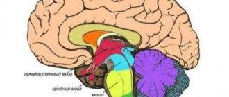
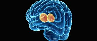
Doctors are still studying it. It consists of 25 billion neurons, which are presented in the form of gray matter. Rice. 1. Sections of the brain.
In addition, this organ of the nervous system is covered with the following types of membrane:
- soft;
- hard;
- arachnoid (cerebrospinal fluid circulates in it - cerebrospinal fluid, which serves as a kind of shock absorber and protects against shock).
The brains of men and women differ in their mass. In representatives of the stronger sex, its weight is 100 g more. However, mental development does not depend in any way on this indicator.
The functions of generator and impulse transmission are performed by neurons. There are ventricles (cavities) inside the brain, from which paired cranial nerves extend to different parts of the human body. There are a total of 12 such pairs in the body.
Structure
The diencephalon is divided into:
- Thalamic brain (lat. thalamencephalon)
- Subthalamic region or hypothalamus (lat. hypothalamus)
- The third ventricle, which is the cavity of the diencephalon
Thalamic brain
The thalamic brain has three parts:
- Optic thalamus (Thalamus)
- Suprathalamic region (Epithalamus)
- Transthalamic region (Metathalamus)
Thalamus
Main article: Thalamus
Thalamus
or
visual tubercle
(lat. thalamus) - a paired ovoid formation - consists mainly of gray matter. The medial and superior surfaces are free, and the lateral-inferior surface communicates with other parts of the brain. The thalamus is the subcortical center of all types of sensitivity (pain, temperature, tactile, proprioceptive). The thalamus is the switching point for all sensory pathways coming from extero-, proprio- and interoreceptors.
Epithalamus
Main article: Epithalamus
Epithalamus
or
suprathalamic region
(lat. epithalamus) is located in the upper posterior part of the thalamus. The epithalamus forms the pineal body (epiphysis), which is attached to the thalamus by means of leashes. The pineal body is an endocrine gland that is responsible for synchronizing the body's biorhythms with the rhythms of the environment.
Metathalamus
Main article: Metathalamus
Metathalamus
or
the post-thalamic region
(lat. metathalamus) is formed by paired medial and lateral geniculate bodies lying behind the thalamus. The medial geniculate body is located behind the thalamic cushion. It is the subcortical center of hearing. The lateral geniculate body is located inferior to the pillow. It is the subcortical center of vision.
Hypothalamus
Main article: Hypothalamus
The hypothalamus or subthalamic region is located below the thalamus. The hypothalamus includes the mammillary bodies, which are the subcortical centers of smell, the pituitary gland, the optic chiasm, the second pair of cranial nerves, the gray tubercle, which is the autonomic center of metabolism and thermoregulation. The hypothalamus contains nuclei that control endocrine and autonomic processes.
The hypothalamus is divided into four parts:
- Anterior hypothalamic part
- Intermediate hypothalamic part
- Posterior hypothalamic part
- Dorsolateral hypothalamic part
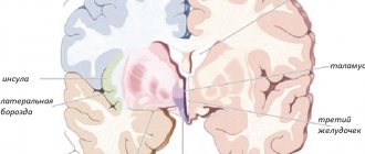
Third ventricle
Main article: Third ventricle of the brain
The third ventricle (lat. ventriculus tertius) is the cavity of the diencephalon. It is a narrow, slit-like space located in the sagittal plane.
The third ventricle has six walls:
- Two lateral walls, medial surfaces of the thalami facing each other
- The lower wall is represented by the subthalamic region and partially by the cerebral peduncles
- The posterior wall is represented by the posterior commissure and the pineal recess
- The superior wall is represented by the choroid of the third ventricle
- The anterior wall is represented by the columns of the arch, the anterior commissure and the terminal plate
It is formed under the influence of four outgrowths of the neural tube (epiphysis, pituitary gland, optic cups) from the outgrowths that serve them. Divisions of the diencephalon: thalamic region (thalamus, metathalamus, epithalamus), hypothalamus, third ventricle.
The ventral part is the hypothalamus, the center of autonomic regulation and endocrine control. Noticeable formations are the funnel, gray tubercle, mastoid bodies, pituitary gland. Anatomically, the hypothalamus also includes the optic nerves and their chiasm ( hiasma
).
Lateral thickenings - visual hillocks or thalamus, switch all sensory information except olfactory. On the outside, the thalami have noticeable formations - the cushion and geniculate bodies (anatomically they belong to the metathalamus). Between the thalami there is a narrow third ventricle and an interthalamic fusion.
The dorsal part - the epithalamus (is a pineal gland connected by leads to the thalamus) is responsible for biorhythms.
The metathalamus consists of the lateral and medial geniculate bodies. The external (lateral) geniculate body is the subcortical visual center. The internal (medial) geniculate body is the subcortical auditory center.
Brain parts and brain
The encephalon is part of the central nervous system, which is located in the cranial cavity, as opposed to the spinal cord. This organ consists of the brain, cerebellum and brainstem.
The diencephalon develops from the forebrain or forebrain, one of the three parts of the brain at the beginning of embryonic nervous system development. The other two initial divisions are the midbrain, which will connect the various parts of the brain, and the rhombencephalon, which will give way to the cerebellum, medulla oblongata, and muscles.
As the fetus grows, the forebrain is divided into the intermediate and telencephalon; from this the cerebral hemispheres, the basal ganglia and the limbic system, including the amygdala, will develop. We will describe the sections of the diencephalon in the next section.
Content
- 1 Anatomical structure 1.1 Connections with other brain structures and peripheral structures
- 3.1 Early brain development
- 4.1 Thalamic brain 4.1.1 Thalamus
Functions of the diencephalon
The area of the brain we know as the diencephalon is made up of various structures. They are connected to each other and to the rest of the nervous system, both at the cortical and subcortical levels.
Its connection with the endocrine system, consisting of glands that secrete hormones into the blood, is also very relevant.
Thalamus
The thalamus functions as a kind of relay core for communication between the cerebral cortex and subcortical structures. It is fundamental for receiving sensory inputs (with the exception of olfactory inputs, which go directly to the cortex) and transmitting them to the lobes of the brain.
This structure also plays a role in regulating consciousness and the sleep-wake cycle, and it influences motor skills through outputs that project from the thalamus to the basal ganglia and cerebellum.
Hypothalamus
The hypothalamus is located below the thalamus. The main functions of this structure include connecting the nervous and endocrine systems and controlling the secretion of hormones from the pituitary gland and other glands.
The hypothalamus directly produces vasopressin and oxytocin, but also stimulates the endocrine glands to produce other hormones. It is also key to regulating the body's homeostasis as it interferes with thirst, hunger, temperature, circadian rhythms, stress and other bodily processes.
- Article on the topic: “Hypothalamus: definition, characteristics and functions”
The pituitary gland is an endocrine gland attached to the hypothalamus. It is very important for growth, kidney regulation, sexual function and reproduction, among other aspects.
It consists of two lobes: the anterior pituitary gland (adenohypophysis) and the posterior pituitary gland (neurohypophysis). While the neuropituitary gland secretes oxytocin and vasopressin synthesized by the hypothalamus, the adenopituitary gland produces and releases corticotropin, growth hormone, prolactin, luteinizing hormone and follicle-stimulating hormone, among others.
- Maybe you're interested in: "Pituitary gland (pituitary gland): connection between neurons and hormones"
Epithalamo
This brain structure is mainly composed of the pineal gland, fundamental in the circadian and seasonal cycles, and the habénula, involved in the function of the neurotransmitters dopamine, norepinephrine and serotonin. The epithalamus connects the limbic system to other areas of the brain.
Subthalamus
The subthalamus is attached to the globus pallidus, one of the main nuclei of the basal ganglia. Because of this, it plays a regulatory role in extrapyramidal and involuntary movements.
The retina develops from the diencephalon and is therefore considered part of the central nervous system. The optic nerve allows information to be transmitted from the eye to the brain through its connection to the diencephalon.
Third ventricle
The ventricles of the brain allow the circulation of cerebrospinal fluid, which performs functions similar to those of blood in the brain and spinal cord, in addition to protecting nervous tissue from shock and injury. The third ventricle is located in the middle part of the ventricular system, below the epithalamus.
- Article on the topic: “Stomachs of the brain: anatomy, characteristics and functions”
TOP 4 articles that are read along with this
- 1.Spinal cord: structure and functions
- 2.Speech
- 3.Spinal cord: structure and functions
- 4.Memory
- two hemispheres;
- trunk;
- cerebellum.
It also has five departments:
- final, constituting 80% of the mass;
- intermediate;
- rear;
- average;
- oblong.
Each section consists of a specific set of cells (white and gray matter).
White matter is presented in the form of nerve fibers, which can be of three types:
- association – connect cortical areas in one hemisphere;
- commissural - connects the two hemispheres;
- projection – connect the cortex with the underlying formations.
Gray matter consists of neuron nuclei, their functions include the transmission of information.
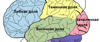
Rice. 2. Lobes of the cerebral cortex.
| Department | Structure | Functions |
| Finite | Located from the occipital to the frontal bone. It consists of two hemispheres, which have many grooves and convolutions. On top they are covered with a bark consisting of lobes. | The right hemisphere is responsible for the left side of the body, and the left hemisphere is responsible for the right side. The temporal lobe of the cerebral cortex regulates hearing and smell, the occipital lobe regulates vision, the parietal lobe regulates taste and touch; frontal – speech, thinking, movement. |
| Intermediate | Consists of the hypothalamus and thalamus. | The thalamus is a mediator in the transmission of stimuli to the hemispheres and helps to adequately adapt to changes in the environment. The hypothalamus regulates the functioning of metabolic processes and endocrine glands. Manages the work of the cardiovascular and digestive systems. Regulates sleep and wakefulness, manages food and drink needs. |
| Rear | It consists of the cerebellum and the pons, which is presented in the form of a white thick cushion located above the oblongata. The cerebellum is located behind the pons and has two hemispheres, the inferior and superior surfaces and the vermis. | This section provides a conductive function during the transmission of impulses. The cerebellum controls the coordination of movements. |
| Average | Located from the anterior edge of the bridge to the optic tracts. | Responsible for hidden vision, as well as the work of the orienting reflex, which ensures the body turns in the direction of the heard sharp noise. |
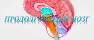
| Oblong | Presented as a continuation of the spinal cord. | Controls coordination of movements, balance, regulates metabolic processes, breathing, blood circulation. Controls the process of coughing and sneezing. |
Rice. 3. Functions of parts of the brain.
The brainstem consists of the medulla oblongata, midbrain, diencephalon and pons. The trunk is the connecting link between the spinal and head sections of the central nervous system. Its functions include controlling articulate speech, heartbeat and breathing.
Embryonic development
Reconstruction of human embryonic peripheral nerves measuring 10.2 mm.
The diencephalon is marked on the left. The diencephalon, or diencephalon, is one of five secondary brain vesicles formed during the embryonic development of the brain of chordates, namely the second in a row, starting from the rostral (head) end of the embryo. It is formed from the posterior part of the anterior (the very first from the cephalic end) of the primary brain vesicle (prosencephalon). From the anterior part of this primary brain bladder, the telencephalon is formed.
Early brain development
At a certain stage of embryonic development (in the human embryo this is the third week), in one of the areas of the ectoderm (outer germ layer), at the future head end of the embryo, the so-called primary neural plaque begins to form - the place where the ectoderm cells begin differ from neighboring ones, and differentiate into the so-called neuroectoderm, or neuroepithelium. Growing in diameter both due to the intensive division of neuroectoderm cells and due to the involvement of neighboring cells in differentiation according to the neuroectodermal type, the primary neural plaque quickly turns into the primary neural plate. Then the ends of the neural plate begin to bend inside the embryo, “dragging” the neural plate from the surface of the head end of the embryo inward. This process is called primary neurulation. As a result of neurulation, the primary neural tube is formed. It quickly extends along the notochord, an embryonic structure that indicates the axis of bilateral symmetry to the cells of the embryo. Subsequently, the lateral ends of the primary neural tube (not fully closed corners of the neural plate) grow together, and the openings at the rostral and caudal ends of the primary neural tube (neuropores) close. This process is called secondary neurulation. A neural tube with already closed neuropores and fused lateral ends is called a secondary neural tube. The notochord serves as the organizer and conductor in the early stages of embryonic development of the central nervous system, and the prototype of the future notochord in lower chordates or the future spine in vertebrates. At the head end of the neural tube, a so-called “head thickening” is formed - a prototype of the future brain. The cavity inside the neural tube forms the prototype of the future central canal of the spinal cord.
Stage of three primary brain vesicles
At this stage, three separate thickenings with cavities inside filled with fluid (a prototype of the future cerebrospinal fluid) are formed in the developing brain of the embryo - three primary brain vesicles. The most anterior one is called the forebrain, or prosencephalon. The middle one is called the midbrain. The posteriormost one, shaped like a diamond, is called the rhombencephalon.
Stage of five secondary brain vesicles
At this stage, two of the three primary brain vesicles—the most anterior, forebrain (prosencephalon) and the most posterior, rhombencephalon (rhombencephalon)—are each subdivided into two secondary brain vesicles. The forebrain is divided into the telencephalon and diencephalon. The rhombencephalon is divided into the hindbrain (metencephalon) and the medulla oblongata (myelencephalon). The middle primary brain vesicle (midbrain, or mesencephalon) does not
is subdivided and enters this stage unchanged.
From the embryonic telencephalon, the cerebral hemispheres are subsequently formed, in particular the cerebral cortex, subcortical white matter and basal ganglia. From the embryonic midbrain, the roof of the brain and, in particular, the quadrigeminal peduncle, the cerebral peduncles, the tegmentum of the midbrain and its constituent structures, such as the substantia nigra and red nuclei, are formed. The pons and cerebellum are formed from the embryonic hindbrain. Further differentiation of the embryonic diencephalon is described below in a separate section.
Further differentiation of the embryonic diencephalon
| Primary brain vesicle | Secondary brain vesicles | Primary prosomers | Secondary prosomers | Further prosomerization |
| Prosencephalon (P) | Telencephalon (T) | T | T1 | |
| Pseudoprosomera T2 | ||||
| Diencephalon (D) | D | D1 | ||
| D2 | Rostral parencephalon | |||
| Caudal parencephalon | ||||
| Synencephalon |
Optic chiasm
chiasma opticum, formed by fibers of the optic nerves (II pair of cranial nerves. On each side it continues into the optic tract, tractus opticus. The optic tract ends with two roots in the subcortical centers of vision.
Posterior to the optic chiasm is a gray tubercle, tuber cinereum, behind which lie the mastoid bodies, and on the sides are the optic tracts. Downwards, the gray tubercle passes into a funnel, infundibulum, which connects to the pituitary gland. The walls of the gray tuberosity are formed by a thin plate of gray matter containing gray tubercles, nuclei tuberdles.
Third (III) ventricle
ventriculus tertius , occupies a central position in the diencephalon, limited by six walls: two lateral, upper, lower, anterior and posterior. The lateral walls of the third ventricle are the medial surfaces of the thalami, as well as the medial sections of the subthalamic region. The lower wall, or bottom of the third ventricle, is the hypothalamus. The anterior wall of the third ventricle is formed by the terminal plate, columns of the fornix and anterior commissure. The posterior wall of the third ventricle is the epithalamic commissure,