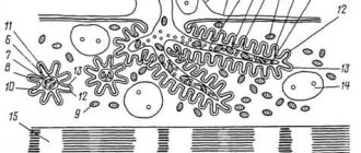
Glia are a structure of the nervous system formed by specialized cells of various shapes that fill the spaces between neurons or capillaries, accounting for 10% of the brain volume.
The size of glial cells is 3-4 times smaller than nerve cells; their number in the central nervous system of mammals reaches 140 billion. With age, the number of neurons in the brain decreases, and the number of glial cells increases.
What is neuroglia
The collection of cells is called neuroglia or glia.
They are considered special cellular structures that are present in the nervous system. They support the brain and spinal cord, as well as the supply of necessary components. With a blood-brain barrier, immune function is thought to be absent. But when foreign substances penetrate the brain or spinal cord, the cell phagocytizes an analogue of the macrophage. The part of the brain from the peripheral tissues works thanks to neuroglia.
Neuroglia are brain structures designed to support the functioning of neurons. The term comes from the phrase “nerve glue.” The principle of its operation was first explained by the Italian biologist Emilio Lugaro in 1907. He proposed that glial cells exchange substances with the extracellular fluid and thus exercise control over the neural environment.
Given this, neuroglia outnumber neurons. It is present in invertebrates and vertebrates, and may differ from neurons in the absence of axons and the presence of only one type of process. Its cells do not form synapses and do not lose the ability to divide throughout their lives. While neurons and neuroglia are in close proximity to each other, there are no direct connections between these components.
Conductive process
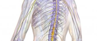
The long process of a neuron is called an axon (ἀξον - axis, Greek), it is also called an axial cylinder. At the site of axon formation on the neuron body there is a mound that plays an important role in the formation of a nerve impulse. It is here that the action potential coming from all dendrites of the neuron is summed up. The axon structure contains microtubules, but almost no organelles. The nutrition and growth of this process is entirely dependent on the body of neurons. When an axon is damaged, its peripheral part dies, while the body and the remaining part remain viable. And sometimes a neuron can grow a new axon. The diameter of the axon is only a few micrometers, but the length can reach 1 meter. These are, for example, the axons of spinal cord neurons that innervate human limbs.
View gallery
development
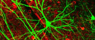
23 week fetus brain culture astrocytes
Most glia are derived from the ectodermal tissue of the developing embryo, particularly the neural tube and crest. An exception is microglia, which are derived from hematopoietic stem cells. In adults, microglia are largely a self-renewing population and are distinct from macrophages and monocytes, which infiltrate the injured and diseased CNS.
In the central nervous system, glia develop from the ventricular zone of the neural tube. These glia include oligodendrocytes, ependymal cells, and astrocytes. In the peripheral nervous system, glia originate from the neural crest. These glial SPS include Schwann cells in nerves and satellite glial cells in ganglia.
Currently, research involving glial cells in the human cochlea suggests that these cells are the common precursor of two mature Schwann cells and satellite glial cells. Additionally, peripheral glial cells located along peripheral processes express NGFR, indicating a phenotype different from peripheral glial cells located along central processes.
Glia retain the ability to undergo cell division into adulthood, whereas most neurons cannot. The opinion is based on a general deficiency of the mature nervous system in replacing neurons after injury such as stroke or trauma, while very often there is a deep spread of glia, or gliosis, near or at the site of injury.
However, detailed studies have found no evidence that "mature" glia, such as astrocytes or oligodendrocytes, retain the capacity for mitosis. Only resident oligodendrocyte precursor cells appear to retain this ability after the nervous system matures.
Glial cells are known to be capable of mitosis. In contrast, the scientific understanding of whether neurons are permanently postmitotic, or capable of mitosis, is still evolving. In the past, glia were considered to lack certain neuronal functions. For example, glial cells are not believed to have chemical synapses or to release transmitters. They are considered passive observers of neural transmission. However, recent studies have shown that this is not true.
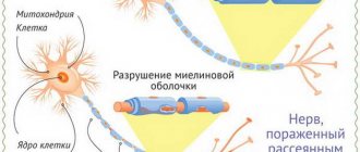
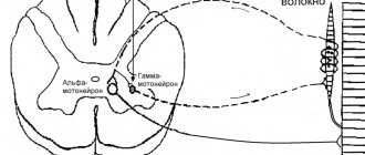
Axon myelination
The shell of the long processes of the neuron is formed by Schwann cells. These cells wrap around sections of the axon, and their tongue wraps around it. The cytoplasm of Schwann cells is almost completely lost and only a membrane of lipoproteins (myelin) remains. The purpose of the myelin sheath of the long processes of neuron bodies is to provide electrical insulation, which leads to an increase in the speed of the nerve impulse (from 2 m/sec to 120 m/sec). The shell has breaks - constrictions of Ranvier. In these places, the impulse, like a galvanic current, freely exits into the medium and enters back. And it is in the constrictions of Ranvier that the action potential occurs. Thus, the impulse moves along the axon in jumps - from constriction to constriction. Myelin is white; this is what served as the criterion for dividing the nerve substance into gray (neuron bodies) and white (conducting pathways).
View gallery
Clinical significance
While glial cells in the PNS often aid in the regeneration of lost nervous system function, loss of neurons in the CNS does not result in a similar response from neuroglia. The CNS regrowth will only occur when the injury was mild, not severe. When severe injury occurs, survival of the remaining neurons becomes the optimal solution.
However, some studies examining the role of glial cells in Alzheimer's disease are beginning to contradict the usefulness of this function, and even argue that it may "aggravate" the disease. Besides affecting the potential recovery of neurons in Alzheimer's disease, scarring and inflammation of glial cells have been further implicated in neuronal degeneration caused by amyotrophic lateral sclerosis.
In addition to neurodegenerative diseases, a wide range of harmful effects, such as hypoxia or physical trauma, can lead to the end result of physical damage to the central nervous system. Typically, when damage occurs in the CNS, glial cells induce apoptosis among the surrounding cell bodies.
Immune activity of the brain
Various biochemical reactions occur in the brain, so it must be protected from humoral immunity. It must be taken into account that neuronal tissue is sensitive to diseases, which is why neuronal recovery occurs partially.
It turns out that the formation of areas in the nervous system where a local reaction occurs causes the destruction of many cells. In the periphery of the body, painful places are filled with new cells. In the brain, a lost neuron is not restored. Thanks to neuroglia, the brain is not affected by the immune system.
Classification
Glial cells are divided into 2 types based on morphology and origin. There are microglial and macroglial cells. The first type has many processes with the help of which solid components are phagosed.
Macroglia are derived from ectoderm. Glial cells are divided according to morphology, and therefore they are ependymal and astrocytic, oligodendrocytes. Each type has its own characteristics.
The peculiarities of the origin of glial elements formed the basis for their division into macroglia (neuroglia itself) and microglia.
Macroglia are heterogeneous in morpho-functional terms. It includes the following types of cells:
- Ependymal;
- Oligodendrocytes;
- Astrocytes.
Moreover, each of the groups also has its own types of cells.
Ependymal cells are represented by ependymal cells of types I and II, as well as tanycytes. They are located in one layer, forming the lining of the pia mater (type I), the inner surface of the ventricles, the cerebrospinal canal (type II) and the bottom of the third ventricle (tanycytes). This structure ensures the barrier function.
Oligodendrocytes are present in the central and peripheral nervous systems. Macroglia are most numerously represented by these cells. Types of oligodendrocytes:
- Central gliocytes;
- Satellites;
- Lemmocytes.
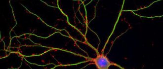
Astrocytes are star-shaped neuroglial elements with numerous processes. Their peculiarities include the fact that they are represented only in the central nervous system, both in the white matter (protoplasmic astroglia) and in the gray matter (fibrous astroglia).
The term “neuroglia” also includes microglial cells or glial macrophages. They have a different structure and origin from macroglia. These special types of multi-process cells are scattered throughout the brain and have the ability to phagocytose (a number of other neuroglial elements also have this feature). The main role of glial macrophages is to protect cerebral structures from pathological agents.
Origin
Glial cells have different origins. Depending on which cells were the precursors of neuroglial elements, macro- and microglia are distinguished.
Macroglia develops from the ectoderm (outer embryonic layer), i.e. has common precursors with neurons. Microglial macrophages are of mesodermal origin (from the middle germ layer). In fact, microglial elements are formed from blood structures (erythromyeloids, primitive macrophages and other cells of the hemocyte lineage), populating the brain in the early stages of embryogenesis. Subsequently, the number of cerebral macrophages is maintained as a result of proliferation.
Properties
Glial cells have a number of distinctive characteristics. Such features create unique conditions for the functioning of neurons. Gliocytes are capable of dividing, but are not able to independently reproduce and transmit nerve impulses. The membrane potential of glia is significantly higher than that of neurons.
This is determined by the concentration of potassium cations in the cytoplasm (glial cells have low permeability for other ions). When exposed to stimuli, glial cells are able to respond only with slow-wave (gradual) changes in the level of membrane potential, whereas in a neuronal response local spikes are typical.
Functions
Laws of axon work
When conducting a nerve impulse along axons, four main laws apply:
- Law of anatomical and physiological integrity. Conduction is possible only through intact neuron processes. This rule also applies to damage resulting from changes in membrane permeability (under the influence of drugs or poisons).
- Excitation isolation law. One axon – conduction of one excitation. Axons do not share nerve impulses with each other.
- Law of unilateral conduct. The axon conducts impulses either centrifugally or centripetally.
- The law of no loss. This property is non-decremental - when an impulse is carried out, it does not subside or change.
View gallery
Peculiarities
Glial cells can change in size, which is their feature. Moreover, this happens rhythmically with the help of a phase of contraction and relaxation. When the processes swell, their shortening is not observed.
Cell activity occurs thanks to the active components: serotonin and norepinephrine. A physiological feature is the effect on the intercellular space. Cells do not have impulse activity like nerve cells, but they do have a charge to generate membrane activity. Its changes occur slowly, which is determined by the activity of the nervous system.
Glial cells can spread, and this happens in 30-60 ms. The development of activity between them occurs with the help of gap junctions. These contacts exhibit low resistance and also create a sphere for current to flow from one area to another. Since glia are located with neurons, the functioning of the nervous system influences the electrical activity in the glial components.
Pathological processes
Due to exposure to pathologies, neuroglial cells are exposed to various negative consequences.
The following changes may occur:
- swelling and swelling;
- hypertrophy and atrophy;
- hyperplasia;
- amoeboid degeneration;
- homogenizing metamorphosis.
This disease, due to which the cellular structure changes, also occurs in histological examination when it is necessary to identify other human diseases. For a long period of time to examine the nervous system, neuroglial substances were considered secondary. Now they are considered the main components of nervous tissue. Pathologies can cause complex diseases.
Impact of neurons and glial cells
They have common properties and structure, for example, a nucleus that contains genetic information. The exchange between them occurs due to signaling molecules that enter through the membrane using a variety of mechanisms. They have the ability to process signals.
To perform their functions, they have processes that work together. Neurons can transmit an electrochemical signal to the axon, which results in an action. They are connected to each other by synapses.
It was previously believed that glia performed minor roles, but it was later determined that they perform major functions. The signals are transmitted by calcium waves that occur slowly. Neuroglia contact neurons with the help of neurotransmitters. In addition, they are considered the area of the brain where GABA and glutamate are formed.
That is why neuroglia are considered an important element necessary for the full development of a person. Their normal functioning ensures thinking and many other brain processes. If any areas are damaged, effective treatment prescribed by a doctor is required.
Dendrite spines
These outgrowths of the dendritic membrane can be found throughout their entire surface in large numbers. These are additional points of contact (synapses) of the neuron, many times increasing the area of interneuronal contacts. In addition to expanding the receptive surface, they play an important role in situations of sudden extreme influences (for example, in case of poisoning or ischemia). Their number in such cases changes sharply towards an increase or decrease and stimulates the body to increase or decrease the speed and number of metabolic processes.
View gallery