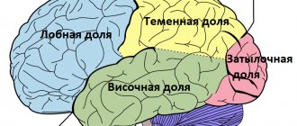
Nervous tissue, consisting of neurons and neuroglia, performs a complex of the most complex and responsible functions: weak electrical impulses arise in it, which are then transmitted to the muscles and organs of humans or vertebrates. The cells of this tissue have a special structure. It ensures both the occurrence of excitation and inhibition processes and their implementation. In neurobiology there is such a definition: dendrites are processes of a nerve cell that perceive and transmit information to the neuron body. In this work, we will familiarize ourselves with modern ideas about the mechanisms of transmission of nerve impulses in the main parts of the nervous system: the brain and spinal cord, and also study the structure of the dendrite as one of the constituent parts of neurocytes.
View gallery
To do this, let us consider in more detail the structural features of a neuron, which is an elementary unit of nervous tissue.
How the structure of a neurocyte is related to its functions
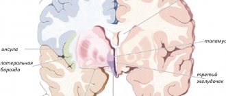
Electron microscopic research methods confirmed the fact of high specialization and complex structure of an open biological system called a nerve cell. It contains a body (soma), one long branch - the axon and many short processes. Each of them is connected to the cytoplasm of the neuron body. This is a dendrite. The structure and appearance of a collection of short shoots resembles the crown of a tree. Through them, bioelectric potentials from other nerve cells enter the neuron body through synapses.
Why is it so difficult to treat cancer?
It is worth noting in advance that the concept “cancer” hides a whole set of a huge number of types of malignant tumors. Some of them are so different that it is extremely difficult to find anything in common between them. Moreover, not all types of tumor diseases can be correctly called cancerous: cancer is only a special case of oncology, which studies both malignant and benign tumors. That is why, most likely, we will not see a universal cure for cancer on the shelves of pharmacies. Due to such a variety of oncological diseases, each patient requires a personalized approach to treatment. But even this personal treatment is often not effective in current practice. The most common methods are chemotherapy, surgery (when possible) and radiation therapy. But, unfortunately, these methods are also not always effective and often carry with them colossal side effects, sometimes incompatible with life.
Tumor cells are similar to healthy ones, like brothers. At the same time, growing up, one brother becomes a conscientious worker, and the other - a parasitic villain. And due to their great similarity, it is very difficult to direct the therapeutic effect specifically to tumor cells. Therefore, traditional therapy has a very low focus, that is, it acts on both bona fide and malignant cells approximately equally.
Currently, many groups of scientists are working to improve the effectiveness of traditional methods of treating tumor diseases. However, it is becoming almost impossible to significantly increase the survival rate of cancer patients using only standard therapy, especially in the last stages, and timely diagnosis is often impossible due to late patients seeking help. One way or another, it’s too early to give up.
Morphology and types
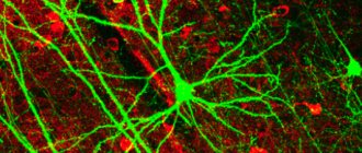
According to modern histological studies, dendrites are the branching ends of a neurocyte that not only receive, but also transmit information encoded in the form of electrical impulses through a multichannel system of anatomically and functionally interconnected nerve cells. They contain a large number of protein-synthesizing organelles - ribosomes. Some types of short processes, for example in pyramidal neurocytes, are covered with special structures - spines.
View gallery
According to the classification proposed by the Spanish neurohistologist S. Ramon y Cajal, two dendrites can extend from the body of a nerve cell in opposite directions (bipolar neurocytes). If there are many dendrites, then they diverge radially from the soma. This structure is characteristic of interneurons. In the cerebellar Purkinje cells, processes extend from the body of the neurocyte in the form of a fan. Each dendrite, whose structure is three-dimensional, differs from neighboring branches in the amount of electrical charges accumulated on it.
What's wrong with these tumor cells?
In the human body, the cellular structure is constantly renewed, old cells die, new ones are born. But along with healthy cells, as a result of mutations (that is, changes in the set of hereditary information under the influence of external or internal forces), atypical cells are formed. Such “eccentrics” most often cannot perform their functions correctly, and in an unfavorable scenario, their appearance leads to the formation of a malignant tumor.
Normally, such atypical cells are destroyed by the immune system, which is a kind of army that confronts the body’s enemies. But the peculiarity of malignant cells is their ability to “escape” immune control. They do this very sophisticatedly and extremely effectively, so that immune scout molecules often cannot detect them (Fig. 1), and killer cells are deactivated due to the expression of blocking factors by tumor cells.
Figure 1. Skillful camouflage of tumor cells.
collage by the author of the article
An additional background for the development of tumor cells is weakening of the immune system as a result of illness, stress, and poor lifestyle. As a result, tumor cells become “special” in the body; they ignore “anti-growth” stimuli, signals that trigger cell death, etc. The characteristics of tumor cells can be correlated with the behavior of an egoistic psychopath; not only do these cells not perform their proper functions, but they also divide uncontrollably and spread throughout the body, consume nutrients in crazy quantities, which they then spend on creating the same “psychopaths.” "(Fig. 2) [1]. Consequently, the metabolism and functioning of body tissues are disrupted, which most often leads to disastrous consequences.
Figure 2. What cancer cells can do.
collage by the author of the article
What is affected by the branching of nerve processes?
The neuron body is a universal transmitting and simultaneously receiving biological object. The volume (primarily of incoming information) is directly proportional to the number of incoming nerve impulses. They are determined by the degree of branching of the dendritic tree. Therefore, dendrites are structures of a neurocyte that play an integrative function.
View gallery
Moreover, the processes expand the area of contact between nerve cells. The additional formation of synapses greatly increases the efficiency of all parts, both the brain and spinal cord, and the nervous system as a whole.
Dendrite structure
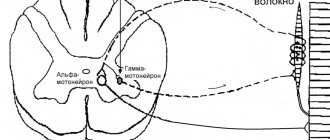
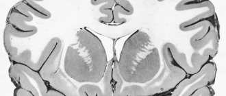
Based on the study of microscopic preparations of nerve cells, it was established that most of the processes are cylindrical in shape. Their diameter averages 0.9 microns. The length of dendrites varies widely. For example, stellate neurons of the gray matter of the cerebral cortex have short (no more than 200 μm) branches of the dendritic tree, while the processes of the motor neuron entering the anterior horn of the spinal cord are about 2 mm.
Special formations - spines, formed on the branches of neurocytes, lead to the appearance of a large number of synapses - slit-like places of contact with the axon, dendrite or soma of another neuron. Synapses can be located on the body of the dendrite and are called stem synapses or directly on its spines. As we already know, dendrites are branched processes of neurocytes that can receive excitation. The transfer of biopotentials occurs in them with the help of molecules of chemical compounds - mediators, for example, GABA or acetylcholine. In the membrane covering the dendrite, ion channels were found that selectively transmit calcium, sodium and potassium cations, which are involved in the passage of nerve impulses through the neuron.
View gallery
But I, cherishing my dream, solved the problem ingeniously...
Thanks to intensive research in the field of immunology, many factors have been discovered that influence the immune response [5]. It became clear that one of the key roles in the play “Immune Response” is played by special processes— dendritic ( DCs ). They were discovered in 1868 by the German scientist Paul Langerhans, who mistakenly mistook these cells for nerve endings with similar processes. DCs were again described in 1973 by Ralph Steinman, who also established their belonging to the immune system [6]. Only 38 years later he was posthumously awarded the Nobel Prize for his work.
In recent decades, there has been a growing trend towards the use of dendritic cells as adjuncts in the treatment of various types of cancer. According to scientists, their systematic use in immunotherapy will achieve its maximum effect.
Dendritic cells are a population of special cells whose function is to present “enemy” antigens to other cells of the immune system. In this way they activate adaptive immunity [6]. Scientifically, such messenger cells are called antigen presenting cells ( APCs ). DCs received their name due to the branched processes of the membrane, reminiscent of the dendrites of nerve cells, which grow in them at certain stages of development. DCs are located mainly in the blood and tissues that come into contact with the external environment. These cells have special mechanisms for recognizing “enemies.” In peripheral tissues, DCs capture antigens through several additional mechanisms [7]. Simply put, they are capable of foreign engulfment, that is, phagocytosis and pinocytosis of antigens, protruding the cell membrane and capturing the enemy particle.
After a “meal”, they move through the bloodstream or through lymphatic vessels to the lymph nodes [8]. Meanwhile, in DCs, protein antigens are converted (processed) and split into peptide pieces, which ultimately bind to molecules of the major histocompatibility complex ( MHC ) located on the surface of DCs [9]. After this, the DC reaches full maturity and, with the help of MHC molecules, presents the enemy antigen to other cells of the immune system.
These “other cells” are “army recruits,” not yet trained T cells that have not previously encountered an enemy antigen. After the encounter, the T cells begin to actively divide and differentiate into special forces, or antigen-specific effector T cells. Special units of special forces - CD4+ T cells - become indispensable helpers or T helpers (Fig. 3). They stimulate chemical soldiers - B-lymphocytes, which produce antibodies. These are special protein molecules that, like antidotes, go to fight specific foreign particles [10]. This chemical defense or immune response involving antibodies is referred to as humoral immunity.
Figure 3. Immune army.
collage by the author of the article
In addition, untrained T cells and T helper cells, through the release of the activating substance interleukin-2 ( IL-2 ), attract the help of sniper T killer cells, which subsequently destroy the infected cells by firing toxic cytotoxins. This is how cellular immunity works.
Some of the “trained” T cells become memory cells; they live in the body for years. Whenever they encounter an old familiar enemy, they quickly launch an immune response.
The type of immune response is partly determined by which DCs present the antigen and the release of which substances they stimulate [11]. Thus, by correctly selecting and processing DCs, it is possible to achieve the development of immune responses that interest us, for example, those that even tumor cells cannot resist.
How information enters a nerve cell
In the process of transfer of electrical charges, which underlies excitation and inhibition, along with the axon, dendrites also participate. These are neuron processes that form synapses with the branches of the dendritic tree of other neurocytes. It has been experimentally established that dendrites are outgrowths of the cell cytoplasm covered with a membrane. It produces weak electrical impulses - action potentials.
Thanks to the system of short processes, one nerve cell receives and transmits several thousand such impulses generated by synapses. This is not the only function of dendrites. They also process and integrate information entering the neurons, which ensures the regulation and control exercised by the nervous system over all organs and tissues of the human body.
There are very few images of the dendritic structure of metals on the Internet, not counting the famous photograph of Chernov’s crystal, and even diagrams from A.P.’s textbook. Gulyaeva. But if you are going to study the structures of metals, you need to know what they look like. In such a matter as metallurgy, no description can replace real images of structures, their consideration, comprehension, and analysis. So, dendrites in metals . First of all, it must be said that dendritic structures are formed, as a rule, during crystallization from a melt.
Crystallization from a liquid begins with the appearance of crystallization centers, i.e. points from which further construction of crystals continues. As a result of this, crystalline formations of various types begin to form from the liquid. In exceptional cases, a crystal is formed that has a geometrically regular shape - a polyhedron or polyhedron. This occurs in cases where external conditions contribute to the full development of the crystal (in all directions). Under normal conditions, crystals of irregular shapes are formed, which are called crystallites. There are two types of crystallites . In one case, the shape of the crystallite approaches multifaceted, or takes on a rounded shape. This formation is called grain. In another case, the crystalline formations have a branched shape with unfilled spaces, resembling a tree. They are called dendrites. Dendrites are the initial stage of crystal formation. The crystal begins to form from the center of crystallization. In this case, dense packing of crystalline groups into one crystal does not result; First, these groups are connected to each other in certain directions, forming the axes of the future crystal. If the crystallization conditions are such that the spaces between the axes do not have time or cannot be filled, the shape of the dendrite is preserved and can be observed. Dendrites (from the Greek δένδρον - tree) are complex crystalline formations of a tree-like branching structure (wikipedia - article “Dendrite (crystal)”). This definition is very apt - dendrites really have a branching structure, similar to a tree. And this can be proven. Figure 1 shows a real dendrite . It was formed in the process of self-propagating high-temperature synthesis in the Ni-Ti-O system.
Figure 1. True dendrite.
A dendrite is a single crystal (i.e. one crystal). The scheme of dendrite formation is presented in Figure 2. First, first-order axes are formed, then second-order axes arise and grow on them. Next - the third.
Figure 2. Scheme of dendrite formation.
As can be seen from the figures below, dendrites in metal are actually “branches” in shape. Sometimes they say dendrite branches .
| Alloy AL25 | Alloy AK12 |
Figure 3. Dendrites in aluminum alloys : dendrites of aluminum solid solution and Al-Si eutectic.
| Austenitic cast iron ChN15D7 | Hypoeutectic cast iron |
Figure 4. Dendrites in cast iron.
In a real crystal, axes of the first and second orders are usually visible, the third - less often (in essence, there is simply not enough time for their formation - crystallization ends). In general, the more orders of magnitude visible, the slower the alloy crystallized. Below, Figure 5 shows a dendrite containing axes of three orders. The third order is not fully formed; in some places the axes of the third order are just emerging. The first order axis is a green arrow, the second order is blue, and the third order is red.
Figure 5. Dendrites of different orders in silumin.
The dendritic structures of different alloys are similar. It is not always possible to understand what kind of alloy it is by looking at the cast structure, especially with low magnification. For example, dendrites in steel, cast iron, copper and oxide systems.
| Steel | Oxygen copper |
| Hypoeutectic cast iron | Oxide system based on Ni-Ti |
Figure 6. Dendritic structure in various alloys at magnifications from 100x to 200x.
Sometimes the dendrite has a shape (usually called “morphology”) characteristic of completely specific alloys. For example, in hypereutectic silumin (aluminum-silicon alloy. Silicon content is more than 11.7%), when cast into the ground, silicon crystals with a dendritic structure are formed. These are the so-called skeletal silicon crystals . Sometimes they say "skeletons" of silicon . At a higher crystallization rate (casting into a metal mold - chill mold), silicon crystals already have a polygonal shape. There are, however, exceptions...
| Hypereutectic silumin, in-ground casting | |
| Hypereutectic silumin, chill casting | Silicon “star” in hypereutectic silumin |
Figure 7. Silicon crystals in hypereutectic silumin.
At higher magnification, the alloy is easier to identify: alloyed silumin (silicon phase dendrite), ferritic cast iron (ferrite dendrites), babbitt ( antimony dendrite ). The fourth pattern is not easy to identify - it is a structure obtained by self-propagating high-temperature synthesis (possibly a dendrite of an intermetallic compound against a eutectic background).
| Alloyed silumin | Ferrite-pearlite cast iron |
| Babbitt | Ni-Ti-O system |
Figure 8. Characteristic dendrites in various alloys.
One might ask: why so much about dendrites?
The fact is that each material is given a certain structure based on practical purposes. For example, cast iron “works” in a cast state (they can also be deformed, but this is not the topic of this article). Steel is usually supplied in a deformed state. Sheet, rod, strip, tape - all these are forms of supply of semi-finished steel products. To obtain such semi-finished products, the initially cast steel undergoes special pressure treatment at elevated temperatures. There should be no cast structure after such processing. Therefore, if it is preserved, then it is a marriage. This is shown in Fig.9. Circumference about in steel. We will return to this topic in the “Anti-production” section.
Figure 9. Remains of the cast structure in P18 steel (product - tap ).
Dendrites should be recognizable not only in the alloys themselves, but also in auxiliary materials, for example, in Wood's alloy. The type of Wood's alloy structure varies. It depends on the composition, and whether it is a “fresh” alloy or a reused one. Figure 10 shows dendrites in a Wood's alloy that has been repeatedly remelted. Naturally, in such an alloy there is quite a lot of “dirt” that got into the alloy during remelting.
| A | b |
| V | G |
Figure 10. Dendrites in Wood's alloy: a - bright-field image; b-d - differential interference contrast.
Ice patterns are always recognizable. Ice is a solid form of water that is formed during the process of crystallization (freezing). Its forms are varied. By the way, ice dendrites can be seen in every freezing puddle (it should be remembered that water in the temperature range from 0 to 1000C is melted ice).
Figure 11. Ice dendrites of various morphologies (photo from glass).
Snowflakes are also dendrites , only in the shape of stars.
But below are dendrites, which we, unfortunately, do not see as much as we feel. These are ice crystals on the surface of paving slabs. at the top there is water. After the frost there came a thaw and it began to rain. The tile did not have time to heat up due to its insufficient thermal conductivity. Some of the rainwater crystallized.
Figure 11. Ice dendrites on the surface of a tile where everyone is falling.
The following photographs are “ dendrites on metals ”. Figure 13 shows the results of washing a beryllium bronze section with ethyl alcohol (instead of water) after etching with a saturated solution of potassium dichromate in sulfuric acid. Washing with alcohol was unsuccessful; the reagent remained on the surface and dried. At various magnifications, crystals of potassium bichromate can be seen on the surface. They have their own characteristic color.
| A | b |
Figure 13. Dendrites of potassium bichromate on a sample of beryllium bronze BrB2.