All convolutions and grooves of the human brain have long been named and described. In neuroanatomical atlases, the same gray matter of the cerebral cortex is painted in different colors. This color map is over a hundred years old. And the very idea that mental functions are localized in different places on the surface of the human cerebral cortex arose at the turn of the 18th and 19th centuries. The German physician Franz Gall (1758–1828) created the so-called phrenological maps of the brain, where he placed the properties of the psyche, which he called “soul abilities.” From the point of view of modern science, Gall's amazing maps are the fruit of conclusions based not on experimental data, but only on his own observations. However, scientists have been struggling to implement his idea for two centuries.
|
Map of projections of body parts onto the postcentral (A) and precentral (B) cortex of the cerebral hemispheres. Sensory (A) and motor (B) homunculus. |
At the end of the 19th century, German physiologists discovered a zone in the cerebral cortex of dogs and cats, the electrical stimulation of which caused involuntary contraction of the muscles on the opposite side of the body. They were able to accurately determine in which parts of this zone different muscle groups are represented. Later, this zone (it was called motor) was described in the human brain; it is located in front of the central (Rolandic) sulcus, which most deeply divides the cerebral cortex in the transverse direction. Here, representations of the muscles of the larynx, mouth, face, arm, torso, and leg are sequentially located, and the area of the cortex does not at all correspond to the size of the body parts. Canadian neurologist Wilder Graves Penfield and E. Baldry, comparing both, drew a funny little man in this place - a homunculus. He has a huge tongue, lips, thumbs, and his arms, legs and torso are very small. The symmetrical homunculus also lives behind the central sulcus, only it is not motor, but sensory. Parts of this area of the cerebral cortex are associated with skin sensitivity of various parts of the body. The motor and sensory areas interact closely with each other, so they are usually considered as a single sensorimotor cortex. Later it turned out that everything was a little more complicated: physiologists found another complete motor representation of a smaller body, responsible for maintaining posture and some other complex slow movements.
| And here is what the “motor” homunculus looks like in volume. |
All sense organs also have their authorized representation in the cerebral cortex. For example, in the occipital region of the human brain there is a visual cortex, in the temporal lobe there is an auditory cortex, and the olfactory representation is scattered across several parts of the brain. In the cortex there are also so-called associative fields, where the analysis and synthesis of information coming from the primary fields of the sensory organs takes place. Associative fields are most strongly developed in humans, especially those located in the frontal lobe; physiologists associate the highest manifestations of the psyche with them - thinking, intelligence. Back in the middle of the 19th century, the French scientist Paul Broca and the German psychiatrist Carl Wernicke discovered two areas in the left hemisphere of the human brain that are related to speech. If Broca's area is damaged - in the posterior third of the inferior frontal gyrus, the patient's speech is impaired, but if Wernicke's area is affected - in the posterior third of the superior temporal gyrus, the patient can speak, but his speech becomes meaningless.
Representations of sensory organs in the human cerebral cortex. 1 - visual zone; 2 - auditory zone; 3 - zone of skin sensitivity; 4 - motor zone; 5 - olfactory zone. |
So today physiologists know a lot about the structure and functions of the brain. But the more they learn, the more mysteries remain. And none of the modern researchers can claim to know how the brain works. In terms of the level of information content, the brain maps that exist today can probably be compared with the geographical maps of the Middle Ages, when the outlines of the continents only vaguely resembled real ones, and the white spots were larger in area than everything else. “And most importantly, knowing approximately the geography, we have no idea what is happening in different “countries.” What do they do, how do they live,” comments the director of the Institute of Human Brain of the Russian Academy of Sciences, corresponding member of the Russian Academy of Sciences Svyatoslav Vsevolodovich Medvedev.
| Broca's and Wernicke's centers. |
The task of removing blank spots from a brain map and increasing its resolution is much more difficult than filling in blank spots in geography. Especially when we are talking about the human brain and the highest manifestations of the human psyche. Is it really possible to project human feelings, the tension of thought, the pangs of creativity onto the surface of the brain? Will it ever be possible to say: this zone is responsible for making decisions, this group of cells is responsible for the feeling of beauty, this is where envy nests, and this is where the love zone begins?
“It’s more correct to talk not about mapping the brain, but about mapping brain functions,” explains S.V. Medvedev. “The challenge is to determine where the neurons that are involved in solving a particular task are located, and to understand how these parts of the brain interact with each other. Finally, the ultimate task for a neurophysiologist—a goal from which we are still very far away—is to correlate events occurring in the brain with what a person thinks, to decipher the codes of higher nervous activity.”
The brain speaks electrical language
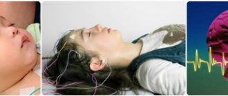
The first data on the localization of higher brain functions were obtained in the era of “clinical and anatomical comparisons,” that is, observations of patients who had damage to some areas of the brain. Then, in the late 1920s, the era of dominance of electrophysiological research began. Physiologists have learned to record the electrical activity of the brain - the electroencephalogram (EEG) of a person through electrodes placed on the scalp (this was first done by the Austrian psychiatrist Hans Berger in 1929). This method became the main one in the study of the functioning of the brain and its diseases - the first electrophysiologists believed that with the help of EEG it was possible to know everything. Indeed, EEG reflects various processes occurring in the brain, but the difficulty is that it records the total electrical activity, summarizes and averages the work of a huge number of nerve cells - neurons. And this is its methodological limitation.
Then other methods of studying the electrical activity of the brain appeared, for example, the method of evoked potentials - these are electrical waves that arise in certain areas of the cerebral cortex in response to specific stimulation. In the visual cortex they appear in response to a flash of light, in the auditory cortex - in response to sound, etc. This method has given a lot to the study of the localization of functions in the areas of the cerebral cortex, and with its help the brain map has been significantly refined. But it also has limitations, especially when studying the human brain.
With the development of microelectrode technology, it became possible to record electrical discharges of individual neurons. This is mainly done, naturally, in experiments on laboratory animals. A breakthrough in human brain research came when it became possible to record the electrical activity of human neurons directly from the brain using implanted subcortical electrodes. Academician Natalya Petrovna Bekhtereva began to use this method in the early 60s. Thin electrodes were inserted into the patient's brain for therapeutic purposes - with their help it was possible to target areas of the brain. But since an electrode is implanted into the patient’s brain, then we need to use this opportunity and get maximum information from him. Such an electrode records the activity of surrounding neurons, and this is a completely different level of resolution than can be obtained from an electrode located on the surface of the head.
A new stage in the study of the brain and neurocomputers
The discoveries of O'Keefe and the Mosers are undoubtedly among the most significant in neuroscience in recent decades. Thanks to their research, scientists became acquainted with a completely new type of neuronal operation, in which cells form a multicomponent network that allows complex cognitive processes to be carried out.
In addition to its fundamental importance, the study of the orientation system of the brain also plays an important role for clinical practice. Some diseases of the nervous system, such as Alzheimer's disease, are accompanied by impaired spatial orientation and spatial memory.
Studying the workings of complex neural structures is important for the rapidly developing field of neurocomputing and robotics, allowing the use of elegant natural solutions as technological discoveries.
Written using materials from the Nobel Committee.
Neurons are “literate” and “creative”
With the help of implanted subcortical electrodes, physiologists from the Institute of Human Brain of the Russian Academy of Sciences managed to learn a lot about how the brain copes with speech. As already mentioned, Broca's and Wernicke's areas related to speech have been known for a long time. “It would be more correct to limit ourselves to the definition of “related to speech” and not to use the expression “speech zone,” emphasizes S.V. Medvedev. — Remember the joke about a cockroach, which, it turns out, has “ears on its legs”? We need to realize that Broca’s and Wernicke’s areas may not be the center of speech, but some kind of interface.”
| Test for distinguishing semantic and grammatical features of speech. A group of neurons that changes electrical activity depending on the nature of the response. |
In a completely different place in the cerebral cortex, researchers found a detector for the grammatical correctness of a meaningful phrase. A group of neurons increases their electrical activity if the phrase the subject hears is grammatically correct, and decreases it when it is grammatically incorrect. If a subject is presented with the phrases “blue ribbon” and “blue ribbon,” these “literate” neurons will immediately notice the difference. Another group of neurons distinguishes between words of the native language, words that are phonetically similar to them, and foreign words. “This means that the neural population almost instantly analyzes the phonetic structure of the word and classifies it into the following types: “I understand,” “I don’t understand, but something familiar” and “I don’t understand at all,” says S.V. Medvedev. In this regard, the question arises whether these neurons work the same or differently in people gifted with innate literacy and in those who have problems with this. Most likely, there are differences, but in order to give an accurate answer, you need to recruit quite a lot of subjects.
“We found groups of neurons that distinguish between concrete and abstract words, neurons that appear to be responsible for counting,” says Svyatoslav Vsevolodovich further. “We have identified areas of the brain that are associated with generalization and decision making. All neuronal systems are characterized by polyfunctionality: this means that the same cells can participate in different functions. The specialization of neurons is relative - depending on the situation, they can take on different responsibilities. For example, when the captain of a ship dies, a navigator or someone else takes his place. Therefore, the brain is a very flexible system.” Neurons lose their ability to be interchangeable over time and become more specialized. A small child cannot walk and talk at the same time; if you call him, he will stumble and fall. The fact is that his entire cortex is occupied by either one or the other. The student should not be distracted during the lesson, otherwise he will not learn the material. Over time, more and more division of brain territories occurs, so an adult can simultaneously drive a car and carry on a conversation, talk on the phone and look at documents, etc.
N.P. Bekhtereva and her collaborators found neurons in the brain that work as an error detector. What is their role? They react to any violation of the stereotypical sequence of actions. “You leave home and on the street you feel: “Something is wrong...” explains S.V. Medvedev. “That’s right - they forgot to turn off the light in the bathroom.” Error detector neurons are located in different parts of the brain - in the parietal cortex of the right hemisphere, in the Rolandic sulcus, in the superior parietal and parietotemporal areas of the cortex, in the cingulate gyrus.
But the method of implanted electrodes also has limitations. Electrodes, of course, are not implanted everywhere where physiologists would like them, but only where necessary for clinical indications. Doesn't this mean that we are looking where it is brighter, and not where we have lost?
A little about history
Many scientists were involved in constructing a map of the surface of the brain: Bailey, Betz, Economo and others. Their maps differed significantly from each other in the shape of the fields, their size, and number. In modern neuroanatomy, Brodmann's brain fields have received the greatest recognition. There are 52 fields in total.
Pavlov, in turn, divided all fields into two large groups:
- centers of the first signal system;
- centers of the second signaling system.
Each center consists of a core, which plays a key role in performing the function of a specific center, and analyzers surrounding the core. It is noteworthy that centers in the cerebral cortex regulate the functioning of organs on the opposite side of the body. This is due to the fact that the nerve fiber pathways cross on their way from the center to the periphery.
Brodmann's brain fields are designated by Arabic numerals, some also have a designation from which the function of a particular field can be understood.
Brain scanner runs on positrons
Traditionally used in medicine, X-rays are not the best method for obtaining a picture of the brain. Completely different possibilities arose with the advent of magnetic resonance imaging (MRI). The Institute of Human Brain of the Russian Academy of Sciences actively uses the positron emission tomography (PET) method. Both methods provide images of the brain. What is the difference between them?
MRI is based on the properties of certain atomic nuclei, primarily the nuclei of hydrogen atoms, when placed in a magnetic field to absorb energy in the radio frequency range and emit it after the cessation of exposure to the radio frequency signal. Depending on the “environment,” that is, on the properties of the biological tissue in which these nuclei are located, the intensity of their radiation changes. Therefore, it is possible to see images of various brain structures. The essence of the PET method is to monitor vanishingly small amounts of a substance labeled with a radioactive ultra-short-lived (half-life of minutes) isotope. The isotope emits positrons, which annihilate with electrons, emitting two gamma quanta, and fly away in opposite directions. If you register these gamma rays with a detector, you can determine the location of the atoms of the labeled substance. The substance is chosen so that its concentration reflects the activity of brain cells. For example, if the concentration of radioactively labeled glucose increases somewhere, this means that neurons are actively consuming it, and therefore are actively working. If at this time the subject performs any task, then we see which areas of the brain are involved in its implementation. The PET method allows the use of short-lived isotopes (O, N, C, F), which are not very harmful to the patient.
Using PET, you can also observe changes in cerebral blood flow during a particular behavior. When any area of the brain is activated, blood actively flows to it. If water labeled with radioactive oxygen is injected into a vein, it enters the vessels of the brain and can be registered. Where there is more labeled oxygen, more blood flows, which means that this is where activity increases.
Second Signal System: Location
The presence of a second signaling system is characteristic only of humans. It is these centers that provide higher thinking, which includes generalization of information, dreams, and logic. In fact, for normal thinking and speech, activation of all Brodmann fields is necessary, but centers can be distinguished that have their own specific functions:
- 44 - located in the posterior part of the inferior frontal gyrus;
- 45 - located anterior to field 44, in the anterior portion of the frontal gyrus;
- 47 - located below the two previous fields, closer to the basal part of the frontal lobe;
- 22 - one of the most anterior ones;
- 39 - located in the posterior part of the superior temporal gyrus.
From grammatical outposts to labyrinths of creativity
Using PET scans, researchers continued to study human speech using the whole brain. They saw where speech information is processed: individual words, the meaning of the text, where it is memorized. They showed that the medial extrastriate cortex is involved in processing the orthographic structure of words, and a significant part of the left superior temporal cortex (Wernicke's area) is probably involved in semantic analysis. Word order is analyzed by the anterior superior temporal cortex. When a person is shown a coherent text without even being asked to read it (they just had to count the number of times a letter appears), cerebral blood flow increases, which means the brain is involved in linguistic work. (If presented with words mixed in random order, the brain does not react this way.)
Brain regions activated when searching for a letter in a connected text (left), compared to perceiving an unrelated sequence of words (right) |
Even the “divine” process of creativity turned out to be decipherable, at least by physiologists in the laboratory of N.P. Bekhtereva came close to this. A person is given a creative task, for example, to compose a story from a set of words, and in real time they see which areas of the brain begin to work actively. It turned out that creative activity is accompanied mainly by changes in connections between different areas of the brain. Most of the new connections appear in the left anterior temporal zone with the anterior zones of the cortex, and with the posterior zones, on the contrary, the connection is weakened. The connections between the parietal and occipital structures are lost. And all this happens precisely when performing a creative task, but if the task is devoid of creative elements, there are no such changes. Local cerebral blood flow increases in the right prefrontal cortex during a more creative task compared to a less creative one. From this, scientists conclude that this particular area is directly related to “creativity.”
| Brain organization of creative thinking. Shows the area of the brain in which local blood flow increases during a more creative task compared to a less creative one (right prefrontal cortex). |
Researchers are also interested in the phenomenon of involuntary attention: for example, a person is driving a car, listening to the radio, talking and suddenly instantly reacts to the knocking of the engine, indicating that something is wrong with the engine. In two laboratories using two different methods: S.V. Medvedev using the PET method and Yu.D. Kropotov, using the method of implanted electrodes, discovered the same zones where activation occurs at such moments - in the temporal and frontal cortex. Activation occurs in response to a mismatch between expected and actual stimuli, for example when the sound from a motor is not what it should be. Another phenomenon is selective attention, which helps a person, in the continuous hum of voices at a cocktail reception, to follow the speech of one interlocutor, the one who is interesting to him. Apparently, the prefrontal cortex is responsible for focusing spatial attention in this case. It tunes either the right or left auditory cortex, depending on which ear receives important information.
When talking about brain mapping, it is important to understand that the brain, strictly speaking, is not divided into clearly demarcated areas, each of which is responsible only for its own function. Everything is much more complicated, since in the process of performing any function, neurons from different areas interact with each other, making up a neural network. Studying how individual neurons are combined into a structure, and the structure into a system and whole brain, is a task for the future.
“PET is a powerful tool for studying almost any function, but it alone is not enough,” says S.V. Medvedev. — The purpose of PET is to answer the question “where?”, and to answer the question “what is happening?”, PET should be combined with electrophysiological methods. Together with British physiologists, we have created a system for parallel analysis of PET and EEG, which complement each other. This approach is probably the future.”
A year ago (the article was published in 2004 - P.Z. ), a group of scientists from six countries announced the creation of a three-dimensional computer map of the human brain, which can be used to determine a person’s predisposition to certain diseases. The creators of the map believe that they can already associate certain diseases, such as Alzheimer's disease or autism, with different parts of the cerebral cortex. Now they are busy clarifying the details of their invention.
Paul Brodmann
- Fields 1 and 2, 3 - somatosensory area, primary zone. Located in the postcentral gyrus. Due to the commonality of functions, the term “ fields 1 and 2, 3
” is used (front to back) - Area 4 - primary motor cortex. Located within the precentral gyrus
- Area 5 is the secondary somatosensory area. Located within the superior parietal lobule
- Area 6 - premotor cortex and supplementary motor cortex (secondary motor area). Located in the anterior sections of the precentral and posterior sections of the superior and middle frontal gyri.
- Field 7 is the tertiary zone. Located in the upper parts of the parietal lobe between the postcentral gyrus and the occipital lobe
- Field 8 - located in the posterior parts of the superior and middle frontal gyri. Includes the center of voluntary eye movements
- Area 9 - dorsolateral prefrontal cortex
- Area 10 - anterior prefrontal cortex
- Area 11 - olfactory area
- Field 12 -
- Field 13 -
- Field 14 -
- Field 15 —
- Field 16 -
- Field 17 - nuclear zone of the visual analyzer - visual area, primary zone
- Field 18 - nuclear zone of the visual analyzer - center of perception of written speech, secondary zone
- Field 19 - nuclear zone of the visual analyzer, secondary zone (assessment of the meaning of what is seen)
- Area 20 - inferior temporal gyrus (center of the vestibular analyzer, recognition of complex images)
- Area 21 - middle temporal gyrus (center of the vestibular analyzer)
- Field 22 - nuclear zone of the sound analyzer
- Field 23 -
- Area 24 - anterior cingulate cortex
- Field 25 —
- Field 26 -
- Field 27 -
- Field 28 - projection fields and associative zone
The brain is the most complex organ in the human body. Its most highly organized part is the cortex. Thanks to its presence, a person is able to read, write, think, remember, and so on. Many scientists have paid attention to the study of the structural features of the cortex. There are many works on the division of the cortex into so-called Brodmann fields. They are what will be discussed later in the article.
The second hypostasis of the gene
In the early 50s of the last century, the idea arose that memory cannot be limited only to electrical processes - for long-term storage of information in the brain, it must be preserved in a chemical form. Although at that time there were still very general ideas about the cell genome, the idea arose that it not only stores hereditary information, but also participates in the storage of information acquired during life.
To test this, we needed to see whether learning triggered the synthesis of nucleic acids and proteins in the brain. After the principle of the genome—DNA → RNA → protein—became known, experiments became more targeted. And this is what turned out. Immediately after animals are taught a skill, RNA synthesis increases in their brains. (In order to detect this, they were injected with radioactively labeled RNA precursors.) This happened with mice that were trained to avoid electric shock in response to a sound signal, and with chickens that developed an imprint on an object, and with goldfish that were trained to swim with a raft attached to their abdomen. And if RNA synthesis is slowed down, then animals make many mistakes or are not able to learn the skill at all.
At the same time, new proteins are also synthesized in the brain - this was also determined by the inclusion of radioactive isotopes. Protein synthesis blockers disrupt long-term memory without affecting short-term memory. From this it becomes clear how genes work: when trained on a DNA template, RNA is synthesized, which, in turn, generates new proteins. These proteins come into action a few hours after the acquisition of information, and they ensure its storage. And the initiators of all these events are electrical processes occurring on the membrane of the nerve cell.
A group of researchers from the Department of Systemogenesis of the Institute of Normal Physiology of the Russian Academy of Medical Sciences, led by Doctor of Medical Sciences, Corresponding Member of the Russian Academy of Medical Sciences K.V. Anokhina set herself the task of finding research methods that would make it possible to simultaneously study the activity of nerve cells throughout the brain in connection with any behavior or cognitive activity. “When we started our work, we were convinced that information from synapses is transmitted to another, deeper level - it penetrates the cell nucleus and somehow changes the functioning of genes,” says Konstantin Vladimirovich. “It remains to find these genes.”
It must be said that a myriad of genes work in brain cells—in humans, half of all studied genes are expressed only there. The task was to find from all their multitude the key ones involved in storing new information. The search was successful in the mid-1980s, when K.V. Anokhin and his colleagues paid attention to the so-called “immediate early genes.” They received this name for their ability to be the first to respond to extracellular stimuli. The role of “early” genes is to “wake up” other - late genes. Their products—regulatory proteins—transcription factors—act on sections of the DNA molecule and trigger the process of transcription—copying information from DNA into RNA. Ultimately, the “late” genes synthesize their proteins, which cause the necessary changes in the cell, for example, the formation of new neuron connections.
First Signal System: Location
The centers of the first signaling system are located in Brodmann's fields, which are present in both animals and humans. They are responsible for a simple reaction to an external stimulus, the formation of sensations and ideas. These centers are present in both the right and left hemispheres of the cerebral cortex. Brodmann fields of the first signaling system are present in humans from birth and normally do not undergo changes throughout life.
These fields include:
- 1 - 3 - are located in the parietal lobe of the cerebral cortex behind the central gyrus;
- 4, 6 - located in the frontal lobe anterior to the central gyrus, they contain Betz pyramidal cells;
- 8 - this field is located anterior to the 6th, closer to the frontal part of the frontal cortex;
- 46 - located on the outer surface of the frontal lobe;
- 41, 42, 52 - located on the so-called Heschle convolutions, on the basal part of the temporal lobe of the brain;
- 40 - located in the parietal lobe behind 1 - 3 fields, closer to the temporal part;
- 17 and 19 - located in the occipital part of the brain, most dorsal from the other fields;
- 11 is one of the most ancient structures, located in the hippocampus.
The most inquisitive gene
Of the entire group of “early” genes, researchers were most interested in the c-fos gene by K.V. Anokhin and his colleagues have been studying the role of this gene in learning since 1987 - in their opinion, it is suitable for the role of a universal probe for brain mapping. “This gene has several unique properties,” explains K.V. Anokhin - Firstly, in a calm cell state it is silent, it has practically no “background level” of activity. Secondly, if any new information processes begin in the cell, it very quickly responds to them, producing RNA and proteins. Thirdly, it is universal, that is, it is activated in various parts of the central nervous system - from the spinal cord to the cortex. Fourthly, its activation is associated with learning, that is, with the formation of individual experience.” To prove the last statement, scientists conducted dozens of experiments, testing under what influences c-fos would come out of hiding and begin to act. It turned out that the gene does not respond to very strong stimulation, such as light, sound or pain, in cases where the effect does not contain elements of novelty. But as soon as the situation is enriched with new information, the gene immediately “wakes up.”
| c-fos gene expression: A) neurons with c-fos protein are identified by immunohistochemical staining; |
For example, in an experiment, mice were placed in a chamber where they had to endure a series of weak but unpleasant electrodermal stimulations. In response, c-fos was highly expressed in several areas of their brains—the cortex, hippocampus, and cerebellum. However, if this procedure is carried out daily, then on the sixth day the gene no longer responds. Mice still react to electric shock, but it is no longer a new event for them, but an expected event. It is possible to reactivate c-fos by placing the mice in the chamber again without subjecting them to the usual procedure. In both cases, the gene marks an event when external stimuli are not consistent with the individual memory matrix. Such a mismatch occurs with any assimilation of new information, and therefore c-fos is an inevitable companion of cognitive processes in the brain.
Another experiment involved newborn chicks, which were divided into four groups. The chicks of the first group were hatched in the dark and never saw light, the second group was luckier - it was kept on a normal 12-hour light cycle, the chicks from the third group were transferred to an enriched visual environment immediately after birth, and the chicks from the fourth group were first kept in a normal environment. conditions, and on the second day they were transferred to an enriched medium. In all experimental chickens, c-fos gene expression was assessed on the second day after hatching. What happened? In the first three groups, despite such different conditions in which they spent two days of their short lives, c-fos did not manifest itself. But in the fourth group, which changed the environment to a visually enriched one, c-fos became more active. It was new to them, while the chickens of the third group had already gotten used to it.
The expression of c-fos increased and in chicks that pecked the bead that interested them, it turned out to be bitter, and the chicks learned to avoid it in the future. But in general it turned out that gene activation does not at all depend on the success of learning and accompanies erroneous actions in the same way. The c-fos gene also reacts simply to a new object - to activate it, simply presenting a new object to the animal for just 10 seconds is enough.
The researchers suggested that c-fos and other early genes are the very bridge through which an animal's individual experience interacts with its genetic apparatus.
Second signaling system: functions
As noted above, Brodmann’s cytoarchitectonic fields of the second signaling system are necessary for the implementation of higher nervous activity. And the main difference between a person and an animal is the ability to speak.
In field 45 is the Broca Center. It is necessary for normal speech motor skills. It is thanks to the presence of this center that a person is able to pronounce words. When it is damaged, a condition called “motor aphasia” develops.
In field 44 there is the center of written speech. Impulses from this area of the cortex travel to the skeletal muscles of the fingers and hand. When it is destroyed, a person loses the ability to write, which is called “agraphia.”
Field 47 is responsible for singing. It is during the normal functioning of this center that a person can pronounce words into a chant.
In field 22 there is the Wernicke center. This is where auditory speech analysis takes place. Thanks to the normal operation of the 22nd field, a person perceives words by ear.
Field 39 is the center of visual speech. The functioning of this field allows a person to distinguish between characters written on paper. When it is damaged, a person loses the ability to read, which is called sensory alexia.
Notes
- Brodmann Korbinian.
Vergleichende Lokalisationslehre der Grosshirnrinde : in ihren Principien dargestellt auf Grund des Zellenbaues. — Leipzig: Johann Ambrosius Barth Verlag, 1909. - Sapin M. R., Bilich G. L.
Human anatomy. - M.:: "Higher School", 1989. - P. 417. - 544 p. — 100,000 copies. — ISBN 5-06-001145-3. - Gerhardt von Bonin & Percival Bailey.
The Neocortex of Macaca Mulatta. — Urbana, Illinois: The University of Illinois Press, 1925. - E. D. Chomskaya.
Neuropsychology, 4th edition. — Peter, 2008.
| Brodmann's cytoarchitectonic fields at Wikimedia Commons |