Functions of the lobes of the brain
The brain is a powerful control center that sends commands throughout the body and controls the progress of their implementation. It is thanks to him that we perceive the world and are able to interact with it. The kind of brain a modern person has, his intelligence, thinking, are the result of millions of years of continuous evolution of mankind, his structure is unique.
The brain is characterized by division into zones, each of which specializes in performing its own specific functions. It is important to have information about what functions each zone performs.
Then you can easily understand why specific symptoms appear in such common diseases as Parkinson's disease, Alzheimer's disease, stroke, etc.
Violations can be regulated with medication, as well as with the help of special exercises and physical procedures.
The brain is structurally divided into:
- rear;
- average;
- front.
Each of them has their own role.
In an embryo, the head develops faster than other parts of the body. In a one-month-old embryo, all three parts of the brain can be easily seen. During this period they look like “brain bubbles”. The brain of a newborn is the most developed system in his body.
Scientists attribute the hindbrain and midbrain to more ancient structures. It is this part that is entrusted with the most important functions - maintaining breathing and blood circulation. The boundaries of their functions are clearly separated.
Each gyrus does its job. The more pronounced the groove became during development, the more functions it could perform.
But the anterior section provides everything that connects us with the external environment (speech, hearing, memory, ability to think, emotions).
There is an opinion that a woman's brain is smaller than a man's brain. Data from modern hardware studies, in particular on a tomograph, have not confirmed this. This definition can easily be called erroneous. The brain of different people may differ in size and weight, but this does not depend on gender.
Knowing the structure of the brain, you can understand why certain diseases appear and what their symptoms depend on.
Structurally, the brain consists of two hemispheres: right and left. Outwardly, they are very similar and are interconnected by a huge number of nerve fibers. Each person has one side that is dominant, right-handers have the left side, and left-handers have the right side.
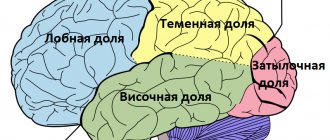
There are also four lobes of the brain. You can clearly see how the functions of the shares are differentiated.
What are shares?
The cerebral cortex has four lobes:
- occipital;
- parietal;
- temporal;
- frontal
Each share has a pair. All of them are responsible for maintaining the vital functions of the body and contact with the outside world. If injury, inflammation, or disease of the brain occurs, the function of the affected area may be completely or partially lost.
Frontal
These lobes have a frontal location, they occupy the forehead area. Let's figure out what the frontal lobe is responsible for. The frontal lobes of the brain are responsible for sending commands to all organs and systems. They can be figuratively called a “command post.” It would take a long time to list all their functions.
These centers are responsible for all actions and provide the most important human qualities (initiative, independence, critical self-esteem, etc.). When they are defeated, a person becomes carefree, changeable, his aspirations have no meaning, he is prone to inappropriate jokes.
Such symptoms may indicate atrophy of the frontal lobes, leading to passivity, which is easily mistaken for laziness.
Each lobe has a dominant and auxiliary part. For right-handed people, the left area will be dominant and vice versa. If you separate them, it is easier to understand which functions are assigned to a specific area.
It is the frontal lobes that control human behavior. This part of the brain sends commands that prevent a specific antisocial action from being performed. It is easy to notice how this area is affected in dementia patients. The internal limiter is turned off, and the person can tirelessly use obscene language, indulge in obscenities, etc.
The frontal lobes of the brain are also responsible for planning, organizing voluntary actions, and mastering the necessary skills.
Thanks to them, those actions that seem very difficult at first become automatic over time.
But when these areas are damaged, the person performs the actions as if anew each time, and automaticity is not developed. Such patients forget how to go to the store, how to cook, etc.
When the frontal lobes are damaged, perseveration can occur, in which patients literally become fixated on performing the same action. A person may repeat the same word, phrase, or constantly move objects around aimlessly.
The frontal lobes have a main, dominant, most often left, lobe. Thanks to her work, speech, attention, and abstract thinking are organized.
It is the frontal lobes that are responsible for maintaining the human body in an upright position. Patients with their lesions are distinguished by a hunched posture and a mincing gait.
Temporal
They are responsible for hearing, turning sounds into images. They provide speech perception and communication in general. The dominant temporal lobe of the brain allows you to fill the words you hear with meaning and select the necessary lexemes in order to express your thoughts. The non-dominant helps to recognize intonation and determine the expression of a human face.
The anterior and middle temporal regions are responsible for the sense of smell. If it is lost in old age, it may signal incipient Alzheimer's disease.
The hippocampus is responsible for long-term memory. It is he who stores all our memories.
If both temporal lobes are affected, a person cannot assimilate visual images, becomes serene, and his sexuality goes through the roof.
Parietal
In order to understand the functions of the parietal lobes, it is important to understand that the dominant and non-dominant side will do different jobs.
The dominant parietal lobe of the brain helps to understand the structure of the whole through its parts, their structure, order. Thanks to her, we know how to put individual parts into a whole. The ability to read is very indicative of this. To read a word, you need to put the letters together, and you need to create a phrase from the words. Manipulations with numbers are also carried out.
The parietal lobe helps to link individual movements into a complete action. When this function is disrupted, apraxia is observed. Patients cannot perform basic actions, for example, they are not able to get dressed. This happens with Alzheimer's disease. A person simply forgets how to make the necessary movements.
The dominant area helps you feel your body, distinguish between the right and left sides, and relate parts and the whole. This regulation is involved in spatial orientation.
The non-dominant side (in right-handed people it is the right side) combines information that comes from the occipital lobes and allows you to perceive the world around you in three-dimensional mode. If the non-dominant parietal lobe is disrupted, visual agnosia may occur, in which a person is unable to recognize objects, landscapes, or even faces.
The parietal lobes are involved in the perception of pain, cold, and heat. Their functioning also ensures orientation in space.
Occipital
The occipital lobes process visual information. It is with these lobes of the brain that we actually “see.” They read signals that come from the eyes. The occipital lobe is responsible for processing information about shape, color, and movement. The parietal lobe then turns this information into a three-dimensional image.
If a person stops recognizing familiar objects or loved ones, this may indicate a dysfunction in the occipital or temporal lobe of the brain. In a number of diseases, the brain loses the ability to process received signals.
How the hemispheres of the brain are connected
The hemispheres are connected by the corpus callosum. This is a large plexus of nerve fibers through which the signal is transmitted between the hemispheres. Adhesions are also involved in the joining process. There is a posterior, anterior, and superior commissure (fornix commissure). This organization helps to divide the functions of the brain between its individual lobes. This feature has been developed over millions of years of continuous evolution.
Conclusion
So, each department has its own functional load. If a separate lobe suffers due to injury or disease, another zone may take over some of its functions. Psychiatry has accumulated a lot of evidence of such redistribution.
It is important to remember that the brain cannot function fully without nutrients. The diet should have a variety of foods from which the nerve cells will receive the necessary substances. It is also important to improve blood supply to the brain. It is promoted by sports, walks in the fresh air, and a moderate amount of spices in the diet.
If you want to maintain full brain function until old age, you should develop your intellectual abilities. Scientists note an interesting pattern - people with intellectual work are less susceptible to Alzheimer's and Parkinson's diseases.
The secret, in their opinion, lies in the fact that with increased brain activity in the hemispheres, new connections are constantly created between neurons. This ensures constant tissue development.
If a disease affects one part of the brain, its functions are easily taken over by the neighboring zone.
Source: //vsepromozg.ru/stroenie/doli-golovnogo-mozga
Diseases
Many researchers have found a decrease in neuronal density in the large temporal lobes of patients who have been diagnosed with schizophrenic diseases. According to research results, the right temporal lobe was larger in size compared to the left. As the disease progresses, the temporal part of the brain decreases in volume. In this case, there is increased activity in the right temporal lobe and disruption of connections between neurons of the temporal and cephalic cortex.
This activity is observed in patients with auditory hallucinations who perceive their thoughts as third-party voices. It was noted that the stronger the hallucinations, the weaker the connection between the temporal lobe and the frontal cortex. In addition to visual and auditory abnormalities, disorders of thinking and speech are added. The superior temporal gyrus in people with schizophrenia is significantly smaller than in the same region of the brain in healthy people.
Occipital lobe of the brain - functions and structure
20.03.2019
It is known that a person receives up to 85% of information about the environment through vision, and only the remaining 15% is hearing and other senses. The occipital lobe is the area responsible for the highest processing of visual signals.
Thanks to it, healthy humanity is able not only to distinguish surrounding environmental objects by their visual characteristics, but also to contemplate the creations of artists, to create themselves.
We can catch the mood of other people by observing the changes in their facial expressions, enjoy the beauty of the sunset, and, finally, choose food based on our favorite color.
- Location
- Functions
- What fields are included?
- Symptoms of the lesion
Location
The occipital lobe is the region of the telencephalon that is located behind the temporal and parietal lobes.
In the occipital lobe of the cerebral cortex there is a central part of the analyzer, namely the visual one.
This region of the brain includes the non-constant lateral occipital sulcus, which separates the superior and inferior occipital gyri. Inside this area is the calcarine groove.
Assigned functions
The functions of the occipital lobe of the brain are associated with the analysis, perception and containment (storage) of visual information. The visual tract consists of several points:
- An eye with its retina. This paired organ is only a mechanical component of vision, performing an optical function.
- Optic nerves, through which electrical impulses directly travel at a certain frequency and carry certain information.
- The primary centers are represented by the visual thalamus and quadrigeminal.
- Subcortical and cortical centers. All of the above structures act as points of elementary perception and delivery of information. The visual cortex, in contrast, plays the role of a higher analyzer, that is, it processes the received nerve impulses into mental visual images.
It is noteworthy that the retina of the eye perceives a set of light waves, each of which has a length and consists of quanta of electromagnetic radiation.
But the cortex, evolving over millions of years, “ learned ” to work with such signals and turn them into something more than a set of energy and impulses. Thanks to this, people have a picture of the environment and the world.
Thanks to this crust, we see the elements of the universe as they appear.
The visual cortex, located on both hemispheres of the occipital lobe, provides binocular vision - the world appears three-dimensional to the human eye.
The human brain is a multifunctional structure, like each area of its cortex - therefore, the occipital lobe of the brain in a standard functional state takes little part in processing auditory and tactile signals. Under conditions of damage to neighboring areas, the degree of participation in signal analysis increases.
The visual cortex, called the association area, constantly interacts with other brain structures, forming a complete picture of the world.
The occipital lobe has strong connections with the limbic system (especially the hippocampus), parietal and temporal lobes.
Thus, a particular visual image may be accompanied by negative emotions, or vice versa: a long-standing visual memory evokes positive feelings.
The occipital lobe, in addition to simultaneous analysis of signals, also plays the role of an information container. However, the amount of such information is small, and most environmental data is stored in the hippocampus.
The occipital cortex is strongly associated with theories of feature integration, the essence of which is that the cortical analytical centers process individual properties of an object (color) both separately, in isolation, and in parallel.
To summarize, we can answer the question of what the occipital lobe is responsible for:
- processing visual information and integrating it into a general attitude towards the world;
- storage of visual information;
- interaction with other areas of the telencephalon and partial succession of their functions;
- binocular perception of the environment.
What fields are included?
The occipital lobe of the cerebral cortex contains:
- 17th field – accumulation of gray matter of the visual analyzer. This field is the primary zone. Consists of 300 million nerve cells.
- 18th field. It is also a nuclear cluster of the visual analyzer. According to Brodmann, this field performs the function of perceiving written speech and is a more complex secondary zone.
- 19th field. This field takes part in assessing the meaning of what is seen.
- 39 field. However, this brain area does not entirely belong to the occipital region. This field is located on the border between the parietal, temporal and occipital lobes. The angular gyrus is located here, and its list of tasks includes the integration of visual, auditory and general sensitivity of information.
Symptoms of the lesion
When the area responsible for vision is affected, the following symptoms are observed in the clinical picture:
Dyslexia is the inability to read written words. Although the patient sees the letters, he cannot analyze and understand them.
Visual agnosia : loss of the ability to distinguish objects in the environment by their external parameters, but patients manage to do this by touch.
Impaired visuospatial orientation .
Impaired color .
Hallucinations are the visual perception of something that does not exist in the real objective world. In this case, the characteristics of photopsia are lightning-fast color perception and various types of flashes.
Visual illusions and - perverted perception of real-life objects. For example, the patient may perceive the world in red colors, or all surrounding objects may seem extremely small or large to him.
When is damaged, loss of the opposite visual fields is observed.
With large-scale tissue damage in this area, complete blindness .
Didn't find a suitable answer? Find a doctor and ask him a question!
Source: //sortmozg.com/structure/zatylochnaya-dolya-golovnogo-mozga-funktsii-i-stroenie
Limbic cortex
Deep in the lateral sulcus in the temporal lobe is the so-called limbic cortex, which resembles an insula. A circular groove separates it from adjacent nearby areas on the side. The anterior and posterior parts are visible on the surface of the island; localized in it The inner and lower parts of the hemispheres are combined into the limbic cortex, including the amygdala nucleus, olfactory tract, areas of the cortex
The limbic cortex is a single functional system, the properties of which consist not only in providing communication with the outside, but also in regulating the tone of the cortex, the activity of internal organs, and behavioral reactions. Another important role of the limbic system is the formation of motivation. Internal motivation includes instinctive and emotional components, regulation of sleep and activity.
Occipital lobe
The occipital lobe occupies the posterior parts of the hemispheres.
On the convex surface of the hemisphere, the occipital lobe has no sharp boundaries separating it from the parietal and temporal lobes, with the exception of the upper part of the parieto-occipital sulcus, which, located on the inner surface of the hemisphere, separates the parietal lobe from the occipital lobe.
The grooves and convolutions of the superolateral surface of the occipital lobe are not constant and have a variable structure. On the inner surface of the occipital lobe there is a calcarine groove that separates the wedge (a triangular lobe of the occipital lobe) from the lingual gyrus and the occipitotemporal gyrus.
The function of the occipital lobe is associated with perceptions, processing of visual information, and the organization of complex processes of visual perception. In this case, the upper half of the retina of the eye is projected in the wedge area, perceiving light from the lower fields of vision; in the region of the lingual gyrus there is the lower half of the retina of the eye, which perceives light from the upper fields of vision.
Island
The insula, or the so-called closed lobule, is located in the depths of the lateral sulcus. The insula is separated from the adjacent neighboring sections by a circular sulcus. The surface of the island is divided by its longitudinal central groove into anterior and posterior parts. A taste analyzer is projected in the island.
Location
The superior lateral parts of the hemisphere belong to the parietal lobe. In front and from the side, the parietal lobe is limited by the frontal zone, from below - by the temporal zone, from the occipital part - by an imaginary line running from above from the parieto-occipital zone and reaching the lower edge of the hemisphere. The temporal lobe is located in the lower lateral parts of the brain and is emphasized by a pronounced lateral groove.
The anterior part represents a certain temporal pole. The lateral surface of the temporal lobe displays the superior and inferior lobes. The convolutions are located along the furrows. The superior temporal gyrus is located in the area between the lateral groove above and the superior temporal groove below.
On the verso layer of this area, located in the hidden part of the lateral sulcus, there are two or three gyri belonging to the temporal lobe. The inferior and superior temporal gyri are separated by the middle. In the lower lateral edge (temporal lobe of the brain) the inferior temporal gyrus is localized, which is limited by the groove of the same name at the top. The posterior part of this gyrus continues in the occipital zone.

