Content
- Introduction: what studies are meant by MRI of the head?
- The most common MRI examinations of the head: MRI of the brain General (survey) examination of the brain
- Brain examination for neurovascular conflict or MRI of the “trigeminal nerve”
- Study of brain neurodegeneration, exclusion of Parkinson's disease
- Study of brain neurodegeneration, exclusion of Alzheimer's disease
- Search for episyndrome using high-resolution MRI
- MRI examination of the sella turcica in the brain
- MRI examination of the pineal gland in the brain
- MRI of the temporomandibular joint
- Magnetic resonance arteriography of the head
The essence of the method
Unlike CT or X-ray, this test relies on high-power nuclear magnetic resonance (NMR) rather than radiation. In this case, atomic nuclei are excited in combination with electromagnetic waves, which change their direction in a constant magnetic field. In medicine, NMR registration is used on the nuclei of hydrogen atoms that are part of the water molecule. Since the body is saturated with this element, even minor changes in its concentration are recorded by the tomograph and reflected in the image. This ability allows you to identify different structures, distinguishing normal tissues from pathological ones (inflammation, tumor). This means that structures with a high water content (soft tissues, discs and cartilage, the brain) are easy to examine, and their changes are not difficult to decipher. As for bones or lungs, an MRI examination will not be informative. That is why MRI is prescribed primarily for diseases of the head and spine.
MRI allows you to obtain high-quality images of internal organs.
The most common MRI examinations of the head: MRI of the brain
2.1. General (survey) examination of the brain
See full details of the study:
Full description of the studyWhat a survey MRI of the brain showsIndications
A general survey study is carried out when there is no clear suspicion of a specific disease.
Since the symptoms are characteristic of different diseases, it is recommended to conduct this study immediately with intravenous contrast - this will allow you not to miss minor changes (for example, tumors or metastases). The frequency of “cuts” in survey research is relatively low.
For comparison, in addition to general MRI studies of the brain, there are high-resolution MRI studies, in which the slice frequency is much higher, but such a study will take 40 minutes or more.
High-resolution studies are used to diagnose some specific diseases, for example, episyndrome.
2.2. Brain examination for neurovascular conflict or MRI of the “trigeminal nerve”
See full details of the study:
More information about the disease Causes and symptoms MRI examination for diagnosis
This is a targeted examination designed to accurately examine the area where the trigeminal nerve exits the brainstem in a magnified manner.
The study is carried out only as prescribed by a doctor.
The reason for the prescription may be pain on one side of the face, as well as numbness of the lip or tongue on the same side.
Sometimes a patient may have a “distorted face,” which is a consequence of paralysis of the muscles on one side of the face.
2.3. Study of brain neurodegeneration, exclusion of Parkinson's disease
See full details of the study:
More information about the disease Causes and symptoms MRI examination for diagnosis
The test may be prescribed by your attending physician if Parkinson's disease is suspected. With this disease, the death of brain neurons and neurodegeneration in the midbrain occurs.
There are so-called “MRI signs”, the presence of which signals the disease.
For example, researchers have found that in the case of Parkinson's disease, MRI images of the brains of affected people lack the characteristic "dovetail" outline of a certain area of the brain in 90% of studies.
There are also other signs of the development of the disease, such as atrophy of the substantia nigra, globus pallidus and caudal nucleus.
2.4. Study of brain neurodegeneration, exclusion of Alzheimer's disease
See full details of the study:
More information about the disease Causes and symptoms MRI examination for diagnosis
Alzheimer's disease is a neurodegenerative disease that causes dementia (acquired dementia) in 60-80% of cases.
An MRI study allows us to evaluate changes in the volume of the temporal lobes of the brain, which are responsible for memory, with a diagnostic accuracy of 87%.
The study will help determine the rate of brain volume loss, as well as identify other pathologies that signal vascular or frontotemporal dementia.
This MRI examination is prescribed by the attending physician.
2.5. Search for episyndrome using high-resolution MRI
See full details of the study:
More details about the diseaseFeatures of research in epilepsyWhat does MRI show in epilepsy?Indications for MRI in epilepsyHow does epilepsy differ from episyndrome?MRI in episyndromeWhat does MRI show in episyndrome?
Epilepsy is one of the most common chronic neurological diseases. It is characterized by the sudden onset of convulsive attacks due to the course of other diseases associated with injuries, tumors, and inflammation of the brain.
Unlike epilepsy, episyndrome is not a chronic disease.
There is a special MRI protocol used to detect episyndrome.
It is important to note that the study must be carried out on a high-field MRI machine using a special algorithm, because Artifacts in the images may be mistaken for a false positive result. This study should be prescribed by the attending physician.
2.6. MRI examination of the sella turcica in the brain
See full details of the study:
Indications for MRI of the pituitary gland and sella turcica What does MRI of the pituitary gland show? In what cases is MRI with contrast required and when is it not needed?
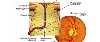
During the sella turcica MRI, the pituitary gland is examined, which is an endocrine gland located in the bony cavity at the base of the skull.
Due to the location, as well as the characteristically small size of the formations (tumors), diagnosing diseases in this area of the brain is extremely difficult.
The size of the pituitary gland can vary depending on hormonal status and age. Because of this, the outlines of the gland in photographs of people of different ages and genders can differ radically. But it is the most minor changes that can be signs of meningitis or metastases.
The above suggests that this targeted study should be carried out on a medium- or high-field device, and the highly qualified doctor responsible for conducting the study and interpreting the images is extremely important. Often the study requires the use of contrast.
2.7. MRI examination of the pineal gland in the brain
See full details of the study:
More details about the disease MRI diagnostics of the pineal glandIndications for examination Why is it better to do it with contrast? General list of diagnosed diseasesTypes of neoplasms
An examination of the pineal gland (also called the pineal or pineal gland) is prescribed by a doctor to specifically exclude tumors, cysts and microcysts. Symptoms can be very different and depend on the severity of the damage to the gland.
The gland is very small in size, but is well illuminated when contrast is administered, since it is located outside the blood-brain barrier.
This targeted study, like the study of the sella turcica, is best carried out on a medium- or high-field apparatus.
It is extremely important that a highly qualified specialist conducts the research and interprets the results.
Preparation and carrying out the procedure
MRI of the spine should be performed on an empty stomach. The patient does not eat for 6 hours prior to the procedure. You do not need to undress for the examination, but you cannot have any metal objects, magnets, watches, or magnetic cards on you - this could result in injury to the patient and damage to the camera.
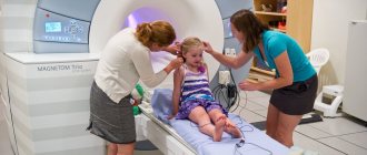
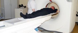
The patient lies down on a mobile table, which is then moved inside the chamber. Contact with the patient is constantly maintained, so that he can report any ailments that arise during the study. While the procedure is taking place, the patient should remain as still as possible, as this affects the quality of the image.
While the MRI scan of the spine is being performed, the patient is placed in a lighted, narrow tunnel and must lie still. He is in acoustic contact with medical personnel at all times. An MRI takes 30-90 minutes depending on its type. Before resonance imaging, the patient must remove all metal jewelry (earrings, brooches, necklaces), as they can disrupt the magnetic field and the operation of the device.
MRI studies of other parts of the head
3.1 MRI of the temporomandibular joint
See full details of the study:
More information about the studyIndications for the studyContraindicationsWhat MRI shows of the temporomandibular joint
The temporomandibular joint is located in the head and connects the lower jaw to the base of the skull.
This MRI study is carried out in cases of impaired mobility of the temporomandibular joint, inflammatory processes in the joint, deformation of the joint (due to destruction), displacement and degeneration of the articular disc, as well as in cases of dislocation or subluxation of the joint.
MRI is the standard method for assessing the state of the ANS. Sometimes the examination is performed in the open and closed position of the mouth to determine abnormalities in the orientation of the joint.
As a rule, the study is carried out on one side.
3.2. MRI of the internal auditory canal (MRI of the ear), petrous part of the temporal bones
See full details of the study:
More information about the studyIndications for the studyWhat MRI of the ear canal shows
MRI of the middle and inner ear is prescribed for a number of pathologies associated with dysfunction and structure of the auditory analyzer.
The study is carried out to identify one of the possible pathologies, including: disruption of the structure and development of the middle ear, destruction or inflammation of tissue, injuries to the eardrum, pathologies of the facial and auditory nerves, tumors of the cerebellopontine angle (the area of the brain between the pons, cerebellum and medulla oblongata), detection of tumors and meningiomas.
All this is not a complete list of possible problems and diseases, so it is very important that the study is prescribed by the attending physician, who, based on the symptoms, will be able to determine exactly what anomalies and processes need to be looked for during an MRI examination of the ear canal.
3.3. MRI of the eye (orbit), MRI of the orbits
See full details of the study:
More information about the studyIndications for the studyContraindicationsWhat MRI of the eyes shows
During this study, the contents of the orbit are examined and signs of pathological processes are established. The examination may reveal foreign bodies in the eye, inflammation of the eye structure and extraocular muscles, as well as tumors.
Visualization of the internal structure of the orbit during an MRI examination makes it possible to identify cranial and intraorbital nerves, as well as to detect formations in the orbit, in the intracranial or pituitary regions.
It is the MRI study that provides the most detailed information about the condition of the eye and orbit, in comparison with other types of studies.
The list of indications for MRI of the eye (orbit) is very large, so it is very important to obtain a preliminary referral from a doctor to clarify the areas to be examined and the pathologies that need to be specifically looked for during the examination.
3.4. MRI of the sinuses (paranasal sinuses)
See full details of the study:
Read more about the studyIndications for the studyWhat MRI of the nose shows What diseases can be detected
MRI of the nasal sinuses (or maxillary sinuses) is a non-invasive and modern diagnostic method that allows doctors to accurately determine the presence and nature of pathological conditions localized within the area under study.
During this study, the following can be examined: the frontal sinus, the ethmoid sinus, the maxillary sinus.
Examination of the sinuses reveals:
- proliferation of the mucous membrane (polyps);
- cavities filled with fluid (cysts);
- inflammation of the sinuses (tumors or sinusitis);
- inflammation of the maxillary (maxillary) sinus;
- frontitis
Also, MRI of the nose provides important imaging information for the surgeon.
A large number of symptoms and diagnosed pathologies require an initial referral from the attending physician to clarify the area of study - this will allow MRI examination of the sinuses to be carried out specifically and as efficiently as possible.
3.5. MRI of the skull base
See full details of the study:
Read more about the studyTargeted MRI studies in the area of the skull baseMain pathologies that MRI can revealTumors of the skull baseOther diseasesMRI of the craniovestebral junction
During this study, pathologies are identified in the ligament of the occipital bone of the skull and the first cervical vertebra. The study is targeted and should be prescribed by the attending physician.
Symptoms of the disease are very different, and, as a rule, depend on the severity of the lesion: infringement of the structures of the occipital bone of the skull and the first cervical vertebra can begin with a buzzing in the ears when turning the head, and end with such serious symptoms as abnormalities in the spine or paralysis of the arms and legs
Where can I do it?
The manipulation is performed by budget and private commercial clinics and medical centers. Judging by the reviews of those who visited them, MRI of the spine is done well in the institutions listed in the table below.
| Megapolis | Name of the medical center, clinic | Address | Telephone |
| Moscow | Hospital RAS (CDB RAS) | Litovsky Boulevard, 1A | (499) 110-57-69 |
| Moscow | Honey | st. Tverskaya, 24 | (499) 110-70-67 |
| Moscow | Medical (K+31) | 1st Kolobovsky lane, 4 | (499) 450-26-57 |
| Moscow | Medical on Tverskaya-Yamskaya | 2nd Tverskoy-Yamskoy lane, 10 | (499) 490-47-65 |
| Saint Petersburg | Medical diagnostic | st. Chapaeva, 5 | (812) 643-33-55 |
| Saint Petersburg | MEDEM | st. Marata, 6a | (812) 643 52 73 |
| Saint Petersburg | Medical | 6th Krasnoarmeyskaya st., 7 | (812) 615-91-94 |
Also, the MRI procedure can be performed at a municipal budget institution, but on a first-come, first-served basis, which can sometimes take several months.
Examination of veins and arteries in the head (in the brain)
4.1. Magnetic resonance arteriography of the head
See full details of the study:
Full description of the study What diseases does it show Indications Contraindications What does MRI of head vessels show
Arteriography is a specialized study and is carried out as prescribed by a doctor.
The study is carried out to diagnose thrombosis (impaired blood flow in the lumen of a vessel), as well as to identify venous plexuses (malformations).
4.2. Magnetic resonance venography of the brain
See full details of the study:
More about the disease
Venography of the brain is a specialized study and is carried out as prescribed by a doctor.
This MRI study is necessary to identify pathological narrowings (stenoses), protrusion of vessel walls with risks of rupture (aneurysms), as well as increased tortuosity and the presence of additional bends of the veins.
If you suspect the above pathologies, it is important to undergo an MRI examination in a timely manner, primarily because insufficient blood supply from the neck to the head can cause serious changes in the brain.
What is needed for the examination
If you were referred by a doctor, you must have the referral written by him with you. When undergoing a second examination for this disease, it is advisable to take with you pictures and descriptions of previous studies (even if this was carried out with other equipment - for example, X-rays or computed tomography.
Food and liquid intake is not limited. When carrying out a procedure with the introduction of a contrast agent, you should remember that if you have already done such a procedure before, then time must pass from the previous one: 3 days for intravenous administration of a contrast agent and 7 days for injection into the spinal canal.
Advantage of the method
Unlike radiography, in one MRI scan, for example of the head, you can view about 20 images at different levels from a certain part of the body and with a slice thickness of 4-5 mm. The thinner these sections, the easier it is to make the correct diagnosis. And unlike CT, on MRI the image is visualized in any projection: frontal, sagittal, axial, that is, you can examine transverse and longitudinal sections of the selected object. The advantage of the method is also the ability to record on electronic media. This can be useful for online consultation and in case the patient has lost or damaged the image. In addition, the method has the following properties:
- good quality;
- high information content;
- safety (the patient is not exposed to radiation);
- the ability to obtain images of almost all tissues of the body.
This research method does not cause discomfort or pain to the patient.
Contraindications to magnetic resonance imaging
Unfortunately, not all patients can undergo this examination. You need to remember this, and also know what contraindications exist. There are absolute and relative indications for magnetic resonance scanning.
| Absolute contraindications: | Relative contraindications: |
|
|
There is a misconception that breastfeeding, menstruation and the presence of an intrauterine device are also contraindications. It doesn't really matter. The radiologist, who interprets the scan results, decides whether or not to conduct an examination, but other specialists can also describe the images obtained.
The presence of a pacemaker is one of the contraindications for MRI.