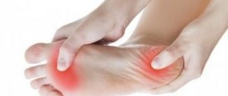
Despite the high level of development of medicine, some pathologies remain not fully understood. One of these diseases is Tolosa-Hunt syndrome. Currently, only the symptoms of this pathology are known, while the causes have not yet been fully disclosed. Most likely, this is due to the fact that the disease is very rare and was discovered not so long ago. In addition, Tolosa-Hunt syndrome is often “masked” as other pathologies and is easily confused. The main symptom is damage to the eyes and cranial nerves. It is known that one of the properties of this disease is that it is amenable to hormonal therapy, after which improvement quickly occurs. However, relapse can occur at any time.
Description of Tolosa-Hunt syndrome
The first mention of this disease appeared relatively recently. It was described in 1961 by the English ophthalmologist Hunt. In addition to the main name, the pathology is also called differently, for example, “chameleon”. This is due to the fact that its symptoms resemble many other ailments. Among them: head tumors, infectious encephalitis, orbital myositis and even diseases of the hematopoietic system.
In addition, Tolosa-Hunt pathology is called a symptom of the superior orbital fissure, which does not accurately reflect the essence of the problem. The clinical picture of this disease depends on which nerves of the brain are affected. Most often, Tolosa-Hunt syndrome is characterized by pain in the orbital area, the inability to move the gaze, and diplopia. With timely initiation of hormonal therapy, a complete cure or stable remission can be achieved.
Pathogenesis
It is very difficult to describe the condition of this disease. If you combine in one anesthesia of the upper eyelid, half of the forehead and cornea with full ophthalmology, you can correlate similar parameters.
This condition usually occurs when the ophthalmic, abducens, and oculomotor nerves are affected. This, in turn, occurs under various conditions - from a small number of pathologies to mechanical damage.
The main group of causes of this syndrome:
- brain tumor located in the orbital area;
- deviations of the arachnoid membrane of the brain of an inflammatory nature - arachnoiditis;
- meningitis in the area of the superior palpebral fissure;
- orbital injury.
The clinical picture manifested in superior palpebral fissure syndrome is characteristic exclusively of this disease.
Symptoms of the disease:
- ptosis of the upper eyelid;
- paralysis of the eye muscles caused by pathology of the optic nerves – ophthalmoplegia;
- low tactile sensitivity in the cornea and eyelids;
- mydriasis – dilation of the pupil;
- dilated retinal veins;
- inflammation of the cornea in a sluggish state.
- exophthalmos - bulging eyes.
Syndromes are not necessarily fully expressed, sometimes only partially expressed. During diagnosis, this fact is always taken into account. If two or more signs coincide, you should immediately contact an ophthalmologist.
Mechanism of disease development, symptoms of the disease
Superior orbital fissure syndrome (another but less accurate name for STX) develops gradually. Initially, a slight pain appears above the eyebrows, behind the eyeball, in the temples and forehead. It gradually intensifies and turns into “tearing” or “burning.” After 14 days, the pain in one half of the face becomes very severe, and double vision is added to the unpleasant sensations.
If left untreated, the eyeball begins to move with great difficulty, and strabismus develops on the side where the pain first appeared. Very rarely, symptoms do not appear gradually, but all at once.
It has been noticed that in about a quarter of patients, the optic nerves are very severely affected, making the eyeball completely immobile. The disease is sometimes accompanied by low-grade fever, swelling of the conjunctiva of the eyeball and its bulging.
Symptoms may worsen over days or weeks. Sometimes remission begins for no apparent reason, but the disease is sure to recur. If treatment is started on time, symptoms disappear within 2-3 days.
At the initial stages, general malaise, febrile temperature, headache, fatigue, and weakness are observed. After a few days, the skin of the auricle, the front part of the tongue and the soft palate become covered with herpetic eruptions in the form of transparent or hemorrhagic blisters. Weakness of muscle tissue is noted on the affected part of the face.
Characteristic features include distortion of the face, smoothing of the nasolabial fold, and drooping of the corner of the mouth. Facial pain appears from the moment the disease manifests itself. They can radiate to the neck, back of the head and area around the eyes. The disease is accompanied by hyperesthesia of the auditory canal, which is replaced by hypoesthesia. In some cases, sensitivity disorders are observed on the entire half of the face.
If the vestibulocochlear nerve is affected, hearing loss and ringing in the ears occur. When the vestibular nerve is damaged, the clinical picture is complemented by dizziness, instability and horizontal nystagmus. Over time, peeling crusts appear at the site of the vesicular rashes. Also symptoms of the disease are swelling, lacrimation and injection of blood vessels in the white membrane of the eyes.
The disease begins without any prerequisites and immediately in an acute form, with pain in the orbital region. Painful sensations appear in the frontal part, in the area of the superciliary arches, and with increasing intensity they spread to the eyes. This is followed by the development of symptoms in increasing order, replacing each other:
- There is a problem with eye movement, diplopia (double vision). It is difficult for the patient to concentrate his gaze on one object, everything is double;
- ophthalmoplegia (impaired mobility of the eyeball). In most cases it is unilateral. The degree of immobility depends on the number of nerves damaged and the intensity;
- exophthalmos (displacement of the eyeball) occurs if no treatment is carried out or with a frequently recurring syndrome;
- strabismus can develop with unilateral nerve damage;
- deterioration of general condition and increase in temperature.
All of these symptoms can increase one after another, gradually causing external changes. But in some cases, with a rapid flow, they can appear almost simultaneously.
Sometimes, for unknown reasons, obvious signs of the disease suddenly disappear on their own. However, without receiving appropriate treatment, relapse is possible. The duration of remission can be completely different, as well as the frequency of relapses.
Classification
Ramsay Hunt syndrome occurs in various clinical variants, which also include combined damage to the facial and vestibulocochlear nerves. Understanding the form of the disease and the stage of its clinical course is necessary when choosing treatment tactics. According to the clinical classification, there are four main forms of the syndrome:
- Hunt I - rashes in the area innervated by the geniculate ganglion are not accompanied by neurological symptoms.
- Hunt II - herpetic rashes occur with paresis of the facial nerve.
- Hunt III - facial paresis and blistering rash are combined with hearing impairment.
- Hunt IV - vestibular disorders are added to the manifestations characteristic of Hunt III syndrome.
There are three clinical periods during the course of the disease:
- General infectious - characterized by general symptoms: weakness, lethargy, fever.
- Skin - the period of appearance of a typical herpetic rash.
- Neuropathic - characterized by the predominance of neuralgic pain syndrome.
Etiology of the disease
Due to the fact that the pathology has various manifestations and begins suddenly, it has not yet been possible to establish the exact etiology. This is also due to the low occurrence of the syndrome. For this reason, doctors do not have the opportunity to properly study this disease. There are several assumptions according to which pathology may develop. The following etiological factors are identified in patients with Tolosa-Hunt syndrome:
- Malformation. This term implies abnormal development of the vascular system of the eyes. As a result of the malformation, mixing of arterial and venous blood occurs, which should not be normal. This disorder is more common among the female population.
- Autoimmune aggression. This factor is the trigger for many diseases. However, it is still not possible to answer the question of why immune cells begin to destroy body tissues. In most cases, “aggression” occurs after suffering stress and long-term infectious processes.
- Various neoplasms of the brain and cranial nerves. These can be either benign or cancerous tumors.
The disease begins suddenly and develops differently for everyone. And its primary symptoms may vary significantly. The disease itself is very rare, so there is not enough material for study. For this reason, doctors have not yet been able to establish exactly what exactly served as the “trigger mechanism”. However, there are a number of factors that are noted in patients who have undergone treatment:
- Malformation. Pathology in the development of the vascular system of the eyes, as a result of which mixing of arterial and venous blood occurs. This type of disorder is mainly observed in women.
- Autoimmune aggression. In most cases, it appears as a result of stress or frequent infectious diseases. It is not completely clear how and for what reason immune cells begin to destroy body tissues. This factor is noted not only in Tolosa-Hunt syndrome, but also in many other diseases of unknown etiology.
- Neoplasms of the brain and orbit. These include malignant and benign tumors, brain metastases, and neuroma.
- Thrombosis, lymphoma, other blood diseases.
Recent studies using brain MRI and biopsy indicate that Tolosa-Hunt syndrome may be caused by a granuloma of the outer wall of the cavernous sinus.
Features of the clinical picture
The symptoms of the disease, depending on its origin, have both common features and distinctive features for each individual
case.
Autoimmune Tolosa-Hunt syndrome develops after infections, nervous stress, and hypothermia. It is characterized by an acute onset, moderate or severe pain in the eyeball area.
The oculomotor nerve is most often affected, limiting inward and inward movement of the eye. Patients experience divergent strabismus, drooping of the upper eyelid, mild exophthalmos, and swelling of the conjunctiva (the membrane of the eye).
Painful ophthalmoplegia, caused by a congenital disorder of cerebral circulation, occurs against the background of persistently elevated blood pressure.
It is distinguished by its acute nature and moderate pain syndrome. The abducens and oculomotor nerves are affected. Bulging eyes and swelling are not observed with this option.
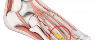
Exophthalmos as one of the symptoms of STX
In the presence of a tumor, the disease begins subacutely. The main symptoms of Tolosa-Hunt syndrome in this case: exophthalmos, significant swelling of the membrane of the eyeball, paralysis of the cranial nerves (one or more).
The syndrome with orbital myositis develops moderately, with intense pain and pronounced bulging eyes, swelling and visual impairment (diplopia).
Causes of Ramsay Hunt syndrome
The pathology is provoked by the third type of herpes virus – viricella-zoster. The disease is transmitted by contact and airborne droplets. Due to the polymorphism and antigenic variability of virions, this pathogen can also cause the development of chickenpox and herpes zoster. As a rule, the virus is activated against the background of reduced immunity.
Insufficient immunological defense of the body may be due to immunosuppressive therapy for oncological and autoimmune processes, long-term use of corticosteroid drugs, chronic pyelonephritis, bronchitis, heart failure, diabetes mellitus, hypothyroidism and severe injuries.
The exact cause of THS is not known, however the disease is thought, and often assumed, to be related to inflammation of the areas behind the eyes (cavernous sinus and superior orbital fissure).
It has been established that the syndrome causes the proliferation of granulomatous tissue on the outer wall of the cavernous sinus. However, what exactly causes this growth is unknown.
Supposed reasons:
- Abnormal structure of blood vessels, in which venous and arterial blood mix (malformation).
- Autoimmune inflammation, in which the body begins to destroy its own tissues, mistaking them for harmful ones.
- Tumors of different parts of the brain, both malignant and benign.
Hantavirus infection
Hantavirus infection is pneumonia and hemorrhagic fevers caused by viruses of the Hantavirus genus of the Bunyaviridae family. Currently, the genus includes the Hanta-an, Dubrava, Puumala, Seoul (causative agents of hemorrhagic fevers with renal syndrome), Prospect Hill, Muerto Canyon and hemorrhagic fever with renal syndrome viruses - all of which cause zoonotic endemic natural focal arboviral diseases in humans. The strains causing lesions in Europe and Asia are significantly different from the hantavirus strains causing pneumonia in the United States. Epidemiology. The reservoir of hantaviruses is rodents (the pathogen is isolated from saliva, urine and feces); Asymptomatic carriage or epizootics are possible. The main reservoirs of pathogens of hemorrhagic fevers are the bank vole, field mouse, gray and black rats; pulmonary lesions - presumably white-footed hamsters (Peromyscus maniculatys) and cotton rats (Sigmodon hyspidus). Fevers are widespread in forested areas; pulmonary lesions - in endemic steppe areas of the USA. A person becomes ill through direct contact with contaminated objects, consumption of contaminated products, or inhalation of viruses. Infections: nutritional (ingestion of contaminated water or food), aerogenic, contact (contact with rodents [through feces or bites] and humans), vector-borne (mosquitoes, fleas or arthropods).
Clinical manifestations
- The term hemorrhagic fever with renal syndrome
- The most common pulmonary disease, also known as four corners disease, is reported in the United States (California, Nevada and the region
- four corners
- — boundaries
- square
- states of Arizona, Colorado, New Mexico and Utah). The incubation period does not exceed 6 weeks; the disease manifests itself as acute respiratory viral infection with the addition of pneumonia, hemorrhagic and renal syndromes. Severity ranges from flu-like symptoms to respiratory failure with possible death.
- Hemorrhagic fevers. Symptomatic and pathogenetic treatment is carried out - replacement of fluid losses (should be carried out carefully due to possible overhydration), dopamine or adrenaline for arterial hypotension. The prognosis is usually favorable
- Pneumonia. Treatment is symptomatic and pathogenetic (prevention of bacterial superinfections is carried out) - oxygenation, monitoring the functions of vital organs, stabilization of hemodynamics. Antiviral therapy is carried out with ribavirin. The prognosis of the disease is quite unfavorable, mortality reaches 60%.
unites a large group of lesions (Korean, Far Eastern, Ural, Yaroslavl, Transcarpathian, Scandinavian and other fevers). The causative agent(s) has several serovars. The incubation period is 7-45 days; diseases begin acutely; Characterized by high temperature (39-40 °C), fever, myalgia, hyperemia of the mucous membranes and sclera. From 3-4 days onwards, symptoms of intoxication (traditionally repeated vomiting) and hemorrhagic syndrome (maculopapular rash, internal bleeding) appear. Oliguria develops almost simultaneously; in severe cases, anuria is possible.
Treatment
Diagnosis of Ramsay Hunt syndrome
The difficulty with CTX is that its symptoms are difficult to distinguish from other diseases. Pain in one half of the face can be caused by:
- Tumors (cranial fossa, pituitary gland, etc.).
- Carotid artery aneurysm.
- Osteomyelitis.
- Meningitis.
- Diabetes and dozens of other diseases.
For this reason, a patient complaining of pain in one part of the face and disturbances in eye movement needs a multifaceted examination. To make an accurate diagnosis, he must undergo:
- examination by an ophthalmologist;
- consultation with a neurologist;
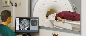
- CT and MRI of the brain;
- CT and MRI of the sella turcica;
- angiography (X-ray examination of blood vessels, which is carried out using contrast agents);
- echography of the orbits.
Most ophthalmologists are confident that if CTX is suspected, a biopsy is necessary. If MRI and CT showed granulomatous inflammation of the outer wall of the cavernous sinus, then this procedure is inevitable.
The patient is consulted by a neurologist and immunologist. To establish and confirm the diagnosis, the doctor collects anamnestic data, analyzes clinical signs, conducts a physical examination and refers the patient for additional examinations. As part of the diagnosis, audiometry, a general blood test, enzyme immunoassay, polymerase chain reaction, and blood testing for immunoglobulin levels can be performed. The pathology is differentiated from trigeminal neuropathy and neuralgia of the IX pair of cranial nerves.
THS is usually diagnosed by exclusion, and as such a huge number of laboratory tests are needed to rule out other causes of a patient's symptoms. These tests include a complete blood count, thyroid function tests, and serum protein electrophoresis. Cerebrospinal fluid studies may also be helpful in distinguishing between TCS and conditions with similar signs and symptoms.
MRI scans of the brain and orbit with and without contrast, magnetic resonance angiography or digital subtraction angiography, and CT scans of the brain and orbit with and without contrast may all be useful in identifying inflammatory changes in the cavernous sinus, superior orbital fissure, and/or orbital apex.
Sometimes a biopsy may need to be obtained to confirm the diagnosis, as this is useful in ruling out a neoplasm.
Differentials to consider when diagnosing THS include craniopharyngioma, migraine, and meningioma.
This disease is compared with other pathologies of the eyes and nervous system. First of all, it is necessary to exclude inflammatory processes in the brain and its membranes, as well as tumors. If meningitis or encephalitis is suspected, a spinal tap is performed. In order to exclude benign neoplasms and cancer, the patient undergoes MRI, CT scan of the brain and X-ray of the skull.
The disease is differentiated from lymphomas, cavernous sinus cysts and its thrombosis. Also, similar symptoms can be observed in systemic pathologies, such as sarcoidosis, orbital myositis, migraines, etc. To make an accurate diagnosis, the patient must be examined by various specialists: an ophthalmologist, a neurologist, a vascular surgeon, an endocrinologist.
What is hantavirus?
Hantavirus under a microscope
Hantaviruses belong to the Bunyavirus family. They are carried by rodents and shrews, especially brown rats, found throughout the world. There are a number of strains of hantaviruses that are transmitted from several species of rodents found in different parts of the world, especially in cities along the coasts of Russia, the United States, parts of Canada, Asia and Mexico.
Researchers refer to hantavirus strains as "New World" or "Old World" hantaviruses. Old World hantaviruses are primarily transmitted from rodents living in Europe and Asia. New World hantaviruses are primarily found in rodents native to the Americas.
How common are hantavirus infections?
Generally speaking, viruses transmitted by rodents are considered rare. But experts still say that "the extent to which Old World hantaviruses pose a threat to public health in developed countries remains unclear and likely varies significantly by region."
Principles of treatment
The only effective treatment for Tolosa-Hunt syndrome is immunosuppressive therapy with hormonal drugs - corticosteroids. They suppress the aggression of the immune system on the body.
The doctor prescribes Prednisolone, Medrol, Cortisone or their analogues, which are also effective in the treatment of other autoimmune diseases. The drug is taken orally or intravenously. The dosage is selected by the doctor at the rate of 1-2 mg/kg body weight or 500-1000 mg per day (solution).
In addition to this drug, symptomatic treatment is prescribed: analgesics, anticonvulsants. It is mandatory to prescribe vitamin complexes.
If all recommendations are followed, the disease responds well to treatment, the symptoms completely disappear. Significant improvement is noted by the patient already on days 3-4. The patient remains able to work. But with frequent relapses, light work or registration of disability is recommended, which happens extremely rarely.
According to the doctor’s decision, in case of persistent damage to the optic nerves (diplopia, decreased visual acuity, impaired movement of the eyeballs), the patient may be prohibited from driving.
Treatment includes THS immunosuppressants such as corticosteroids (often prednisolone) or steroidal diuretic agents (such as methotrexate or azathioprine).
Radiation has also been suggested.
The main treatment for Tholos Hunt syndrome is immunosuppressive therapy. This means that the patient is prescribed drugs that suppress the “wrong” immune reactions of the body. For STX, corticosteroids are prescribed. Cortisone, Hydrocortisone and analogues cope well with the problem: improvement occurs within 2-3 days.
To treat symptoms, analgesics and anticonvulsants are prescribed. General strengthening complexes and vitamins are included in the additional program.
Similar therapy can help with other diagnoses, but improvement occurs much more slowly. Therefore, a good response to steroids is a criterion for making an accurate diagnosis of CTX.
It is important to remember: timely consultation with a doctor will relieve unpleasant symptoms already on the 3rd day. The disease can be completely eliminated.
In the acute phase, etiotropic antiviral therapy is carried out with acyclovir or valacyclovir hydrochloride. If the patient is confirmed to have immunodeficiency, thymus preparations and human immunoglobulin are prescribed. The drug regimen is supplemented with anticonvulsants, B vitamins, anticholinesterase and sedative drugs, as well as agents that improve microcirculation.
Treatment for Tolosa-Hunt syndrome involves prescribing medications that suppress the immune system. For this purpose, hormone-containing medications are used: Prednisolone, Hydrocortisone. These drugs also have an effect in other autoimmune pathologies, but with this disease the symptoms disappear after 3-4 days.
Forecast
The prognosis of THS is generally considered good. Patients typically respond to corticosteroids, and spontaneous remission may occur, although eye muscle movement may remain impaired. Approximately 30-40% of patients treated with TCS experience a relapse.
In most cases, with timely treatment, Ramsay Hunt syndrome has a favorable prognosis for recovery. Incomplete elimination of the virus during antiviral treatment causes individual relapses of the disease. The persistent course of prosopalgia is observed mainly in old age.
Specific prevention involves vaccinating children against chickenpox. An additional measure that can prevent Hunt syndrome is active antiviral treatment of patients with chickenpox. A healthy lifestyle, which allows you to maintain a high level of immunity, is of no small preventive importance.
Prevention of Tolosa-Hunt syndrome
There is no way to prevent the development of Tolosa-Hunt syndrome as such, since it develops transiently and for an unknown reason. However, if primary symptoms appear, such as double vision, pain in the eye area, forehead, or orbit, it is necessary to consult a doctor for an examination.
When the syndrome is in remission, it is important to carry out secondary prevention: take hormonal medications, vitamins, strengthen the immune system, and avoid stressful situations. In addition, it is important to avoid hypothermia, infectious and inflammatory diseases.
It is impossible to predict the development of Tolosa-Hunt syndrome in advance, so there is no primary prevention. If symptoms such as pain in the eyes and forehead, diplopia are present, then a thorough diagnosis should be carried out. For Tolosa-Hunt syndrome, secondary prevention is necessary. This includes timely hormonal therapy and immune support. You also need to avoid stressful situations and inflammatory processes.
Children are vaccinated against chickenpox. To maintain a high level of immunity, it is recommended to adhere to the principles of proper nutrition, exercise and harden yourself.
General information
Isolated inflammation of the peripheral nerve ganglion in neurology is designated by the term “ganglionitis”.
Ganglionitis of the geniculate ganglion of herpetic etiology was named Ramsay Hunt syndrome in honor of the North American neurologist who first described it in 1907. Along with herpetic trigeminal neuralgia, Hunt's syndrome refers to postherpetic prosopalgia (facial pain), which develops in 10-20% of cases of acute herpes zoster infection. Ramsay Hunt syndrome
Precautions if you think you are infected
Experts warn that people who have potentially had some exposure to rodents and are experiencing signs or symptoms of hantavirus, including fever, deep muscle aches and severe shortness of breath, should get emergency medical help right away. When hantavirus infection is suspected, the patient should tell their doctor that they have been exposed to rodents. This way, the doctor can test him for a rodent-borne viral infection and provide proper treatment.
Final Thoughts on Hantavirus
- Hantaviruses belong to the Bunyavirus family. These viruses are transmitted from rodents around the world to humans through droppings, urine and bites.
- Preventing rodent infestations is very important. The only risk factor for rodent-borne viruses, including hantavirus, is rodents and their droppings in and around your home.
There are no treatments for hantavirus symptoms, such as fever and severe breathing problems. But there are ways to help keep your home safe; boost your immune system with herbal remedies and supplements; and treat symptoms such as dehydration, difficulty breathing, pain and low blood pressure.
Tags: hantavirus
« Previous entry