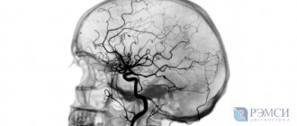
To study the condition of the vessels of the brain and skull, a modern method is used - cerebral angiography. This is an X-ray instrumental examination method, the essence of which is the introduction of a contrast agent into the vascular bed of the brain, followed by radiography.
Thanks to the contrast agent, the image produces a vascular pattern in the form of an impression, on which changes in the walls, places of narrowing of the lumen of the arteries or the presence of an obstruction to blood flow, anomalies and protrusion of the walls are visible. This study is invasive, that is, one in which a puncture (puncture) of a skull vessel is performed, followed by the introduction of a contrast agent into it.
How can you see the blood vessels of the brain?
Cerebral angiography is an x-ray method for visualizing cerebral vessels, which consists of staining the vascular bed with previously injected contrast. This is a highly effective and modern diagnostic method that allows you to make an accurate diagnosis. The method of visualizing blood vessels using a contrast agent has been known to medicine for about a century. Back in 1927, a neurologist from Portugal began using this method, and it came to Russia in 1954. Despite such a long use, cerebral vascular angiography has changed significantly over this time, becoming more sophisticated.
Classic angiography
X-ray angiography is a method of obtaining information about vascular disorders using X-rays and a contrast agent.
The rays pass through different tissues and are reflected differently. As a result, the silhouette of the organ is displayed on film. Standard x-rays cannot see soft tissue. To isolate them, an iodine-containing substance capable of reflecting radiation is used.
Technique
Classic angiography of cerebral vessels is performed in a hospital. The subject is placed on a regular couch. If necessary, intramuscular sedatives and antihistamines are given. Lidocaine is injected subcutaneously to numb the puncture site. To prevent spasms and reduce irritation, Novocain is administered.
The puncture site is disinfected. The drug itself is injected into the vessel being tested or through a catheter inserted into the vertebral, carotid or femoral artery. After its introduction, the study begins.
It is permissible to administer a contrast agent 2-3 times. Upon completion of the procedure, the catheter is removed, the injection site is clamped, and a sterile bandage is tightened. It can be removed only when the bleeding stops completely.
Types of cerebral angiography
According to the established classification, this diagnostic technique is divided into 2 types:
- Selective - targeted, local. When performing selective cerebral angiography, an iodine-containing contrast agent is injected into an arterial vessel that supplies one of the brain regions.
- Survey is an extended research method. A contrast agent is injected into the area of the main artery, which is responsible for the general cerebral blood supply and nutrition. Using this technique, a specialist can carefully examine all cerebral vessels.
The optimal type and method of implementation is determined by a specialist individually, taking into account the characteristics and severity of the clinical case.
Methods of conducting research
There are several different types of cerebral angiography.
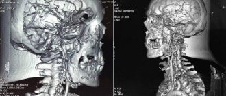
Angiography using computed tomography (CTA) provides a detailed image of the vessels and shows patterns of blood flow. In this case, intravenous contrast enhancement is used.
After CTA, image reconstruction is performed.
A definite positive side of this method is the reduced radiation exposure to the patient’s body.
CT angiography is often performed for stenosis, thrombosis, aneurysms, and vascular developmental defects.
Contraindications are allergies to the contrast agent, diabetes, pregnancy, obesity, thyroid problems, myeloma, heart disease, uncontrollable arrhythmia and tachycardia.
The study is carried out on an outpatient basis. Approximately 100 ml of contrast agent is injected into a venous catheter, which is inserted into the cubital vein. The patient lies on the CT scanner table.
X-rays scan the area under study in parallel with the introduction of a contrast agent.
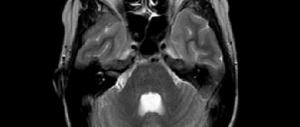
Magnetic resonance angiography (MRA) allows you to study the functions of blood flow and its anatomical features.
The basis of magnetic resonance imaging is to track energy changes in tissues, their structure and chemical composition. Contrast agents are practically not used in MRA (occasionally gadolinium-based to obtain high-precision images).
MRI angiography of cerebral vessels is used to diagnose aneurysm dissection, congenital heart defects, and vasculitis.
Contraindications include installed implants, pacemakers, nerve stimulators, blood-restoring clips, insulin pumps, prosthetic heart valves, heart failure, pregnancy, claustrophobia.
Cerebral angiography is a kind of “gold standard” for studying cerebral vessels.
The author of this method is Egas Monitz, who performed angiography for the first time in 1927.
The method is of the highest value because it allows you to accurately detect aneurysms, narrowing of blood vessels or the site of their blockage, and brain tumors.
The catheter is inserted into the vessel through the femoral artery and directed to the carotid artery. A contrast agent is injected into the blood vessels and X-rays are taken to determine the state of the inflow and outflow of venous blood.
During cerebral angiography, surgical intervention is possible. The information content of the method is much superior to CTA and MRA.
Arteriography
Arteriography involves the introduction of a contrast agent into the lumen of the vessel, which makes it possible to determine the presence of tumors located close to blood vessels, arterial pathologies and other circulatory disorders.
Most often, this method is used to study the limbs.
Arteriography is relatively simple, performed on an outpatient basis, but is painful because the contrast moves quite quickly through the arteries.
X-ray contrast agents (about 30-40 ml) are injected through a catheter or directly into the artery under strong pressure in the direction of the blood flow (less often against the blood flow).
This method allows you to diagnose changes even in the deepest arteries, which are monitored using the screen of an X-ray machine.
Venography
Another name for venography is phlebography. The essence of the method corresponds to its name.
Venography allows you to see the distribution of veins; it is actively used for varicose veins and thrombosis, as well as arrhythmia. The patient is advised to breathe calmly and relax during the procedure.
This is a simple and painless method, but in rare cases, your health may deteriorate after the procedure, and phlebitis may appear - inflammation at the site of contrast injection.
Venography involves the use of small amounts of a contrast agent that is injected directly into a vein (direct venography). After the procedure, an injection is made using 60 ml of saline to cleanse the vessels.
It is most justified to use venography before surgery on the veins.
Indirect venography can be performed in three ways:
- contrast is injected into the artery and then enters the veins through the capillaries;
- contrast is introduced into the tissue of the affected organ that needs to be examined, and the pictures show veins draining blood from the organ;
- contrast is injected directly into the medullary space.
What is the danger of post-traumatic encephalopathy of the brain - treatment and prevention of the disease. What is the most common cause of a retrocerebellar arachnoid cyst of the brain and what to do if there is a suspicion of this formation.
Lymphography
Lymphography is a method of studying the lymphatic system also using a radiopaque agent.
The study is carried out in three projections and the data is studied immediately after the administration of contrast (early lymphogram) and after 1-2 days (late lymphogram).
Early lymphograms make it possible to examine the condition of the lymphatic vessels, late lymphograms - the lymph nodes.
This method allows you to identify changes in the external and common iliac, inguinal, supra- and subclavian, lumbar, axillary lymph nodes; identify the presence of tumor processes and optimize cancer treatment.
Indications
Indications for cerebral angiography are pathological conditions that cause disturbances in the functioning of the brain. Hemorrhagic circulatory disorders:
- aneurysms;
- diverticulum;
- angioma.
Ischemic circulatory disorders:
- cerebral atherosclerosis;
- blood clots;
- arterial deformation.
Tumors leading to changes in the vascular pattern, as well as the lack of results after other methods for diagnosing brain diseases in the presence of the following symptoms:
- constant dizziness not related to blood pressure;
- epileptic seizures;
- confusion of consciousness;
- increased intracranial pressure;
- previous stroke or suspected micro-stroke;
- intracranial hematomas caused by head trauma;
- chronic headache of unknown origin;
- nausea accompanied by dizziness and headaches;
- noise in ears.
It is also advisable to perform cerebral angiography to plan upcoming surgery and to monitor the patient’s recovery after brain surgery.
Diagnosis using selective angiography
Angiography is a method of fluoroscopic examination of body systems, namely blood and lymphatic vessels, veins and capillaries. There are general and selective (selective) angiography, using one or another type, guided by the goals set by the doctor. General angiography examines all the main vessels of the area, a selective procedure examines individual vessels.
Diagnostics involves the introduction of radiocontrast agents (organic iodine compounds) into the vessels under study using catheterization or puncture.
The phases of angiography are distinguished by the stages of passage of the radiopaque substance through the arterial system:
- Arterial.
- Capillary.
- Venous.
The rate of disappearance of the radiopaque substance characterizes the hemodynamics in the organ under study.
This method successfully diagnoses various abnormalities in the functioning of the vascular system, as well as various tumors, parasitic and other organ lesions.
With its help, the topographical features and functional states of blood vessels, the paths of the roundabout circulation, and the speed of blood flow are studied.
Types of angiography depending on diagnostic purposes
Depending on the disease and the goals and objectives set during diagnosis, angiography can be classified into:
- Cerebral selective angiography is an angiographic study of cerebral vessels. In general and selective angiography of cerebral vessels, immediately after the invasion of the organic compound of iodine, several angiograms are made, which display the capillary, arterial and venous phases of blood flow. For selective cerebral angiography, both indirect and direct (carotid and vertebral) angiography are used. The most common type of examination is the carotid one, in which a radiopaque contrast agent is injected into the carotid artery. During a vertebral examination, iodine is injected into the vertebral artery. Indirect diagnosis involves the injection of contrast through a catheter into large vessels, for example, on the thigh.
- Arteriography - allows you to diagnose blockage or narrowing of the lumen in the artery, disturbances in the blood flow, as well as neoplasms near the examined blood vessels.
- Lymphography - studies the state of the lymphatic system. The study is in demand in oncology for the diagnosis of malignant tumors of the ovaries and uterus. The procedure is necessary to assess the stage of the disease and the effectiveness of chemotherapy.
- Venography is successfully used to diagnose problems in the venous circulation of the extremities. Angiograms show both deep and superficial veins, their length and location. Research using this method is effective for varicose veins and various degrees of thrombosis, even diagnosing heart pathologies and heart failure.
- Cardiac angiography - used to study the chambers and arteries of the heart muscle, identifies vascular pathologies (blockages, thrombosis, pathological narrowing and dilatation), assesses the pressure in the chambers of the heart and arteries, and diagnoses the contractile function of the myocardium. This type of study provides a more informative picture of heart disease than venography.
- Fluorescein angiography is an angiography method for examining the ocular vessels and fundus. Used in the diagnosis of glaucoma, dystrophic and inflammatory processes in the retina and membrane of the eye, diabetic retinopathy. In this study, fluorescein sodium salt 10% is used as a radiopaque composition; administration occurs through a vein on the elbow.
Preparation and performance of selective angiography
Before examining a patient using angiography, the doctor must familiarize himself with the test results (general urine and blood tests, biochemistry), conduct a medical examination, and obtain intelligible answers to the following questions:
- Do you have a history of allergies?
- Chronic and past infectious diseases.
- What medications are being used at the time of the study.
- If the patient is a woman, make sure there is no pregnancy.
It is important! Some time (the doctor will determine it exactly) before the angiography procedure, you will need to adjust your diet, and possibly stop some of the medications you are taking. This is necessary to minimize errors in the study results.
A catheter with a contrast agent is inserted under local anesthesia. Sometimes, before or during the procedure, if indicated, a sedative is administered to help the body relax. The procedure itself is practically painless. However, there may be a feeling of some discomfort during the insertion of the catheter and a feeling of nausea when the X-ray contrast agent is administered.
The examination can take from 10 minutes to several hours, it all depends on the scale of the study. After the angiography, the doctor will suggest that you stay in the hospital for some time to monitor your condition, thereby reducing the risk of possible complications.
It is important! Accurate information about whether the patient is allergic to X-ray contrast agents, as well as medications and seafood will help reduce the risk of complications.
Upon discharge, the doctor:
- will write down recommendations for the care and treatment of the catheter insertion site;
- will recommend drinking a lot of water to remove the contrast agent from the body;
- writes instructions for taking medications;
- will note the need to reduce physical activity for 24 hours after the procedure.
Even if the angiography passed without visible consequences, the patient should be attentive to his health and, if one of the symptoms appears, immediately consult a doctor:
- temperature increase;
- pain, swelling, or discharge at the catheter insertion site;
- changes in the functioning of the gastrointestinal tract;
- pain in the chest or organ being examined;
- numbness or pallor of the hands and feet;
- labored breathing;
- weakness of facial muscles, problems with speech or vision.
Source: https://VashNevrolog.ru/metody-diagnostiki/diagnostika-metodom-selektivnoj-angiografii.html
Contraindications to the procedure
As with any other procedure, there are contraindications for cerebral angiography. They are associated both with the procedure itself and with the contrast agent that is injected into the bloodstream. Iodine compounds are used as the administered substance. The amount of the substance depends on the volume of the examination; it can be 5-10 ml.
Cerebral angiography is not done in the following cases:
- allergic reactions to iodine-containing contrast agents,
- individual intolerance,
- acute or chronic renal failure, which does not allow the use of a contrast agent,
- exacerbation of chronic diseases,
- pregnancy or lactation,
- diseases accompanied by blood clotting disorders,
- myocardial infarction,
- age up to 2 years,
- mental illness.
When and to whom is angiography prescribed?
Angiography can be prescribed for such types of damage and vascular malformations as:
- aneurysm;
- atherosclerosis;
- defects and damage to internal organs;
- malformation;
- thrombosis;
- tumor processes.
However, this procedure also has its contraindications.
Angiography is not performed if the following problems exist:
- allergy to iodine-containing drugs;
- mental disorders;
- acute inflammatory processes and infectious diseases;
- heart, kidney, liver problems;
- thrombophlebitis;
- serious condition of the patient.
What do angiography indicators mean?
The amount of radiation that will penetrate the veins and other brain tissue is determined by their density. It is expressed in various color shades. The bone in the image will be white, and the cerebrospinal fluid will practically not appear on the resulting images. Other brain substances have different colors and densities. Using them, doctors evaluate the internal structure. The doctor will provide a detailed transcript of the received images.
Preparation for the procedure
Before the study, the patient should not eat for 10 hours and not drink for 4 hours. He needs to remove all metal objects. If surgical intervention is required to administer contrast, the following is prescribed:
- iodine allergy test;
- urine and blood tests;
- ECG;
- kidney function testing;
- consultations with an anesthesiologist and therapist.
Before performing an MRI or CT scan of the brain with angiography, you must adhere to a diet for several days. Eliminate from your diet carbonated drinks, refined sweets and sweet fruits, dishes made from legumes and other products that cause increased gas formation in the gastrointestinal tract.
Methodology for cerebral angiography
The entire procedure of this study can be divided into several stages:
- preparing the site for puncture - treating the skin with an antiseptic solution to destroy microorganisms and local anesthesia;
- direct puncture of the vessel with a special needle and insertion of a flexible catheter;
- introduction of a contrast agent into the vascular bed of the brain through a catheter;
- performing a series of X-ray images of blood vessels; during CT cerebral angiography, a series of layer-by-layer images is taken;
- interpretation and interpretation of the received images with a conclusion - is carried out by a radiologist together with a vascular surgeon.
Considering the paramount importance of proper cerebral circulation, adequate and correct diagnosis of vascular problems is the basis for their further successful treatment.
CT angiography
CT angiography or computed tomography angiography shows pathologies in blood vessels and allows one to study the pattern of blood movement through their internal cavity.
Indications for the use of CT angiography are:
- the presence of stenosis or thrombosis of the vessel;
- the presence of an aneurysm in the vessel;
- suspicion of another vascular disease or congenital pathology.
Before undergoing a diagnostic procedure, it is necessary to exclude contraindications, which differ in some ways from the limitations inherent in other angiography methods. Among them:
- hypersensitivity to substances that are included in the contrast;
- development of renal failure;
- development of diabetes mellitus in severe form;
- pregnancy period (due to possible teratogenic effects);
- presence of severe general condition;
- overweight and obesity;
- disruption of the endocrine system;
- development of multiple myeloma;
- presence of acute heart failure.
The technique requires special training. Thus, before the study, possible contraindications are excluded, in particular, an allergic predisposition to the injected contrast agent. In order to reduce the risk of a corresponding reaction, take an antihistamine before the test.
The essence of the diagnostic procedure is as follows:
- The patient is placed on a special table.
- A catheter is inserted into the cubital vein, through which an iodine-based contrast agent is passed.
- Next, multiplanar and three-dimensional computer reconstruction is carried out with interpretation of the resulting images.
In some cases, computed angiography causes complications, including contrast extravasation. This negative consequence is the penetration of the substance into soft tissues that are located outside the vessel. As a rule, the volume of contrast entering the tissue does not exceed 10 ml. If it spreads to a larger extent, it causes severe damage to the subcutaneous tissue.
Factors that increase the risk of extravasation include a history of multiple punctures of one vessel and a weakened immune system. Characteristic symptoms are pain and swelling in the area where the needle was inserted. Treatment consists of ensuring an elevated position of the injured area and applying cold compresses.
Other negative consequences of computed angiography include intolerance to the contrast agent, the symptoms of which in most cases appear suddenly. Clinical manifestations of allergies are rash, itching, burning, hyperemia of the skin, swelling, feeling of lack of air.
As a result of a computed tomography scan of the cerebral vessels, a three-dimensional image of them is obtained. It shows detailed structure, tissue condition and blood movement. To perform the procedure, a contrast agent is injected.
A variation of the method is multislice computed tomography. MSCT makes it possible to obtain 160-320 images at a time and track the slightest changes.
Differences from classical radiography:
- Less radiation exposure.
- It is carried out on an outpatient basis.
- The contrast agent is administered intravenously, which is less traumatic compared to arterial administration.
- The result is sharper images.
- The patient can quickly return to normal life.
Technique
In order for the image to be clear, the scanner is adjusted to the heart rate of the subject. The pulse should be smooth, no higher than 65 beats per minute, so before the examination the patient is given medications aimed at reducing tachycardia. If necessary, vasodilator drugs are administered intravenously.
The patient lies down on a movable table, his chest and limbs are secured with straps, and an iodine-containing substance is injected intravenously. Next, the couch is moved into the tomograph. There he and the patient move smoothly, stopping at the moment of filming. At these moments, the diagnostician may ask you to hold your breath. The device reproduces the reconstruction of the received information into three-dimensional images.
You cannot move during the examination. Two-way intercom allows you to inform the diagnostician about discomfort. Time: 30-60 minutes.
Possible complications
Despite the high level of safety for patients of different ages, angiography may result in the development of negative consequences for the patient. The most commonly observed conditions are:
- release of a radiopaque substance from the vascular bed into the surrounding tissues. This situation can lead to inflammatory changes of varying severity;
- allergic reactions to the contrast agent or its individual intolerance. In such cases, the patient may experience itching, urticaria, Quincke's edema and other allergy-specific symptoms;
- acute renal dysfunction, as a complication of the examination, is observed in patients with their diseases.
To prevent complications of the procedure, it is necessary to ensure a comprehensive examination of the patient before the study.
Related posts:
- Sexual Aversion Disorders Aversion to sexual activity is a disorder defined as persistent or...
- Argyrosis or blue skin Argyrosis is a persistent bluish-gray discoloration of the skin caused by deposits in…
- Numbness of the hands - dangerous or not? Constantly chilly fingers, frequent feeling of numbness in the hands, especially...
- Morton's neuroma (foot pain) Morton's neuroma is a fairly common disease characterized by thickening of the nerve sheath...
Risks and complications
There is a very small risk of an allergic reaction when contrast material is used. Patients with severe kidney disease may develop nephrogenic systemic fibrosis associated with gadolinium administration. Mothers should not breastfeed their babies for 24 to 48 hours after receiving contrast.
Despite its relative safety, cerebral angiography can have the following negative consequences:
- anaphylactic shock due to an allergic reaction to the administration of an iodine-containing substance;
- inflammation or necrosis of the tissues surrounding the vessel due to contrast getting there (extravasation);
- acute renal failure.
Allergy is the main and most common problem during angiography. Since an allergic reaction to iodine substances is characterized by a sudden and rapidly developing course, it may have the following manifestations:
- edema;
- hyperemia (redness);
- itching;
- hypotension (low blood pressure);
- weakness and loss of consciousness.
The use of modern non-ionic radiopaque agents can significantly reduce the risk of developing anaphylactic shock.
Omnipaque solution belongs to a new generation of radiopaque agents that do not cause allergies.
Extravasation is usually a consequence of improper technique for puncturing the arterial wall. In this case, the artery is punctured through, and the contrast penetrates into the soft tissue surrounding the artery, causing inflammation and, in rare cases, necrosis.
Acute renal failure occurs in cases of pre-existing dysfunction of the renal apparatus. Since the removal of contrast from the body is carried out primarily by the kidneys, they are subject to intense negative effects, which results in ischemia of the parenchyma and progression of renal dysfunction.
Despite the fact that angiography of cerebral vessels is not, in the usual sense, a surgical intervention, it is a rather complex invasive procedure that involves a serious burden on the body. In this regard, the patient, after diagnosis, should be under the supervision of a doctor to prevent the development of complications. In this case, systematic temperature measurement and inspection of the puncture site should be included in the mandatory list of postoperative measures.