Brain tumors come in two types: tumors and cysts. Their origin, development, structure, prognosis and treatment are radically different from each other.
A cyst is a benign formation, which is a cavity that is filled with a liquid, watery or jelly-like mass. They can be congenital or acquired. According to localization, there is a retrocerebellar cyst (intracerebral), which is located in the thickness of the brain, and an arachnoid (cerebrospinal fluid), which forms between the meninges.
In this article we will talk about the first type of cyst. We list its possible symptoms:
- headaches (can be of varying intensity and nature, depending on the type, location and size);
- dizziness;
- nausea, vomiting;
- movement coordination disorders;
- oculomotor disorders, decreased vision;
- changes in hearing sensitivity, hearing loss;
- sleep disorders;
- fainting;
- tremor of the limbs;
- convulsive seizures.
What is this
A retrocerebellar cyst of the brain is an accumulation of fluid in areas of dead gray matter . The problem can disturb people of different age groups, so no one is immune from it. Only timely diagnosis will allow achieving a positive result, because the correct treatment will be started. It is extremely important to determine what triggers cell death in order to prevent the development of pathology.
A head cyst can form in any area of the brain where the process of cell death has begun. Of course, this process cannot be called normal, so medical consultation is required. Just treating a retrocerebral cyst is wrong, because this will not correct the problem itself. You definitely need to take care of eliminating the root cause, and then there is a chance to avoid complications.
At the moment, there are several types of scalp cysts, and each of them is dangerous in its own way. It will be important for the doctor to diagnose the specific variant so that individual and effective treatment can be prescribed. To better understand the disease, it is worth familiarizing yourself with the types of the disease.
Varieties:
Arachnoid cyst . This is one of the most common types, in which the tumor is located between the meninges. It is filled with cerebrospinal fluid, and can occur due to trauma to the skull, inflammation of the brain, and also in case of increased pressure inside the head.
CSF cyst . It is less common than the previous type, and is distinguished by a certain fluid content inside the retrocerebellar cyst. Appears with hemorrhage in the brain, in the case of an inflammatory process, trauma and surgery.
Congenital retrocerebral cyst . As the name suggests, it exists in the child at the moment of birth. That is, pathology develops even when the baby is in the mother’s womb.
Purchased . It appears in adulthood due to various factors. For example, a retrocerebellar cyst may occur due to a serious head injury or a person suffering from inflammation of the brain.
The neoplasm comes in different sizes, and its symptoms, as well as health hazards, depend on this. If the pathology is small, then it may not even manifest itself. Such diseases are often not treated, because they do not interfere with life.
If the retrocerebellar arachnoid cyst continues to grow, then treatment will definitely be required. Without treatment, complications may occur, including complete brain death.
Only a specialist can definitely say whether a specific pathology needs to be treated. Retrocerebellar arachnoid cysts may need to be examined to determine their condition. Based on the test results, it will be possible to choose the right treatment that will help eliminate the problem.
be careful
Headache is the first sign of hypertension. In 95% of cases, headaches occur due to impaired blood flow in the human brain. And the main reason for impaired blood flow is blockage of blood vessels due to poor nutrition, bad habits and an inactive lifestyle.
But what to do? How to treat if there is deception everywhere? Doctor of Medical Sciences Bokeria L.A. conducted his own investigation and found a way out of this situation. In this article, Leo Antonovich told how to FREE of charge protect yourself from death due to clogged blood vessels, pressure surges, and also reduce the risk of heart attack and stroke by 98%! Read the article on the official website of the World Health Organization.
Within the federal program, every resident of the Russian Federation can receive a remedy for hypertension for FREE:
Causes
A retrocerebellar cyst of the brain appears only when there are specific reasons. Most often, it indicates that a person has serious health problems. A healthy body will not suffer from this pathology if it is not congenital.
Reasons for tumor formation:
- Post-stroke condition.
- The occurrence of encephalitis and meningitis.
- Various head injuries.
- Cerebral ischemia, which disrupts normal blood flow.
- Hemorrhage into the organ during surgery.
- Genetic abnormalities, such as Marfan syndrome or missing septa.
- Degenerative brain condition.
- Problems arising during intrauterine development. They can appear due to poor ecology, as well as due to the fact that the mother took a number of medications.
When diagnosing an arachnoid retrocerebellar cyst, it is important to determine the reason for the pathology. The effectiveness of therapy will depend on this, because it may be necessary to treat not only the tumor, but also the factor that provoked it.
What is a retrocerebellar cyst of the brain?
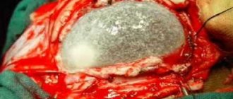
With a disappointing doctor’s conclusion, patients often wonder about the diagnosis of a head tumor. It is a cavity containing a capsule with liquid exudate. Pathology is formed at the site of necrotic nerve cells in any part of the brain. In most cases, MRI shows the location of the tumor in the posterior fossa behind the cerebellum.
Retrocerebellar cysts are asymptomatic for a long time, but over time the pathology progresses, compressing neighboring organs of the brain, which is often dangerous due to serious complications and symptoms of neurological deficit.
Main symptoms
There are a number of signs that indicate the formation of a retrocerebellar arachnoid cyst. Symptoms can manifest themselves in different ways - they depend on the size of the formation, as well as on the characteristics of the person.
For some, characteristic signs may be completely absent if the cyst develops slowly and is capable of dying. In such situations, the diagnosis is made by chance, when a preventive examination is carried out, or an examination is performed to identify another disease.
However, a retrocerebellar arachnoid cyst can manifest itself clearly, which is a reason to consult a doctor. It is important for a person to undergo diagnostics so that the presence of the disease can be confirmed or denied. If pathology is present, then it will be necessary to determine its stage, as well as the tendency to progression.
In order to contact a specialist in a timely manner, you need to know what symptoms the pathology may manifest:
- Frequent and intense headaches that are practically not eliminated with the help of medications.
- A feeling of nausea that does not lead to vomiting.
- Difficulty sleeping, possible insomnia.
- Deterioration of hearing and vision.
- Impaired skin sensitivity.
- The occurrence of paralysis of the arms or legs.
- Deterioration of muscle tone.
- Psycho-emotional disorders. A person may suffer from depression, apathy, or causeless aggression.
- Hydrocephalus. It increases intracranial pressure, which causes nervousness and drowsiness. Without proper treatment, death can occur.
- Balance problems.
- Fatigue, hallucinations.
- Losing consciousness for no reason.
Of course, a retrocerebellar cerebrospinal fluid cyst will not necessarily manifest itself with all of the listed symptoms. Only a few signs may be present, but they should also alert you.
You should not treat yourself, because you first need to carry out a professional diagnosis. Once the disease is identified and its type is determined, therapy can begin.
Treatment methods
The symptoms of a tumor clearly depend on its location, the reasons for its appearance and the size of the tumor.
It should be taken into account that growing cysts mean the presence of clearly defined symptoms, while cysts that have stopped growing often do not cause unusual sensations. The growth of a cyst is influenced by autoimmune processes in the body, circulatory disorders, the presence of infectious processes, multiple sclerosis, and neuroinfections. Quite often, retrocerebellar cysts appear suddenly during an unscheduled examination, in the absence of any symptoms.
Symptoms that may indicate that the patient has a retrocerebellar cyst:
- A feeling of pulsation, a feeling of “bloating.”
- Regular headaches.
- Decreased concentration.
- Hallucinations.
- Fainting.
- Convulsive seizures.
- Partial (sometimes complete) paralysis of the limbs, their numbness.
- Deterioration of vision (lack of a clear outline of objects, their bifurcation, the appearance of spots before the eyes).
- Tinnitus, hearing loss.
- Tremor of the limbs.
- Regular attacks of loss of consciousness.
If the nature of such symptoms is clearly regular, then a visit to a neurologist is extremely necessary as soon as possible.
To make an accurate diagnosis of “retrocerebellar cyst of the brain” based on anamnesis, it is necessary to conduct a number of serious examinations, which include computed tomography and magnetic resonance imaging of the brain. The images can reveal the presence or absence of the disease.
To further distinguish a cyst from a tumor, a contrast agent is injected into the cavity, which a malignant tumor tends to accumulate, while a benign formation behaves differently.
The next stage involves finding out the cause of the cyst. To do this:
- Heart examination (electrocardiogram) to identify cardiovascular insufficiency and the presence of possible defects.
- Doppler examination of the vessels of the head and neck, which helps to identify vasoconstriction and, accordingly, a lack of blood supply to the brain. This study reveals narrowing of blood vessels that provide blood flow to the brain.
- Screening a patient for autoimmune or infectious diseases using a blood test.
- Blood test for coagulability and cholesterol. Inflated values indicate the possibility of vascular blockage.
- Monitoring blood pressure, periodic increases in which can cause strokes with the subsequent appearance of tumors in the brain.
A retrocerebellar cyst causes symptoms in children that are similar to the main symptoms of brain tumors: pain in the head, a feeling of pulsation, numbness in the limbs. In newborns, the disease can lead to divergence of bone sutures.
The most common cause of retrocerebellar cysts in the brain in children is meningitis. The disease can also be caused by cerebral hemorrhage or other inflammatory processes in the brain.
If a child is suspected of having a cerebral cyst, he needs to undergo an ultrasound examination through the large fontanelle, and, if necessary, a course of intensive therapy immediately after birth. Surgery includes bypass surgery or endoscopy.
The diagnosis implies immediate contact with a doctor.
In some cases, treating a cyst does not involve removing it. In the absence of dynamic growth of a brain tumor, pronounced symptoms, and its small size, constant monitoring, drug treatment and observation by a neurologist are sufficient.
Knowing what a retrocerebellar cyst of the brain is, drug treatment is carried out in several areas:
- Normalize blood pressure.
- Prescribed drugs to improve blood clotting rates
- Drug treatment is carried out and a therapeutic diet is prescribed to reduce blood cholesterol levels.
- Prescribe drugs that improve blood circulation.
If the cause of the tumor is a brain infection, then drug treatment will be powerless and surgical intervention will be required.
Separately, it is worth mentioning the homeopathic treatment of cystic neoplasms. This method of control is effective only as an addition to the main treatment. It is impossible to reduce the size of the cyst using such means alone.
If the disease is infectious, as well as when the size of the cyst increases while taking medications, mandatory surgical intervention is indicated, since the enlargement of the tumor negatively affects brain activity.
- Endoscopic surgery is a microsurgical treatment method and is the most modern means of cyst removal. The operation involves inserting an endoscope into the area of the brain where the retrocerebellar cyst is located, and using instruments, fluid is drained. The possibility of performing this operation depends on the location of the tumor and its accessibility. This type of treatment has established itself as the most modern and relatively safe, because the operation takes place under the constant visual control of the surgeon.
- Removal of the formation by opening the skull. The most difficult, but also the most effective operation, which is successfully carried out by foreign clinics in Israel and Germany.
- Neurosurgical surgery (bypass surgery) is performed in the case of a constant influx of fluid against the background of existing hydrocephalus. Fluid is drained from the cyst using a shunt. It is worth noting that this operation is traumatic.
After a particular surgical intervention, it is necessary to strictly follow the doctor’s recommendations; it is mandatory to take multivitamins and medications that strengthen the walls of blood vessels and improve blood circulation.
As for the surgical treatment of arachnoid cysts, it is similar to the treatment of the retrocerebellar type of tumor, but differs in the choice of medications. The neurologist prescribes drugs to resolve adhesions, normalize metabolism in the brain, and antioxidants to make cells more resistant to increased intracranial pressure.
In case of late diagnosis of “retrocerebellar cyst” or incorrectly performed surgery, the following symptoms are possible:
- The appearance of epilepsy attacks.
- General cerebral syndrome, which implies the appearance of pain in the head, increased intracranial pressure, motion sickness in a car, bus, inability to stay in stuffy rooms).
- Focal syndromes resulting from damage to certain areas of the brain (speech disorder, movement disorders, impaired nervous function, hearing loss, vision impairment).
- Astheno-neurotic syndrome (with the appearance of a feeling of general malaise and decreased immunity).
- Developmental delay.
- Hypermobility syndrome (manifests in children who have undergone surgery to remove a retrocerebellar tumor, reveals itself in developmental delay).
The retrocerebellar type of the disease implies not only timely attention to alarming symptoms, but also regular examination after its treatment. A patient who knows firsthand about a retrocerebellar cyst in the brain who has undergone surgery, just like a patient after drug treatment, needs to be regularly monitored by a neurologist and undergo prescribed examinations to prevent relapses.
Retrocerebellar and arachnoid cysts of the brain are not a death sentence. Attentiveness to the symptoms that appear and timely seeking medical help will be the key to successful treatment.
The size of this formation can be different; experts divide them into the following types:
- the cyst at the initial stage of development has a thickness of no more than two millimeters;
- cyst of moderate severity - dimensions increase to 10 millimeters;
- a large cyst with a severe course - the thickness of the formation is more than 10 millimeters and the length is more than 8, but less than 13 centimeters.
There is no clear trend that would contribute to an increase in the size of the cystic formation. A retrocerebellar arachnoid cyst may not change its size for a long time and be at the initial stage of development. However, there is also a variant of rapid growth of pathological formation.
Rapid growth is observed in cysts that have developed in connection with previous infections, but their increase cannot be called rapid. There is also a pathology that does not change in size. It usually does not require treatment, but it is necessary to monitor it.
The severity of symptoms depends on a number of factors:
- the first place is occupied by the size of the cyst and the rate of its growth: the larger the formation, the greater the pressure on the brain tissue;
- localization of the cyst, in this case the presence of pathological symptoms will depend on which part of the brain is affected;
- the presence of infection in the organ;
- autoimmune pathology – multiple sclerosis.
General, neurological, focal and convulsive symptoms are noted. If the cyst grows, the following general and neurological signs of the disease appear:
- Headaches that may come and go or be constant. Many patients complain of throbbing, unilateral headaches, which are characteristic of migraines. In this case, the pain exhausts the person; he cannot find a comfortable position. The spasm is not relieved by conventional analgesics.
- Increased blood pressure.
- Pulsation in the temporal region.
- General weakness, fatigue.
- Single vomiting is noted.
Convulsive symptoms:
- Presence of seizures.
- Seizures of the epilepsy type.
- Paralysis of the upper and lower limbs.
1. Visual impairment:
- blurred image, spots before the eyes;
- double vision;
- the silhouettes begin to blur.
2. Changes in speech, it may become slurred, the person drags out, “chews” words.
3. Hearing disorders:
- noise in ears;
- feeling of stuffiness;
- the patient notes that he began to hear poorly.
4. Impaired coordination of movement, gait becomes unsteady.
The presence of this pathology in a child can lead to serious consequences. The larger the brain tumor, the more severely the baby suffers the pathological symptoms. It is especially worth noting small and infant children, whose brain functions are in their infancy and active development.
- Hearing disorders.
- Numbness of arms and legs.
- Paresis and paralysis of the upper and lower extremities may occur; they can be complete or partial.
- Coordination of movements may be severely impaired.
- Magnetic resonance imaging helps to clarify the location and size of the tumor. Normally, no cavities should be found.
- Computed tomography is another modern and highly accurate diagnostic method that provides detailed information about the pathology.
- A biochemical blood test is necessary to detect elevated cholesterol levels and increased blood clotting.
- A blood test to detect infections in the body that may cause the disease.
- Angiography of cerebral vessels. Using this study, it is possible to differentiate malignant neoplasms from brain cysts. In the presence of a cancerous tumor, the contrast agent accumulates in its body, but with a cyst this sign is not observed.
- Monitoring changes in blood pressure levels.
- Ultrasound Dopplerography (Doppler ultrasound) allows you to assess the condition of the vessels and their patency.
- Diagnosis of pathology of the cardiovascular system. This is necessary, since developed heart failure is a pathological factor in the formation of brain cysts.
How to detect a cyst
Of course, just knowing the symptoms is not enough to identify an arachnoid cyst of the quadrigeminal cistern. Such neoplasms require careful diagnosis so that they can be distinguished from other pathologies. It is also important to determine the size of the cyst and its tendency to progress. Only after this will it be possible to decide how exactly to treat the disease.
Diagnosis of an arachnoid retrocerebellar cyst includes a number of examinations that the doctor will prescribe. If characteristic symptoms appear, it is worth visiting a neurologist, because communication with him is the first stage in determining the disease and its cause.
It may be necessary to carry out a number of actions that will identify the disease . First of all, the specialist will interview you about what negative manifestations are troubling the person. You will also need to study your medical history and find out about the health of close relatives.
The neurologist will assess the function of vision and hearing and measure the volume of the head. It may be necessary to obtain information about the patient's gestation period as well as when the patient was born. There may have been congenital pathologies or birth injuries.
The diagnosis is not limited to this, and the person will have to undergo a series of examinations. These procedures are needed to confirm or refute the presence of a dangerous pathology in the brain. The specialist can send you for a computed tomography or MRI of the head, an ECG, ultrasound, as well as daily blood pressure monitoring.
It is often necessary to undergo electroencephalography, as well as to conduct a study of blood vessels using a contrast agent. In addition, a general blood test is prescribed, which can also tell a lot about a person’s state of health.
After all studies have been completed, a retrocerebellar arachnoid cyst may be identified. Based on her condition, the doctor will decide on treatment options. There are many treatment options, so it is useful to consider the varieties.
Diagnostics
The disease is diagnosed using digital brain imaging methods.
Magnetic resonance imaging shows expansion of the fourth ventricle, which communicates with the cyst. The cerebellar vermis is also displaced. MRI also shows signs of increased intracranial pressure.
As an alternative, computed tomography is prescribed. The method provides information about the zone of insufficient signal intensity, which has defined contours. also provides information about the density of the cyst. However, it is difficult to predict the course using computed tomography. Unlike CT, magnetic resonance imaging makes it possible to differentiate malignant and benign neoplasms. Therefore, if a cyst is suspected, MRI has greater diagnostic value.
If there is a suspicion that the cyst has increased in size, the patient is prescribed magnetic resonance imaging with contrast. Typically, negative dynamics (enlargement of the tumor) are combined with arteriovenous malformation or Arnold-Chiari malformation.
When examining the fetus of a pregnant woman, the disease may be discovered by chance if the diagnosis was carried out in the brain area using neurosonography. The same method confirms the presence of a cyst in children one and two years old.
How to treat
If a retrocerebellar cyst of the brain in children and adults does not have symptoms and does not grow, then therapy will not be carried out. However, a person will have to be constantly monitored by a doctor to prevent the progression of the pathology. If the tumor begins to grow, treatment will be required.
At first, if the disease is at an early stage, doctors will try to stop the development of the pathology with the help of medications. To do this, they will use drugs that eliminate inflammatory as well as infectious processes. This is relevant for cases where the cyst has formed, for example, due to meningitis.
It happens that a retrocerebellar cyst begins to grow rapidly, which causes the patient’s condition to significantly worsen. In this case, there is no longer any point in trying to be treated with medications. A specialist may recommend an operation that will involve intervention in the cranial cavity.
What measures will need to be taken:
- Bypass surgery . It makes sense to carry it out in a situation where a regular flow of fluid to the tumor has been noticed. During the operation, the injured vessels will be connected to healthy arteries, which will normalize the outflow of fluid and improve cerebral circulation.
- Trepanation . One of the most effective and at the same time complex operations. During surgery, the tumor and adjacent tissue will be removed.
- Endoscopy . They will make several punctures in the skull, and special equipment will be inserted through them to eliminate the pathology.
After the operation, rehabilitation will be required, because the person will not be able to immediately return to normal life. In the future, it will be necessary to check every six months to prevent the recurrence of retrocerebellar arachnoid cysts.