Decoding the results
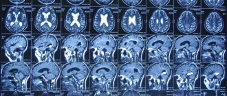
After processing the X-rays, the doctor places them on a backlit surface and examines them. Blood vessels and cerebrospinal fluid are black. They stand out clearly against the background of white bone tissue and gray brain matter.
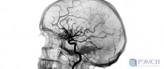
Smooth lines of blood vessels are considered a sign of health. They smoothly narrow and expand, and also branch, giving the blood network a resemblance to a tree. The contrast agent should fill the vessels evenly.
To identify pathology, the doctor compares the X-ray image of the examined person with that of a healthy person. If the image is unclear or the doctor has doubts, he may order a repeat angiogram.
Deviations from the norm
A sharp narrowing or dilation of a vessel is considered a deviation from the norm. Aortic aneurysm and other types of this pathology cause protrusion of the wall of the blood vessel. If intracranial hemorrhage has occurred, the doctor will find dark spots surrounded by ring-shaped stripes on the image.
The tumor can be seen on the x-ray. Neoplasms displace blood vessels to the side, disrupting the uniformity of vessel branching. Tumors can compress blood vessels, impairing blood circulation in certain areas of the brain. Areas where tissues are starved of oxygen appear lighter in color. On them, the blood vessels are narrowed or not visualized.
Risks
Angiography, like any invasive diagnostic method, is associated with certain risks. But they are very small, most often the likelihood of disease progression is much higher than the complications of the examination. The doctor recommends doing it after assessing the risk-benefit ratio.
The possibility of complications arises both from the need to inject a contrast agent, which can cause an allergic reaction, and from puncturing a blood vessel, which can then bleed. A solution of iodide compound is most often used as a contrast agent; an allergic reaction is prevented through premedication, i.e. by administering medications that reduce allergies.
The contrast agent is excreted by the kidneys. If this organ becomes weakened, there is a risk of impaired excretion, so it is important to tell your doctor about any kidney problems.
The most common complication of angiography is bleeding at the puncture site, the formation of a hematoma that resolves within 2-3 weeks. Rarely, other problems may arise as a result of manipulation of the vascular bed, such as thrombosis or embolism (i.e. blockage of a vessel), but this risk is much lower than not treating the existing disease.
During angiography, blood vessels are visualized using X-rays, so the patient is exposed to a small dose of X-ray radiation.
Possible complications
Despite the relative safety of angiography, scanning can lead to a number of undesirable reactions, including:
- the appearance of an allergy to the administered contrast agent, leading to anaphylactic shock;
- the formation of an inflammatory process (neurosis) of tissues surrounding the vessel, which develops due to the penetration of contrast into it;
- development of acute renal failure.
An allergic reaction is the most common of the rare unwanted deviations. The reaction to iodine-containing drugs appears unexpectedly and progresses sharply. Possible manifestations of this adverse reaction include:
- swelling;
- redness of the skin;
- itching;
- low blood pressure;
- lethargy;
- fainting.
The use of non-ionic contrast agents will help prevent the occurrence of anaphylactic shock.
Extravasation is the result of poor technique for puncturing the arterial wall. Under such circumstances, the artery is punctured through - the contrast drug enters the soft tissue near the artery, provoking an inflammatory process or, less commonly, necrosis syndrome.
Acute renal failure manifests itself with previously diagnosed malfunctions in the functioning of the renal apparatus. This adverse reaction occurs when contrast is used.
Since the kidneys are responsible for removing the auxiliary agent, the organs undergo significant stress. The result of this is parenchymal ischemia and renal dysfunction.
To reduce the intensity of the load on the excretory organ and speed up the process of removing contrast from the body, after diagnosis the patient is advised to drink a lot.
What is angiography
The term angiography comes from two Greek words: Greek. ἀγγεῖον (angillon) - vessel, vase and γράφειν (graphein) - write, write. Thus, angiography is an image of blood vessels reproduced on a monitor screen or on film, obtained in the following ways:
- radiographic;
- fluoroscopic;
- computed tomography;
- MRI;
Of all the mentioned methods for studying blood vessels, MRI angiography is the safest and most informative. Considering that blood vessels penetrate a person from head to fingertips, angiography is divided into several sections:
- cerebral MRI angiography examines the vessels of the head and brain;
- fluorescent MRA studies the microvessels of the eye and is used in ophthalmology;
The aorta is examined using the aortography method.
- aortography examines the condition of the aorta with all its branches;
- Portography observes and records the condition of the portal vein and vessels penetrating the liver.
- Phlebography and peripheral MR angiography examine the vessels of the lower and upper extremities.
In addition, magnetic resonance angiography of intracranial arteries located in the brain and extracranial vessels, that is, located between the head and heart, are considered. MRI angiography of the vessels of the neck and chest shows the condition of the extracranial vessels.
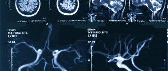
The essence of the procedure is as follows. Under the influence of a powerful magnet that forms a force field, the spins of elementary particles in hydrogen nuclei change their state and are placed along the axis of the magnetic field, which, together with RF pulses, causes protons to rotate relative to new axes. The force field does not act constantly and the protons return to their original position. But during the new rotation, energy is released and absorbed, and new magnetic fields are formed inside the hydrogen atoms. These changes are recorded by an MRI scanner and show individual sections of organs, blood vessels, and tissues.
An MRI machine takes detailed pictures of the vascular system
In images presented in black and white, compactions and solid formations are highlighted in white, and liquids in black. Thus, the darker the fabric structure, the more watery, thinner its structure. Based on changes in shades on organs and tissues, the specialist judges the condition of the organ being examined.
MR angiography can be performed without contrast agents, but gadolinium-based drugs improve image quality and show the smallest details.
X-ray angiography
Angiography is divided according to the organ in which the vessels are contrasted. For example, angiocardiography visualizes the atria and ventricles with large vessels, and angiopulmonography visualizes the vascular network of the lungs. The procedure requires access to a vessel as close as possible to the affected area, the continuation of which is the artery feeding the tumor. A catheter is inserted into the artery and guided under X-ray control to the desired vessel. A contrast agent is passed through the catheter and pictures are taken at a certain time - the phase of passage of the contrast containing iodine.
With an X-ray examination, it is possible to contrast not only arteries, but also veins, and, if necessary, obtain an image of lymphatic vessels, therefore, in principle, this study is called vasography - an image of blood vessels. The study of arteries is called arteriography, venography or phlebography is the image of veins and lymphography, respectively, contrasting the lymphatic vessels. The study not only provides a picture of the vessels, but based on the “picture” of its blood supply, it allows us to identify pathological conditions of the organ and its functions that affect the state of the blood supply.
When deciding on the surgical treatment of cancer, the surgeon’s preliminary understanding of the state of the arterial bed in the area of the tumor is very important, since it allows one to determine the boundaries of tumor growth and the involvement of large-diameter arteries with damage. Blood flows through the arteries under high pressure, so during the operation, a fairly large volume of blood will be lost from the vessel damaged by the tumor until the bleeding is completely stopped. Blood flows through the veins at low speed and low pressure; a large vein damaged by a tumor can easily be compressed without any significant blood loss.
Angiographic information obtained before surgery allows you to prepare for the unexpected, for example, the installation of a prosthesis instead of a vessel affected by cancer. With X-ray angiography, you can immediately carry out a therapeutic measure - install a stent in the vessel narrowed by the tumor. Malignant tumors induce increased blood clotting with the formation of blood clots, the length of which reaches tens of centimeters. With angiography, the thrombus is clearly visible and can be removed immediately.
Book a consultation 24 hours a day
+7+7+78
Advantages of MSCT angiography of brain and neck vessels
All types of computed tomography involve examining the diseased organ using x-rays. The ability of tissues with different structures to absorb radiation differently allows us to obtain a clear picture of the part of the body being examined. During a CT scan, images are taken layer by layer, which makes it possible to create a three-dimensional image.
Multislice computed tomography (MSCT) differs from traditional one in the two-dimensional arrangement of detectors. Due to the high speed of the procedure, it is suitable for examining critically ill patients; increased image clarity is achieved by using contrast.
MSCT of head vessels is especially relevant. Unlike classic X-ray angiography, this method is less dangerous, as it shortens the irradiation period. At the same time, MSCT allows you to assess in detail the condition of the brain, walls and lumens of the vascular system, identify pathologies and outline treatment options.
MRA - angiography
Using this method, accurate information about the blood circulation of the brain, head, and neck is obtained. The peculiarity of this method of examination from the previous ones is that X-rays and radiation are not used. Instead, a magnetic field is used, as well as a high-frequency pulse. By detecting the speed and movement of blood in vessels and arteries, information is provided through images in 2D or 3D format. There are three types of MRA studies:
Angiography of neck vessels
- Venography. Reveals the speed of blood circulation and the condition and health of the veins.
- Time of flight. This involves scanning the arteries of the brain and neck. The scan is performed perpendicular to the bloodstream.
- Performs scanning and examination of any dynamic vessels.
The choice of examination method is made by the doctor performing the procedure. Depending on the needs, characteristics of the body, and the expected disease, the appropriate method is selected.
Survey process
This diagnostic method does not require any special training. The procedure is performed without contrast. The patient is placed on a special movable couch, legs and arms are secured with belts. During the study, body movements are contraindicated. Therefore, before the procedure, the patient is prescribed to take sedatives to calm the nerves and relieve tension.
There is a built-in microphone for the ability to contact medical personnel in case of complications. This type of examination does not entail complications or side effects. No time is required for adaptation and recovery. After the diagnostic procedure, the patient can return to normal life. If necessary, in some cases it may be necessary to use a contrasting liquid. But the standard procedure is carried out without it. Having received the examination results, the doctor deciphers the indicators and passes them on to the attending physician or directly to the patient.
Contraindications to cerebral angiography are pregnancy and breastfeeding, pathologies in the heart, and fear of confined spaces. Also, if metal plates, pacemakers or various implants are installed, it is recommended to undergo another type of diagnosis. The examination cannot be carried out if you have thyroid disease, poor blood clotting, or mental illness.
Carrying out examinations for children
The process of angiography in children is easier than in mature adults. In childhood, blood vessels and veins have increased elasticity. Diagnosis is carried out under anesthesia, because children are characterized by increased excitability and activity. The activity of movements will disrupt the clarity of the examination results. The indication for the procedure is a suspicion of a congenital disease of the cerebral blood flow.
Angiography of the brain in children can detect disturbances in cerebral blood flow and contribute to quality therapeutic treatment
The procedure is trusted to a highly qualified doctor and is performed with extreme caution. Documents consenting to diagnostics are signed by parents
Modern developments and types of computer examination facilitate the research process. The procedure is performed with or without a contrast agent. Depending on the characteristics of the body and symptoms, the attending physician prescribes the type of angiography that will reveal the disease with the least negative consequences for the patient
It is important to follow your doctor's recommendations before undergoing the examination.
Contraindications for examination
Angiography may lead to a deterioration in the patient’s physical condition or cause negative health consequences. If there are possible complications or deterioration in the patient’s physical health, the study may not be performed. Main contraindications for prescribing angiography:
- The presence of inflammatory processes and infectious diseases
- Various types of mental illnesses. This indicates the patient’s ability to correctly respond to all the doctor’s instructions and adequately analyze his or her well-being during diagnosis.
- Heart failure. Throughout the study, both increases and decreases in blood pressure are possible.
- Kidney failure. The entry of contrast into the body can cause irritation to the kidneys, and the inability to properly remove contrast from the body along with urine causes its retention, which can lead to negative consequences
- Decompensated liver failure. The presence of contrast may cause hepatic coma
- Allergy to iodine-containing medications. Allergic reactions of varying severity are possible: toxic bullous dermatitis, Quincke's edema, anaphylactoid shock
- Blood clotting disorders. With reduced blood clotting, the risk of bleeding increases; with increased blood clotting, there is a possible risk of blood clot formation.
- Thrombophlebitis. When a vein is inflamed, the contrast agent enhances inflammatory processes, which can lead to blockage of blood vessels or blood clot rupture
- Pregnancy. X-ray irradiation has a negative effect on the gestating fetus.
If you are contraindicated for angiography, then it is possible to replace it with MRI angiography or vascular ultrasound.
It is also necessary to take into account the individual characteristics of the body and therefore angiography is prescribed individually for each patient.
What allows you to diagnose SCA?
SCA allows you to diagnose various circulatory disorders and accurately determine their location.
Let's consider the main pathologies of cerebral vessels, symptoms, treatment with traditional methods, as well as effective folk remedies.
List of the most important disorders in the circulatory system:
- atherosclerosis;
- stenosis;
- arteriovenous malformation;
- aneurysm;
- carotid-cavernous anastomosis;
- dural fistula.
Atherosclerosis
What is cerebral atherosclerosis, its symptoms and methods of treatment.
The formation of fatty and cholesterol deposits on the inner surfaces of arteries, thickening of the vessel walls and narrowing of the lumen, up to complete disappearance, is called atherosclerosis.
Initial signs of the disease:
- headache;
- noise in ears;
- memory loss;
- sleep disturbance;
- psycho-emotional changes.
As the disease progresses, objective symptoms appear:
- movement coordination disorder;
- impaired skin sensitivity;
- ischemic stroke.
In the treatment of atherosclerosis, lipid-lowering drugs are used to help cleanse blood vessels of cholesterol plaques.
All these funds are divided into three main groups:
- Reducing the absorption of cholesterol in the intestines.
- Lowering cholesterol levels in the blood.
- Correcting cholesterol synthesis in the body.
A specialized diet and periodic cleansing of cerebral vessels with folk remedies play an important role in the prevention of atherosclerosis.
Vascular stenosis
Stenosis is a narrowing of the vascular lumen. It often develops as a consequence of atherosclerosis, leading to a significant decrease in blood flow speed, restriction of blood supply to certain areas of the brain, stroke or heart attack.
Symptoms that suggest stenosis of cerebral vessels, diagnose and prescribe treatment:
- dizziness and headaches;
- paresis and paralysis of the limbs;
- motor coordination disorders;
- intermittent blindness.
Treatment consists of stenting the deformed arteries. In this case, a metal wire tube is inserted into the affected vessel, inflated with a special balloon and expands the walls of the vessel.
Arteriovenous malformation
Arteriovenous malformation (AVM) is a pathological interweaving of intracranial arteries and veins. Characterized by neurological disorders and epileptic seizures.
The walls of the vessels of the malformation are abnormally thin; their overflow with blood in 50% of cases causes rupture and hemorrhage into the surrounding brain tissue.
Treatment is surgical and consists of radical removal of the AVM or complete endovascular exclusion of it from the bloodstream; Radiosurgical treatment is also possible.
Aneurysm
An aneurysm is an enlargement of a small section of a blood vessel.
By appearance they are distinguished:
- saccular;
- lateral;
- fusiform.
Based on the size of the aneurysm, it is considered:
- small if its diameter is less than 11 mm;
- medium - up to 25 mm;
- giant - over 25 mm.
The danger of an aneurysm is that in the almost complete absence of preliminary symptoms, there is a high probability of its rupture and hemorrhage in the brain.
Treatment is surgical. The aneurysm is switched off from the bloodstream in one of the following ways:
- clipping - pinching the neck with a spring clip;
- occlusion - blockage of a cavity with special microspirals or a flow redirection stent.
Carotid-cavernous anastomosis
Carotid-cavernous anastomosis is the flow of arterial blood into the venous system of the cavernous sinus.
More often it has a traumatic origin - the carotid artery is damaged by sharp fragments of bone tissue during a traumatic brain injury. As a result, the venous blood of the sinus mixes with the arterial blood, the pressure in the sinus increases sharply, and venous stagnation occurs.
Treatment is endovascular occlusion of the anastomosis.
Dural fistula
A dural fistula is an abnormal connection of an artery and vein of the dura mater.
The exact etiology is unknown. The following factors are considered as reasons:
- traumatic brain injury;
- inflammation of the epidural space;
- thrombosis of the venous sinuses.
A fistula is dangerous due to vascular thrombosis, rupture and hemorrhage.
Treatment is endovascular embolization.
Indications for the use of angiography
An appointment for the procedure is issued by the attending physician if the appropriate symptoms are present. The blood vessels are under a lot of stress. Often, under the influence of a number of factors, the passage narrows, which adversely affects a person’s well-being. To determine the cause of the illness, you need to undergo diagnostics.
Indications for angiography:
- Prolonged pain in the head area that becomes permanent.
- Frequent fainting without reason.
- Painful spasms in the cervical spine.
- Attacks of nausea that occur spontaneously.
- Frequent dizziness, accompanied by extraneous noises and ringing in the ears.
- The presence of an aneurysm on the walls of blood vessels.
- Diagnosis of benign or malignant neoplasm in the neck area.
- The presence of atherosclerotic disorders.
- Pathologically thick blood with a risk of thrombosis.
- Poor circulation in a specific area of the neck.
- Previous stroke or micro-stroke.
- Diseases of an epileptic nature.
- High predisposition to the formation of malignant tumors in the head area.
- Traumatic brain injury with the formation of an internal hematoma or internal hemorrhage in the brain.
- There is a lack of brain activity, which negatively affects a person’s well-being.
- Surgery is required on the vessels of the head or neck.
- In preventive measures after surgical procedures or drug therapy to evaluate effectiveness.
The diagnostic method is absolutely safe for humans, but the procedure requires careful preparation and is carried out by a highly qualified doctor. The patient is under the supervision of a whole team of doctors. This is required to eliminate possible complications.
Indications for the purpose of the study and types of diseases that can be diagnosed
Catheter angiography is used to study blood vessels in key areas of the body, including a number of organs.
- Brain.
- Neck.
- Heart.
- Breast.
- Belly, for example, kidneys and liver.
- Pelvic organs.
- Limbs.
Angiography is used for a number of conditions and suspicions when it is necessary to carry out certain actions.
- Let's say we need to identify anomalies, such as an aneurysm in the aorta , both in the chest and abdomen, or similar pathologies in other large arteries.
- Find atherosclerotic plaques in the carotid artery in the neck, which can restrict blood flow to the brain and cause a stroke.
- Identify small aneurysms or arteriovenous malformations —abnormal connections between blood vessels inside the brain or other parts of the body.
- Detect atherosclerosis , which has narrowed the arteries in the legs, and help prepare for endovascular intervention or surgery.
- Detect disease in the kidney arteries or visualize blood flow to help prepare for a kidney transplant.
- Direct interventional radiologists and surgeons to perform repairs in the affected blood vessels, such as implantation of stents or post-implantation evaluation of the site.
- Detection of damage to one or more arteries in the neck, chest, abdomen, pelvis or extremities in patients after trauma.
- Assess the arteries supplying the tumor before surgery or other procedures such as chemoembolization or selective internal radiation therapy.
- Identify damage or separation in the aorta in the chest or abdominal area - its main branches.
- Show the extent and severity of the consequences of coronary artery disease and organize a plan of action for surgery such as coronary artery bypass grafting and stenting.
- Examine the pulmonary arteries in the lungs to look for pulmonary emboli—blood clots, such as those that travel from leg veins, or pulmonary arteriovenous malformations.
- Conduct investigations for congenital abnormalities in the blood vessels , especially arteries in children, such as malformations in the heart or other vessels due to congenital heart disease.
- Assess other obstructions to blood flow in the vessels.
Possible consequences
Despite the risk of complications, angiography is considered an informative and safe diagnostic method.
- Extravasation of contrast agent. Extravasation is the accidental entry of an iodine-containing substance into the subcutaneous tissue surrounding the vessel. This can happen when the vein wall is damaged or when the vein cannot withstand the increased pressure when contrast is administered. Extravasation of up to 10 ml of the substance is usually not accompanied by any complications, but if its amount exceeds 10 ml, an inflammatory process in the fatty tissue may develop.
- Allergy to iodine. In most cases, an allergic reaction to iodine occurs spontaneously (anaphylactic type reaction). Itching, redness of the skin and swelling of the tissues appear at the site of drug administration, then suffocation, weakness, and a drop in blood pressure occur, which can result in anaphylactic shock. Therefore, if such symptoms appear, the procedure is immediately stopped, after which the person is provided with emergency assistance. Currently, a new generation of contrast agents without iodine has appeared, due to which the list of contraindications for angiography has decreased.
- Acute renal failure. Before angiography, a kidney examination is required, since the contrast agent is eliminated from the body through the kidneys. In the presence of renal pathology, the introduction of a large volume of the substance can lead to rapid progression of kidney dysfunction, up to acute failure and the need for dialysis (hardware purification of the blood from metabolic products).
- Changes in the diameter of the vessel and its lumen. It may be a consequence of developing arteriosclerosis, the presence of an atherosclerotic plaque, arteriovenous malformation or fistula, as well as vascular spasm.
- Depletion of blood flow. Most often it indicates the development of intracranial hypertension in the patient.
- Displacement of blood vessels. Allows us to talk about the presence of a neoplasm, cerebral edema or disturbances in the outflow of cerebrospinal fluid. An image of the vascular network that supplies the tumor with blood makes it possible to clarify its location and origin, as well as to judge the possibility or impossibility of surgical intervention.
- Changes in the general structure of the external carotid artery and the relationship of its structures. It is a sign of extra-cerebral neoplasms (in particular, meningioma), which is located outside or within the cerebral hemispheres.
- Protrusion or expansion of the vessel wall. A similar phenomenon is observed in the presence of an aneurysm (the image obtained as a result of angiography allows one to measure the extent of the damaged area and other parameters).
The most common complication is extravasation (leakage) of the contrast agent beyond the vascular tract into the surrounding soft tissue. With a large volume of extravasate, damage to the skin and subcutaneous tissues is possible. A serious complication is an allergy to the administered contrast agent. The number of complications during the examination does not exceed 0.1%.
Vascular angiography is an effective, informative diagnostic method that gives the doctor a detailed understanding of the pathological processes occurring in the circulatory system of the brain. Timely diagnosis makes it possible to effectively treat vascular diseases of the central nervous system and prevent serious complications and consequences.
Reviews from doctors
Considering the reviews about cerebral angiography left by doctors, it is worth noting the high information content of the procedure. However, doctors say that such an examination is prescribed as a last resort. The fact is that even modern examination methods have a number of contraindications.
The contrast procedure is not performed on patients who are allergic to iodine or other X-ray contrast agents. It is also strictly forbidden to perform such an examination during pregnancy. This statement is true for classical and CT angiography. In this case, it is permissible to perform only cerebral angiography without contrast. In this case, the patient should not have specific contraindications.
If you have certain mental illnesses, the procedure is also not performed. There are pathologies of this type in which a person cannot help but move during the procedure. Also a contraindication is the presence of an infectious disease or an inflammatory process in the acute stage. In this case, the risk of complications increases significantly.
Before the examination, a coagulogram of the patient is provided. If there are abnormalities in blood clotting (both upward and downward), the procedure is prohibited. If the patient's condition is assessed as serious, the procedure is also not performed. In other cases, if appropriate symptoms are present, the examination is prescribed as one of the most informative.
Features of CT and MR angiography
Both methods of angiodiagnosis are equivalent in terms of information content, but differ in the technique of execution and indications.
MR angiography
- This type of study is performed on an MRI scanner in angiography mode.
- Changes in the vessels of the brain and surrounding soft tissues are visualized.
- MR angiography does not carry radiation exposure, so it is performed without harm to the health of people who have a contraindication to exposure to radiation, including pregnant women and young children.
- Diagnostics are not carried out for people who have metal objects in their bodies - implants, steel plates, pacemakers.
- The duration of the procedure is 30-40 minutes, during which the patient must remain motionless in a confined space.
CT angiography
- Images of the studied vessels are taken x-ray, after which, using modern computer equipment, they are displayed on the monitor as a three-dimensional image.
- The method allows you to study the state of the brain vessels in the context of each layer.
- During CT angiography, the contrast agent is administered only intravenously, which eliminates the development of all possible complications during puncture.
- Ionizing radiation during CT angiography is significantly less than when undergoing a classical type of diagnosis.
- The information content of this type of diagnosis is several times higher compared to the classical angiography method.
Angiography using magnetic resonance and computed tomography allows one to clearly visualize the vascular network, determine the degree of blood patency in the main and auxiliary vessels, and also identify foci of pathology if present, while minimizing the development of side effects and complications.
Is something bothering you? Illness or life situation?
Describe your problem to us, or share your life experience in treating the disease, or ask for advice! Tell us about yourself right here on the site. Your problem will not go unnoticed, and your experience will help someone!
Computed tomography angiography (CTA)
This version of angiography does not require introduction into the body, there is no need for special preparation or an operating room; you just need to inject a contrast agent, also containing iodine, into the patient’s vein and take pictures with a computed tomograph with the necessary software. The study is fast; not only visualization of the vascular bed is possible, but also various reconstructions of the blood supply at any time. Only with CTA can one obtain an image of the lumen of the vessel “in its natural form” and evaluate its narrowing at 100%. Difficulties arise when calcium is deposited in an atherosclerotic vessel, since calcium is not permeable to x-rays.
Computer tomographs are classified by the number of slices - x-ray images in one projection, which the device takes in one second. Today, 16-slice, 32- and 64-slice tomographs, and even 256- and 320-slice tomographs are used. Each picture is taken at a certain distance; the more slices, the more accurate the final “picture” will be. With an increase in the number of slices, the image improves, the negative effect of the patient’s breathing on the image is “extinguished,” and the amount of contrast agent is reduced, which is important for health. But a very large number of sections is required for research work; in clinical practice, 16 sections are quite sufficient.
Limitations for tomographic angiography:
- Contraindications include intolerance to iodine preparations and severe renal impairment, since the contrast agent is toxic to the kidneys. In a typical 16-slice CT scan, approximately 100 ml of contrast is injected.
- After administration of contrast to a nursing mother, milk should not be used for feeding during the day.
- It is not possible to perform CT angiography on a very obese or very active patient.
- X-ray exposure is dangerous for children and pregnant women, and they should be offered alternative vascular testing.
During a CTA examination, therapeutic manipulations with blood vessels are impossible; a simple visual picture of the vascular lesion is obtained. Therefore, CTA could not replace conventional radiocontrast angiography.
Types of angiography
Cerebral angiography, as a type of examination of cerebral vessels, is classified according to several criteria.
According to the localization of blood vessels in individual brain structures, the procedure is:
- Vertebral - the vessels of the cranial fossa are examined by puncturing or inserting a catheter into the bed of the vertebral artery.
- Carotid – calving of the cerebral hemispheres is studied. The contrast agent is administered by puncturing the carotid artery or using a catheter inserted through the femoral artery.
Depending on the area covered, angiography of cerebral vessels can be:
- General is the most complex research method, during which the entire vascular network of brain structures is examined and visualized.
- Selective - a selective method with which the vessels of a separate area of the brain are examined.
- Superselective is a detailed type of diagnosis in which all branches of the arteries of the brain are examined by alternately introducing a contrast agent through a catheter into each of them.
Depending on the method of administration of the contrast agent, angiography can be:
- puncture - the drug is injected into one of the main arteries through a puncture made with a special needle;
- catheterization - a contrast agent is supplied to the site of examination of the vessel with a catheter previously inserted into the bed of the femoral artery.
According to the technique of the procedure, angiography is divided into:
- classical – involves monitoring the condition of blood vessels using radiography;
- CT angiography – allows you to obtain layer-by-layer X-ray images using computer processing;
- MR angiography – visualizes the smallest changes occurring in the vessels of the brain and the condition of the soft tissues located near the arteries being studied.
The research method is determined by the angiographer based on the indications for the procedure and the individual characteristics of the patient’s body.
POSSIBLE SIDE EFFECTS
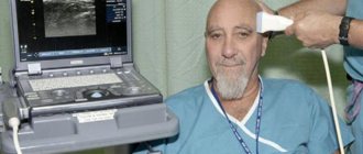
Side effects depend on the chosen technique and area of study. Let's look at pathogenic manifestations using specific examples. During angiography of the vessels of the brain/extremities, the patient is given anesthesia. This is necessary to numb the insertion site of the catheter and introducer. The patient will not feel pain during the advancement of the catheter, since the inner vascular wall is devoid of pain receptors. After administration of an iodine-containing substance, the patient may feel a metallic taste in the mouth, fever, or an increasing feeling of warmth that gradually covers the entire body. Most often, the symptoms go away on their own within a few minutes.
During angiography of the coronary vessels, a feeling of heat may occur. The highest temperature is recorded in the face area. If the catheter touches the heart walls, the heart rhythm is disrupted and blood pressure decreases, so the patient may experience mild malaise, dizziness, coughing, or nausea. These side symptoms need to be corrected. Tell your doctor how you feel and follow the specialist's instructions.
In medical practice, there are rarely any side effects after angiography. The main thing is to track contraindications, identify risk groups and carry out diagnostics only on the direction of a doctor. Normally, an iodine-containing drug is eliminated from the body naturally. Excretion occurs through the kidneys and takes 1 - 1.5 days (average figure, which may vary). Experts say that the process can be accelerated by simply increasing the amount of fluid consumed. Immediately after angiography, the patient needs bed rest. The recovery period may vary for each group of patients. Additional information should be obtained from your attending physician.
Angiography has been known since the 20th century, but entered into everyday practice only a few decades ago. Now the method helps to identify, prevent and select quality treatment for diseases of the vascular system. The cost of diagnostics varies depending on the technique, the area being examined and the chosen medical institution. The examination is prescribed by the attending physician, based on the patient’s condition and the body’s needs. Follow the therapeutic course strictly and stay healthy.
Sources: https://foodandhealth.ru/diagnostika/angiografiya/ https://oserdce.com/diagnostika/sosudov/angiografiya.html https://www.polismed.com/articles-angiografija-chto-takoe-angiografija-pokazanija .html
Varieties of diagnostic method
There are several types of examinations, which differ in the methodology, purpose of the study and the quality of the images obtained. Diagnosis is sometimes carried out without contrast, but more often a contrast solution is used. It improves the quality of images and allows you to visualize the state of the blood vessel at high resolution.
The solution is injected using a syringe if the blood vessel is near the surface. When the arteries and veins being examined are located deep, a catheter is used to inject the solution. The drugs used during the examination contain ionizing iodine.
Types of neck angiography:
- The traditional method involves the use of a solution that is injected into the vessels using a catheter. Diagnosis takes place by taking many pictures to determine the condition of the arteries and veins. The image is obtained using x-rays. The doctor makes a description and interpretation immediately after the procedure.
- CT angiography is considered an improved type of traditional examination. The images are of high quality, which makes it possible to identify the disease in the early stages and begin timely treatment. Iodine is administered using a syringe. The tomograph takes readings from the vessels layer by layer. The method helps to identify abnormalities in the structure of veins and arteries that are difficult to detect with ultrasound. The method is characterized by low stress on the body.
- Spiral computed tomography (SCT) is based on the computed tomography method, but with certain distinctive features. The difference between the method lies in the rotational actions of the emitter and the table - this happens simultaneously. This movement makes it possible to explore the entire area. The speed of the table should allow high-quality and detailed images of diseased vessels to be made. When administering the solution, a vein in the ulnar part of the arm is used.
Angiography procedure
- Multislice computed tomography (MSCT) uses small doses of radioactive radiation to produce high-resolution images. Often used to examine the small capillaries that fill the cervical spine. This method is an improved method of spiral computed tomography. MRI scanners are capable of obtaining images of the neck in one rotation with minimal deviation. The clarity of the images is higher with minimal radiation exposure.
- MR angiography is used to study vascular structure. Magnetic resonance imaging (MRI) is characterized by electromagnetic effects on the body to diagnose the state of the blood supply to the head and neck. It is safe. It is used in cases of serious suspicion of the formation of malignant tumors. A doctor may prescribe a procedure after a severe traumatic brain injury to rule out disruptions in the integrity of the arterial and venous structures. Helps identify atherosclerotic problems.
CT angiography
This is a non-invasive angiographic method for studying blood flow. In this case, the most common access point is a peripheral vein, often in the elbow joint.
There is a difference between CT angiography of arteries and CT venography (venography).
Arterial CT angiography
A CT angiogram of blood vessels is performed in the arterial phase, when the artery is better filled with contrast agent. To correctly determine the scanning time, different methods are used:
- Bolus tracking automatically starts the study when the required lumen density is reached. In practice, modification is often used to visually monitor changes in arterial lumen density, and if this is sufficient, the scan is started manually.
- “Bolus timing” – after the injection of a small bolus of contrast agent, the density curve of the artery lumen is determined, from which the most appropriate time to start scanning is derived.
- Random is the fastest technique, with the greatest risk of suboptimal timing.
CT phlebography
There are CT scans in the portal phase (portography), late venous phase (veins), and venous prephase (peripheral veins).
Preparing for angiography
Angiography requires careful preparation of the body. This will allow the procedure to be carried out without serious complications and will increase the accuracy of the analysis. The results of the readings depend on many factors taken into account in advance. An analysis with incorrect indicators will lead to incorrect treatment, which will complicate the patient’s condition.
Preparation for angiography includes a number of activities:
- 7-14 days before the procedure, you should avoid consuming medications and products containing iodine.
- Alcoholic beverages should not be consumed 7 days before angiography.
- A test is carried out to check for the possibility of an allergic reaction.
- The doctor issues a referral for the procedure.
- Additionally, it is required to undergo a general blood and urine test to exclude inflammatory processes.
- The condition of the kidneys and liver is checked using ultrasound.
- In the morning before the study, you should not drink or eat.
If there is nervous tension, the doctor will sometimes allow you to take a sedative drug to calm the nervous system. This will help the person to calmly postpone the event, eliminating the reason to interrupt the diagnosis.
What to do before the procedure
Before an angiography is performed, the doctor asks the patient about current illnesses and medications taken. Due to the administration of a contrast agent, it is very important to be aware of the risk of allergies (especially to iodine) or asthma, kidney disease, thyroid disease, and diabetes. The doctor also asks about bleeding disorders or a tendency to increase bleeding. Of the medications, drugs that affect blood clotting (Warfarin, Pelentan, etc.) are especially important; before the examination, they should be replaced with injectable Heparin. The difference in the effect of these drugs is not significant, but there are differences in the reaction to the contrast agent.
The day before the examination, a blood sample is taken for clotting. On the day of the examination, you must arrive on an empty stomach, i.e. do not eat for at least 4 hours. During this time, you can only drink a small amount of water. On the contrary, it is advisable to drink more fluids than usual the day before the examination. Increased fluid intake is also recommended after the examination - it helps to improve the removal of the contrast agent.
Before the procedure, the patient usually receives blood thinners and, in the case of allergies, medications to prevent an allergic reaction (eg, Prednisone). Patients with diabetes usually receive glucose and insulin infusions. The last step before angiography and venography is shaving the injection site (groin).