Magnetic resonance imaging, gradually developing, has taken a leading position in the diagnosis of brain diseases. Using the properties of the electromagnetic field to change the response impulses of hydrogen atoms in cells, MRI allows you to create a detailed three-dimensional image of all brain structures. The effect on the body is carried out non-invasively, without violating the integrity of tissues and without causing harm to the body.
Magnetic resonance imaging of the brain
The diagnostic ability of magnetic tomographs is increasing every year. The first tomographs could produce an electromagnetic voltage of about three tenths of a tesla. Currently, tomographs with a field of one and a half, two Tesla, and sometimes eight are widely used, which significantly increases diagnostic performance.
The tomograph carries out sequential scanning of brain structures in steps of several millimeters, taking pictures - sections. The scans can reflect both horizontal and vertical surfaces, recreating a three-dimensional image of brain structures. Some types of pathology require the use of contrast agents, which increase the clarity of images by filling blood vessels and accumulating tissue.
Who evaluates the research results?
The radiologist who conducted the study interprets the MRI results. He consistently, without missing anything, analyzes all the images received and makes a conclusion. The patient receives a study protocol, where the radiologist describes the shape, size and condition of all the structures being examined. At the end of the protocol, the doctor makes a conclusion, which reflects deviations from the norm, if any.
Along with the research protocol, a person receives a film with images and an electronic version of tomograms recorded on a disk or flash card. Some institutions provide electronic results only at the patient’s request.
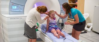
A doctor analyzes an image of the brain
The radiologist, making a description and conclusion to the images, does not have the right to analyze the significance of the identified pathology; he can only indicate the type of deviations and their location. The conclusion about the significance of the identified deviations, as well as about the method of their treatment, is made by the attending physician (usually the one who sent for the examination), so the results of the study must be referred to him. The radiologist can advise the patient which doctor should be contacted with the identified pathology if the person came for an MRI without a referral, at his own request.
Operating principle
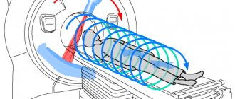
Magnetic resonance imaging is an absolutely safe diagnostic method for the body. Despite what many people think, MRI has nothing to do with harmful radiation exposure. The mechanism of action of the tomograph is the physical phenomenon of nuclear magnetic resonance. The word “nuclear” means a hydrogen nucleus, since all our organs are saturated with hydrogen.
The human body emits radio signals that are picked up by a tomograph. The equipment organizes them and reads them from the desired part of the body. Healthy cells emit the same signal, but pathologically altered ones emit a completely different signal. These signals are processed by an ultra-sensitive and high-speed computer, which produces an image in 3D projection. In addition, you can obtain cross-sectional images of the organ being examined. Therefore, the doctor does not need to perform surgery to evaluate the situation.
The accuracy of this method is very high, since it can be used to diagnose the inflammatory process with an accuracy of 4 mm. And thanks to this, the doctor is able to accurately determine the cause of the patient’s illness.
In order to correctly diagnose the pathological condition of an organ, the doctor sets the necessary parameters - the main one is the width of the cut. Thanks to this, the specialist will understand what the damage is: fatty tissue replacement, a neoplasm with fluid or pus.
Who treats brain diseases?
The attending physician for a patient with brain pathology may be a neurologist or neurosurgeon. As a rule, doctors in this specialty can independently interpret tomograms. A neurologist or neurosurgeon, having performed an independent analysis of tomograms, may not agree with the description and conclusion of the radiologist, since in addition to MRI images, he also analyzes the results of examination and other examinations of the patient.
If the doctor has doubts and disagreements with the radiologist, as a rule, a medical consultation is held, and a repeat study is performed.
Who interprets magnetic resonance imaging scans?
Interpretation of MRI scans takes from thirty minutes to several hours. Private diagnostic centers provide the service of sending image results by e-mail and recording the results on optical media.
Radiodiagnostic doctors have a diploma in general medicine after graduating from medical university.
In some European countries, there are paramedics who perform a significant part of the work - providing first aid, delivering the patient to a medical facility. Such specialists do not have a diploma of higher medical education.
After completing residency, doctors who have received the specialty of radiologist, if they wish to study magnetic resonance imaging, take advanced training courses at the Academy of Postgraduate Education.
Completion of internship/residency/specialization in radiology makes it possible to choose other professions in the field of radiology diagnostics:
- Proton emission tomography;
- Ultrasound examination;
- Scintigraphy;
- Computed tomography (CT).
Refresher courses take three to five weeks.
The MRI scans are interpreted by a radiology doctor, but confidence appears only after two years of practice. When choosing a diagnostic clinic, you should find out how long the specialists work. A competent professional is usually formed after 2 years of work.
The interpretation of the MRI is done not only by the person who conducted the examination itself, but also by the attending physician who referred the patient for examination. This procedure is quite labor-intensive and requires high qualifications. In addition to anatomical knowledge, the specialist must understand various pathologies and the tomograph’s reaction to them.
Thus, a particular disease is determined not only by the echogenicity of the tissue, size, deformation, but also by the shade in the image. Therefore, the interpretation of the MRI obtained by the radiologist is supplemented by the conclusions of the attending physician. Now you understand that you should entrust the interpretation of MRI, especially of the brain, to a competent specialist.
What can be seen on tomograms?
Different parts of the brain differ in color in the image: from white to black. Contrast depends on the degree of vibration of hydrogen protons in the tissue, the impulse from which is picked up by the MRI machine.
In the picture you can see the state of the gray and white matter of the brain, the ventricles (cavity formations within which intracerebral fluid circulates). All subcortical centers and formations responsible for various functions of the body are visible. The meninges and the space between them, the bones of the skull, eye sockets, ear canals and nasal sinuses are also available for analysis.
Which specialist does an MRI?
A diagnostician (radiologist) monitors the progress of the magnetic resonance imaging procedure. The doctor also carries out the preparatory stage:
- Reviews the patient's chart;
- Detects the presence of contraindications to MRI;
- Looks at laboratory tests for creatinine;
- Determines the degree of renal failure;
- Analyzes radiographs;
- Before planning a contrast examination, controls the conduct of a provocative test for gadolinium;
- Eliminates fear of closed spaces and muscle tremors.
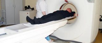
The progress of the tomography is controlled by a radiology doctor through a special X-ray protective window. The patient holds an “emergency button” in his hand. In case of any discomfort, the product helps to call the staff.
Before the procedure, the following contraindications should be excluded:
- Metal devices;
- First trimester of pregnancy;
- Individual characteristics;
- Hypersensitivity to gadolinium contrast agent;
- Muscle cramps.
Prescribing sedatives before a tomography procedure requires consultation between the attending physician and a diagnostician.
Small children are given anesthesia before the scan to prevent the baby from immobilizing. To perform the examination, diagnostic centers must have an anesthesiologist-resuscitator on staff. Preparations for putting a child into medical sleep have a short-term effect. The duration of action of the substance is calculated based on the time of magnetic resonance imaging.
What does a healthy brain look like?
The radiologist describes a healthy brain in the research protocol as follows:
All brain structures should be developed correctly, the intensity of the magnetic resonance signal should be normal. The ventricular system is developed correctly, of normal size, neither dilated nor reduced. The perivascular and subarachnoid space are normal. The sulci and gyri are normal. The brain structures are of normal size and not displaced. Sella turcica, pituitary gland without pathology. The eye sockets, nasal sinuses, and ear canals are of normal size and shape, without signs of pathology. Brain tissues do not have focal or diffuse changes. If contrast was used, the brain vessels are developed correctly and are evenly filled with contrast agent.
Brain MRI: Introduction
Magnetic resonance imaging of the structures and membranes of the brain is a modern diagnostic method that allows medical personnel to visualize the state of anatomical structures in the area under study, identify pathologies, diffuse and focal tissue lesions, determine not only the location, but also the nature of neoplasms, and study the functional activity of the brain.
All this is completely harmless and safe for the patient, does not cause pain or discomfort. MRI of the brain is prescribed for complaints of memory dysfunction, sharp decrease in vision, dizziness and headaches of various localizations.
This type of study is recommended to be carried out using a contrast agent. Gadolinium salts are administered intravenously before manipulation, and, thanks to their physicochemical properties, improve the permeability of tissue structures, making the resulting images graphically clearer and objectively more informative.
What do pathological changes look like on the pictures?
Tumor
In the picture it looks like a lighter, asymmetrical spot with jagged edges (since the picture is a negative, doctors call it a darkening). The formation can deform and displace other structures (ventricles, subcortical centers). Tumor growth, as a rule, is accompanied by the rapid development of additional vessels in the affected area, so contrast is used to analyze volumetric processes.
Stroke
With the development of an ischemic stroke, a zone of brain hypoxia appears; on MRI it will appear lighter in color, but unlike a tumor, the spot will correspond to the area of innervation of one of the cerebral arteries. With contrast, blood supply to the stroke area will be reduced. The smoothness of the furrows and convolutions above the affected area can be determined.
Differences between ischemic and hemorrhagic stroke
Hemorrhagic stroke is accompanied by rupture of a vessel and the formation of an intracerebral hematoma. MRI images reveal a dark cavity with a ring-shaped strip along the periphery, the thickness of the ring decreases over time.
Vascular diseases
Vascular diseases are detected by administering a contrast agent.
This group of pathologies includes:
- Atherosclerosis of cerebral vessels (manifested by reduced contrast of blood vessels due to narrowing of the lumen by atherosclerotic plaques.
- Hypertensive angiopathy is accompanied by expansion of the perivascular spaces (edema). The image shows criblures - round cavities with a clear boundary, located near the vessels.
- An aneurysm is an expansion and thinning of the vessel wall.
- Malformation is a congenital pathology of the vascular bed; MRI shows radially located vessels that converge towards the center (head of the medusa).
Multiple sclerosis
It is characterized by the formation of zones of white matter - nerve fibers that lose their myelin (outer) layer. The tomogram shows focal formations that accumulate contrast differently depending on the stage of the disease.
The lesions appear as light areas and can be located in all parts of the white matter. Their number can vary greatly: from one to dozens. In the initial stage of the disease, if only one lesion is detected, it can be mistaken for a small space-occupying formation. Unlike cancer, lesions in multiple sclerosis do not deform surrounding structures.
Hydrocephalus (dropsy)
Normal and hydrocephalus
Expansion of the cavities of the ventricles, subarachnoid, perivascular space. Severe forms may be accompanied by compression and atrophy of the cortex and subcortical formations of the brain.
Congenital malformations
Serious abnormalities in brain development, as a rule, are detected in the prenatal period or in the first months of a child’s life. If the anomaly does not cause a significant loss of any body function, then it can only be detected by chance, during an MRI, which is planned for another purpose.
Indications for MRI of the brain
- Severe headaches of a chronic nature with unknown etiology.
- Dizziness, worsening with changes in body position.
- Dark spots before the eyes that occur when moving, or a feeling of double vision.
- Convulsive syndromes of various etiologies.
- Epilepsy.
- Complete or partial loss of memory, its deterioration.
- High blood or intracranial pressure.
- Chaotic way of thinking.
- Weakness and numbness in the limbs, loss of coordination.
- Previously identified neoplasms of a tumor nature, of any location and nature.
- Primary and secondary metastasis occurring in the tissues and structures of the brain.
- Congenital anomalies and pathologies (for example, underdevelopment of the corpus callosum or pathology of the development of the rhombencephalon).
- Traumatic effects (traumatic brain injuries of varying severity, penetrating injuries).
- Vascular pathologies: cerebral aneurysms, arterial or venous malformations, vascular tumors (angiomas).
- Strokes of various origins (cerebral infarction or ischemic stroke; hemorrhagic stroke; transient ischemic pathology).
- Parkinson's disease and other degenerative diseases.
- Multiple sclerosis and its consequences.
- Encephalomyelitis (inflammation of the membranes of the brain of viral etiology) and other demyelinating diseases.
- Encephalitis caused by pathogens.
- Brain abscess.
- Hydrocephalus (dropsy) of the brain.
- Cysts, including arachnoid cysts.
- Intracranial hematomas.
- Neurocutaneous syndrome (Bourneville syndrome, phakomatosis).
Magnetic resonance imaging is also prescribed to patients before and after surgery.
In the first case, this is done to coordinate the actions of the surgeon, in the second - to exclude the possibility of re-development of the tumor process within the area under study and to monitor the effectiveness of treatment.