Technique
So, how is a lumbar puncture, or, in other words, lumbar puncture performed? The technique is as follows: the patient lies on his side or sits, leaning forward. The area of the back is disinfected and locally anesthetized, after which a long needle is inserted into the space between the 3rd and 4th or between the 2nd and 3rd lumbar vertebrae to collect a small amount of cerebrospinal fluid (approximately 5-10 ml).
The whole procedure takes no more than 15 minutes. Next, the collected material is examined for the content of glucose, proteins, and other cells and substances. Cultures may be performed to detect infection.
After a lumbar puncture, you should remain in a supine position for some time. The patient is asked to drink plenty of fluids. After an hour, you can get up and go about your business. However, in the next 2-3 days it is recommended to rest more and not overload the body.
Lumbar puncture: technique
Lumbar puncture is performed with the patient sitting or lying down. The supine position is used mainly in severe condition of the patient with increased cerebrospinal fluid pressure in the cerebral fluid reservoirs and during operations, as well as in children. To perform the puncture correctly, it is necessary for the patient to take a position in which the maximum distance of the spinous processes can be achieved.
Lumbar puncture (photo) in a sitting position
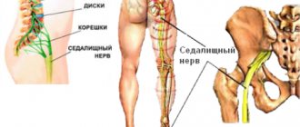
In a sitting position, the patient's head is strongly tilted forward, and in a lying position, he lies on his side, his chin is pressed to his chest, his legs are bent at the knees and hip joints and pressed as much as possible to the stomach. A Quincke line is mentally drawn, which connects the crests of both iliac bones. It passes between the III and IV lumbar vertebrae, that is, in the place where the puncture is most often made. Lumbar puncture can be performed between L3-L4; L4—L5; L5-S1 and with special care between L2-L3. The puncture site is disinfected with ethyl alcohol.
It is worth noting that alcohol solutions of iodine can cause aseptic inflammation of the meninges, and therefore are not currently used in practical medicine.
Anesthesia is very rarely used.
When the dura mater of the brain is pierced, a special feeling of “falling through” occurs, after which the cerebrospinal fluid begins to flow drop by drop. The needle is advanced to a depth of 5-7 cm in adult patients, 2-5 cm in young children and 1-2 cm in infants. Lumbar puncture lasts 1-5 minutes.
If damage to a specific location of the spinal cord or disc herniation is suspected, a puncture is made above and below the suspected “focus” (floor puncture).
Lumbar puncture needle
Reusable needles for lumbar puncture
The puncture is performed in a strictly sagittal direction, using a special hollow needle (figure above). The length of the needle is 10-12 cm, and the thickness is 0.5-1 mm. The top of the needle is cut at an angle of 45. There are needles measuring 20-22 and very rarely 25-26 mm. At the moment, the medical equipment market offers a wide variety of needles for lumbar puncture, which minimize complications of manipulation.
Purpose of the procedure
Carrying out a lumbar puncture can have two purposes: therapeutic or diagnostic.
For medicinal purposes, the procedure is performed to remove cerebrospinal fluid and normalize its circulation; to control conditions associated with hydrocephalus; for the sanitization of cerebrospinal fluid during meningitis. In addition, using lumbar puncture it is possible to administer medications, for example, antiseptics, antibiotics and others.
For diagnostic purposes, a puncture is made to examine the cerebrospinal fluid. After collecting the cerebrospinal fluid, it is analyzed, that is, the color, transparency, composition are determined, the biochemical properties are studied, microbiological tests and culture are carried out. During the procedure itself, cerebrospinal fluid pressure, spinal cord patency are measured, and compression tests are performed.
In addition to the above, lumbar puncture allows the introduction of painkillers into the membrane of the spinal cord during epidural anesthesia, as well as radiopaque agents for some special X-ray studies to determine the intervertebral hernia.
1.86. Standard
Target
: diagnostic (for studying cerebrospinal fluid) and therapeutic (for administering antibiotics, etc.).
Indications
:meningitis.
Prepare
: sterile: syringes with needles (5 ml, 10 ml, 20 ml), puncture needle with mandrin, tweezers, napkins and cotton balls, tray, nutrient medium, test tubes, gloves; manometric tube, 70% alcohol, 5% alcohol iodine solution , 0.5% dissolved novocaine, adhesive plaster, KBU.
Action algorithm:
- Inform the patient about the upcoming procedure and obtain consent.
- The puncture is performed by a doctor under conditions of strict adherence to aseptic rules.
- Take the patient to the treatment room.
- Lay the patient on his right side closer to the edge of the couch without a pillow, tilt his head forward to his chest, bend his legs as much as possible at the knees and pull them towards the stomach (the back should arch).
- Place your left hand under the patient's side, and with your right hand hold the patient's legs to fix the position given to the back. During the puncture, another assistant fixes the patient's head.
- The puncture is made between the III and IV lumbar vertebrae.
- Decontaminate your hands at a hygienic level, treat them with a skin antiseptic, and put on gloves.
- Treat the skin at the puncture site with a 5% iodine solution, then with a 70% alcohol solution.
- Fill a syringe with a 0.5% solution of novocaine and give it to the doctor for infiltration anesthesia of soft tissues, and then a puncture needle with a mandrel on the tray.
- Collect 10 ml of cerebrospinal fluid in a tube, write the directions and send to the clinical laboratory.
- Collect 2-5 ml of cerebrospinal fluid in a test tube with nutrient medium for bacteriological examination. Write a referral and send the biological material to the bacteriological laboratory.
- Give the doctor a manometric tube to determine the cerebrospinal fluid pressure.
- After removing the puncture needle, treat the puncture site with a 5% alcohol solution of iodine.
- Place a sterile napkin over the puncture site and cover with adhesive tape.
- Place the patient on his stomach and take him on a gurney to the ward.
- Place the patient on a bed without a pillow in the prone position for 2 hours.
- Monitor the patient's condition throughout the day.
- Take off your gloves.
- Place syringes, cotton balls, gloves in the KBU, place used instruments in a disinfectant solution.
- Wash and dry.
1.87. Standard “Preparation of the patient and medical equipment for sterile puncture”
Target
:diagnostic: bone marrow examination to establish or confirm the diagnosis of blood diseases.
Indications
: diseases of the hematopoietic system.
Contraindications
: myocardial infarction, attacks of bronchial asthma, extensive burns, skin diseases, thrombocytopenia.
Prepare
: sterile: tray, syringes 10 - 20 ml, Kassirsky puncture needle, glass slides 8 - 10 pieces, cotton and gauze balls, forceps, tweezers, gloves, 70% alcohol, 5% alcohol solution of iodine; adhesive plaster, sterile dressing material, KBU.
Action algorithm:
- Inform the patient about the upcoming study and obtain his consent.
- Sternal puncture is performed by a doctor in the treatment room.
- The sternum is punctured at the level of the III – IV intercostal space.
- The nurse assists the doctor during the procedure.
- Invite the patient into the treatment room.
- Invite the patient to undress to the waist. Help him lie down on the couch, on his back without a pillow.
- Decontaminate your hands at a hygienic level, treat them with a skin antiseptic, and put on gloves.
- Treat the front surface of the patient's chest, from the collarbone to the gastric region, with a sterile cotton ball moistened with a 5% iodine solution, and then 2 times with 70% alcohol.
- Perform layer-by-layer infiltration anesthesia of soft tissues with a 2% solution of novocaine up to 2 ml in the center of the sternum at the level of the III – IV intercostal space.
- Give the doctor a Kassirsky puncture needle, placing the limiter shield at 13 - 15 mm of the needle tip, then a sterile syringe.
- The doctor pierces the outer plate of the sternum. The hand feels the failure of the needle; after removing the mandrin, a 20.0 ml syringe is attached to the needle and 0.5 - 1 ml of bone marrow is sucked into it, which is poured onto a glass slide.
- Dry the slides.
- After removing the needle, treat the puncture site with a 5% alcohol solution of iodine or a 70% alcohol solution and apply a sterile bandage and secure with an adhesive plaster.
- Take off your gloves.
- Dispose of used gloves, syringes and cotton balls in the CBU.
- Wash your hands with soap and dry.
- Show the patient to the room.
- Send the slides to the laboratory after the material has dried.
Note
: Kassirsky's needle is a short, thick-walled needle with a mandrel and a shield that protects the needle from penetrating too deeply.
Source: https://studfile.net/preview/6352731/page:46/
Indications for puncture
There are absolute and relative indications for this procedure. The first includes suspected infection in the central nervous system. This could be meningitis, ventriculitis, encephalitis.
Also indicated is damage to the membranes of the spinal cord and brain due to cancer; hydrocephalus; diagnosis and detection of cerebrospinal fluid fistulas; study of subarachnoid hemorrhage if computed tomography is not possible.
Relative indications for the study: fever of unknown origin in children under 2 years of age; vascular embolism; inflammatory neuropathic syndromes; lupus erythematosus; demyelinating processes.
Contraindications
If the patient has a mass or signs of cerebral edema, hydrocephalus, or intracranial hypertension, there is some risk of axial herniation during the procedure, and the risk increases with the insertion of a thick needle or the removal of large amounts of cerebrospinal fluid. In such conditions, puncture is done in extreme cases, when it is not possible to replace it with another study. The amount of liquor removed should be minimal.
Less significant contraindications are infectious processes in the sacrum and lumbar region, blood clotting disorders, and taking anticoagulants. The lumbar puncture is performed with caution if a ruptured cerebral aneurysm or hemorrhage is suspected, as well as with a blockage of the subarachnoid space in the spinal cord.
Lumbar (spinal) puncture
1. Diagnostic:
1) suspicion of infection of the central nervous system, especially inflammation of the spinal membranes (cerebrospinal meningitis) (absolute indication);
2) autoimmune disease of the central nervous system;
3) metabolic disease of the central nervous system, especially leukodystrophy;
4) some neuropathies;
5) suspicion of subarachnoid hemorrhage in those patients in whom hemorrhage is not confirmed on CT;
6) other diseases of the central nervous system in which examination of the cerebrospinal fluid (CSF) can help in diagnosis, for example, inflammation of the spinal membranes due to a neoplasm;
7) the need for intrathecal or subarachnoid administration of a contrast agent.
2. Medicinal:
1) intrathecal administration of drugs: antibiotics for CNS infections, cytostatics for the treatment of malignant neoplasms of the CNS, anesthetics;
2) immediate removal of a certain amount of CSF in order to reduce its pressure (eg, with hydrocephalus).
Contraindications
1. Absolute: significantly increased intracranial pressure - swelling or swelling of the brain - especially in the back of the skull (in this case, a CT scan of the head should be performed first).
2. Conditional: skin and tissue infections in the area of the planned puncture, malformations of the spine and spinal cord (eg, dysraphism), coagulation disorders (INR >1.5 or APTT more than twice the normal value, or platelet count blood <50,000/μl); suspected subarachnoid hemorrhage (in this case, a CT scan of the head should be performed first).
Complications
1. Post-puncture syndrome
1) headache - usually not severe, appears within 24-48 hours after puncture, often in the frontal or occipital areas; increases in a vertical position and decreases in a supine position; may be accompanied by nausea, vomiting, dizziness, tinnitus, visual disturbances and meningeal symptoms; goes away on its own within 24 hours (sometimes after a few weeks). Prevention: puncture with a finer needle (eg, 22 G instead of 18 G), directing the bevel of the limb of the needle in the lateral direction of the spine (bevel of the needle should be in the sagittal plane) (so that the fibers of the dura are not crossed, but only separated), or application atraumatic needle. Prolonged stay of the patient in a supine position after puncture does not prevent headaches. Treatment: supine position, oral analgesics (paracetamol, paracetamol with caffeine, opioids; do not use NSAIDs or drugs that interfere with blood platelet function).
2) back pain at the puncture site;
3) radicular pain - most often radiating to the lower extremities; if they occur when the needle is inserted, this indicates irritation of the nerve roots (you must immediately pull back the needle and change the direction of the puncture).
2. Others (rare): paresis of the lower extremities (caused by epidural hematoma; usually in patients taking anticoagulants shortly before or after puncture); wedging of the cerebellar tonsils into the foramen magnum (in patients with cerebral edema, tumor or massive subarachnoid hemorrhage; leads to death); subarachnoid or epidural hemorrhage; damage to the spinal ligaments or periosteum of the vertebrae; acute purulent inflammation of the vertebrae; abscess; epidermal tumor.
Patient preparation
1. Informed voluntary consent of the patient, if he is conscious.
2. Assess the number of blood platelets, INR and APTT, in case of deviations, correct them; if the patient is taking anticoagulants, they should be discontinued → table. 2.34-7.
3. Exclude the presence of increased intracranial pressure (cerebral edema or tumor) based on fundus examination (signs of edema and congestion of the optic nerve head) or CT, which should be performed in case of: immunodeficiency, history of central nervous system disease, recent episode of epileptic seizure ( seizures), swelling or congestion of the optic nerve head, impaired consciousness, the presence of focal neurological symptoms.
4. Position the patient on his side, close to the edge of the operating table, with his back to the doctor who is performing the procedure; the lower limbs are bent at the knee and hip joints, the knees are brought to the stomach; the head is bent as much as possible in the direction of the knees (Fig. 24.13-1), avoid excessive flexion of the spine, which should be in the same plane throughout; the back and shoulder line are in a plane perpendicular to the table.
Place of puncture
Intervertebral space, preferably between the spinous processes of L4 and L5 or L3 and L4, not higher than the gap between L2 and L3, along the midline, which passes through the apices of the spinous processes of the vertebrae or slightly lateral to it. The line connecting the upper points of the iliac crests of the femur crosses the lumbar spine at the level of the spinous process of the L4 vertebra (Fig. 24.13-1).
Security
1. Set for preparing the surgical field → section. 24.2 and, possibly, local infiltrative anesthesia →section. 24.3.
2. Sterile disposable puncture needle with mandrel 22 G or 20 G, length 8.75 cm (traditional cutting Quincke type or atraumatic, eg Sprotte or Whitacre type). You can also use smaller diameter needles that are inserted through a short, larger diameter needle (called an introducer needle).
3. Apparatus for measuring cerebrospinal fluid pressure.
4. Sterile tubes.
Technique
1. Prepare the surgical field →section. 24.2. If necessary, apply local anesthesia to the skin and subcutaneous tissue, e.g. EMLA cream or infiltration with 1% lidocaine solution (or other suitable local anesthetic) → section. 24.3 (excessive in unconscious patients).
2. Insert the needle with the mandrel slowly, pointing it at an angle cranially, in the direction of the navel. Direct the beveled cut of the tip of the cutting needle upward (towards the spine). As the ligamentum flavum and dura mater pass through, resistance is felt to be overcome, which is accompanied by a “failure” (in adults, the intrathecal space is at a depth of 4–7 cm). After overcoming the resistance of the hard shell, remove the mandrin; CSF should drip from the needle. If the patient is conscious, ask him to slightly relax his lower limbs (reduce flexion in the hip joints). If the cerebrospinal fluid does not flow out, reinsert the mandrin and smoothly move the needle forward or turn it 90° along the axis, then remove the mandrin again. Do not use excessive force to overcome resistance. The reason for the absence of CSF may be that the needle extends beyond the subarachnoid space. CSF stained with blood may indicate damage to a vein in the spinal canal during puncture; then the CSF quickly clears itself, and if this does not happen, then perform a puncture one intervertebral space higher.
3. For a more detailed assessment (not always necessary), hold the needle with one hand and connect the other to a device to measure CSF pressure (normal is 7–15 [<20] cmH2O; this corresponds to a typical flow of CSF from the needle at a rate of 20– 60 drops/min; the test result is reliable if the patient lies calm and relatively relaxed).
4. After assessing the pressure, draw CSF into several sterile tubes for the purpose of conducting the necessary studies (usually 3-5 ml, after ruling out cerebral edema, max. 40 ml).
5. After obtaining CSF, insert the mandrin into the needle, remove the needle, and apply a sterile dressing to the skin.
After the operation,
the patient must remain in a supine position for about an hour.
Side effects
The most common consequence of a lumbar puncture is headaches. They affect up to 50% of patients. In most cases, pain occurs 2-3 days after the procedure, varies widely in severity, and is worse when the person sits or stands, but is relieved when lying down. In addition, headaches may be accompanied by nausea, ringing in the ears, blurred vision and dizziness.
As a rule, no special treatment is required. The pain is tolerable, neutralized by taking conventional painkillers and goes away on its own after a few days. The important point is that to eliminate it you should not take Aspirin, since it is an anticoagulant that thins the blood, which is undesirable after a puncture.
The following conditions require urgent consultation with a doctor: the appearance of severe fever and chills, a feeling of tightness in the neck area. All this may be a sign of infection and inflammation of the spinal cord membranes.
Contraindications to lumbar puncture
This manipulation is not particularly difficult for specialists. But since there is a possibility of possible negative consequences, there are also contraindications. For diagnostic purposes, only 5 ml of cerebrospinal fluid is taken, and about 700 ml is produced per day. When a contrast agent is injected into the needle, about 10 ml of fluid enters the spinal space. Infections may enter through the needle, and blood vessels may also be injured. Based on the above, the procedure should not be carried out:
- with intracranial hematoma, infringement of the brain stem, its edema, abscess, space-occupying formation and other brain modifications;
- with traumatic shocks;
- with severe damage to the spinal tissues and bedsores in the place where the puncture is made;
- with hemorrhagic diathesis;
- with pathologies of the spinal canal and impaired circulation of cerebrospinal fluid;
- with occlusive form of hydrocephalus.
One of the unpleasant and common consequences is headache after a spinal puncture. It often occurs in patients of all ages. As a rule, the pain intensifies when standing up; on the contrary, it decreases when lying down. Smaller diameter needles reduce the incidence of headaches. Often the symptom goes away on its own and spontaneously. Also, to get rid of it, bed rest, drinking plenty of fluids, analgesics and caffeine are used.
Possible complications
Lumbar puncture, like other invasive interventions in the body, may carry a risk of infection. However, it is very small and amounts to approximately 0.0001%. If the patient has inflammatory skin diseases at the intended puncture site, puncture is not performed, as this significantly increases the risk of infection.
Bleeding is possible when blood accumulates in the epidural space. A so-called epidural hematoma occurs.
It is extremely rare that compression or displacement of the brain stem occurs. This can occur with increased intracranial pressure or an existing tumor in the brain. To avoid such a complication, the patient undergoes a computed tomography or magnetic resonance imaging scan before the puncture.
What are the dangers of spinal cord puncture - complications?
Due to the specific nature of the examination, patients ask doctors a lot of questions. Many people are concerned about whether it is dangerous to perform a spinal cord puncture and what complications there may be.
Some specialists allow you to move around immediately. But after the procedure, the appearance of general cerebral symptoms when trying to stand up cannot be ruled out. Vomiting, headache, and unsteadiness when moving may occur.
The most common consequences of spinal cord puncture are:
- through the fault of the doctor as a result of violation of asepsis, failure to comply with all technical aspects and lack of instructions to the patient on how to behave during and after the manipulation;
- due to the patient's fault;
- due to patient intolerance to the procedure.
Complications after lumbar puncture include post-puncture syndrome, direct trauma, teratogenic factor, and changes in cerebrospinal fluid. The clinical picture of postpuncture syndrome is determined by disruption of the dura mater by the needle. The leakage of CSF into the epidural space causes pain in the occipital and frontal region, which does not go away for several days, rarely longer.
Spinal cord puncture is dangerous due to hemorrhagic complications. These include spinal subarachnoid, chronic and acute intracranial subdural hematoma. Injury to blood vessels provokes bleeding in people with poor blood clotting or thrombocytopenia.
When inserting a needle into the subarachnoid space, spinal puncture is fraught with damage to the roots, IVD injury, and complications if sterility is violated. Due to the penetration of skin fragments into the brain canal, tumors can form, which manifest themselves years after the intervention with increasing pain in the spine and limbs, disturbances in posture and gait.
Features of the procedure in children
Lumbar puncture in children is used for neoplastic diseases, as well as to confirm meningitis, encephalitis, hemorrhages and diagnose degenerative and vascular changes.
In newborns, lumbar puncture is possible in an upright position, since in the supine position in babies, ventilation is often reduced and perfusion is impaired, which can lead to respiratory arrest.
The doctor, as a rule, performs a lumbar puncture with an assistant, who calms and distracts the child, and also keeps him motionless at the moment the needle enters.
Repeated punctures treat intracranial hypertension after hemorrhage or meningitis in children under 2 years of age.
What influences the results?
It is very important to remain stationary during the procedure, otherwise it will be difficult to obtain the correct result.
In addition, proper puncture is difficult if the patient suffers from obesity, dehydration, or arthritis.
So, the article examined such a therapeutic and diagnostic measure as lumbar puncture, indications and contraindications for its implementation. Currently, this procedure is a very informative method for studying processes occurring not only in the spinal cord, but also in other body systems. If you follow all the rules and have a competent doctor’s approach, the risks of complications after a puncture are minimized.