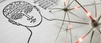
Diseases characterized by changes in muscle contractility are quite common in both adults and children. Electromyography (EMG) is a modern method for studying neuromuscular transmission, which allows us to identify the location of the lesion and, based on this, select the most effective treatment. It is important to note that only a specially trained doctor should conduct the examination; he will also interpret the results, passing them on to the attending physician.
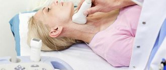
Stimulation electromyography of the upper limbs
History of the discovery of electrocochleography
A famous auditory neurophysiologist named Glen Wever discovered ECoG in 1930. He had a major influence on auditory science, including the work done later by Hallowell Davis. As you know, Hallowell Davis is a famous physiologist and otolaryngologist, the father of the physiology of hearing and the inner ear. At Princeton, Wever and Bray recorded auditory activity in an anesthetized cat. Using an oscilloscope, amplifiers and electrodes, they were able to record activity in the cochlea and in the auditory nerve.
According to this story, they substituted the speaker's voice for the cat during the recording. The words went from a loudspeaker into the cat's ear, and through electrodes placed on the auditory nerve, they were able to decipher some of what was said through the loudspeaker. In other words, the cat's ear functioned almost like a microphone, hence the term cochlear microphone potential (MP).
Shortly after the discovery of MP in cats, the work was published in the scientific journal Nauka, one of the most prestigious journals in the scientific arena at the time. People all over the world immediately read this work and began to reproduce their methods on people. Hallowell Davis in particular found that for him there was more to it than just microphone potential and auditory nerve activity. There was another answer, which he called the summation potential (SP) of the cochlea. Doctors began to realize that MP and SP were quite interesting concepts, but the biggest discovery recorded in humans was the action potential (AP) of the auditory nerve.
By the late 1950s and early 1960s, audiologist Chuck Berlin and otolaryngologist Bob Reuben of Johns Hopkins University said they could perform electrocochleography in the operating room on children who could not be tested any other way. They called it an electrophysiological, objective hearing test. Today, unlike our small computers, they had heavy racks of equipment that they brought into the operating room. To place the electrode near the ear, they had to perform a myringotomy. Myringotomy is a procedure in which a small incision is made in the eardrum. For a child who definitely had significant hearing loss, it was necessary to make an accurate diagnosis of the severity of the loss.
In the 1960s, doctors began to realize that electrocochleography recordings were not that difficult if they used a special transtympanic needle, which is a sharp needle placed through the eardrum. The procedure is invasive and the eardrum is immediately restored. If we place the electrode on the promontorium (promontory), we can still record these responses without using a myringotomy.
Al-Coates was an otolaryngologist at Baylor College of Medicine from the 1960s to the 1980s. He is tasked with assembling the first electrocochleography (ECoG) test battery. He was well versed in vestibular research, but also did a lot of work on this research. He showed that these responses could be recorded less invasively from the ear canal. Other scientific doctors around the world began to realize that this research technique was of great value in diagnosing Meniere's disease. Many people in the late 80's and early 90's began to use these procedures in the operating room with great success. More recently, electrocochleography has been used very widely, including in the diagnosis of auditory neuropathy. She became popular and then disappeared. When otoacoustic emissions (OAE) was introduced in the early 1970s, most audiologists completely forgot about electrocochleography. But today she is probably back for a long time, with too many options to diagnose hearing loss.
How the research is carried out
Electrocochleography can be performed in two ways: invasive and non-invasive:
- The invasive method involves the use of thin transtympanic needles, which are passed through the eardrum and placed on the walls of the middle ear. It gives more accurate and understandable results, since the electrodes are placed in close proximity to the current source (cochlea).
- The non-invasive, or extratympanic, method does not cause any discomfort to the patient and does not require the introduction of needles, anesthesia, or subsequent medical supervision. However, the result is noisier and less accurate. In this case, the electrode is placed on the eardrum or on the wall of the external auditory canal.
In addition to the main electrode, another electrode is located on the lobe of the other ear, in the mastoid region or in the external auditory canal. The patient puts on headphones through which sounds with certain characteristics are presented. By measuring changes in the resulting potentials, the doctor determines how well the cochlea and auditory nerve are functioning.
No preparation for the study is required. It is better not to use decorative cosmetics, remove earrings, and prepare a hair clip.
For invasive examinations, local anesthesia is usually not used, as it increases the risk of eardrum injury. The patient is warned that he will feel short-term pain as the needle penetrates the membrane. In children over 8 years of age, local anesthesia can be used by instilling a lidocaine solution into the ear. In younger children or uncooperative patients, intravenous analgesia may be used.
Stages of electrocochleography:
- inspection and cleaning of the walls of the external auditory canal;
- placing the patient on the couch with the ear being examined;
- irrigation of the eardrum with sterile saline;
- needle insertion with control under a microscope or otoscope;
- fixation of the needle using a foam rubber insert placed in the external auditory canal;
- installation of a second electrode in the form of a silver-plated cup on the opposite mastoid process;
- installation of a grounding electrode on the back of the neck;
- applying a series of stimuli using a special apparatus to an electrode located in the ear under study, and recording the electrical response to stimulation.
The study takes about an hour. Then the patient, after a short rest and feeling well, can leave the clinic.
Why do we need such diagnostics?
ECoG is intended for the diagnosis of Meniere's disease and, in particular, swelling of the inner ear. It may also be abnormal with a perilymphatic fistula. A common feature linking these diseases is a pressure imbalance between the endolymphatic and perilymphatic spaces of the inner ear.
The test can also be used to show that the cochlea is normal in individuals who are deaf. The microphonic potential of the cochlea may be normal in auditory neuropathy, as well as other disorders in which the function of the cochlea is preserved, but the auditory nerve is damaged. Finally, this examination is also used as an indicator of the temporary threshold shift that may occur with acoustic ear trauma.
Decoding the results
The examination results are a curved line with recorded changes (peaks and valleys) of electrical impulses. The doctor analyzes the ratio of the summation potential to the auditory and microphone potential, as well as violations of the norms of electrophysiological indicators.
The interpretation of the results is carried out by an otolaryngologist; on the electrocochleogram he can see disturbances in the conductivity of frequency waves, a decrease or increase in their amplitude, a change in the microphone potential (its norm is 80 dB NHL). Thus, in Meniere's disease, its diagnostic sign is the ratio of SM (summation potential) to AP (action potential of the auditory nerve), which exceeds the norm (more than 0.5 units). With central auditory disorders, AP peaks change, and a decrease in amplitude or loss of electric waves often are a sign of neuropathy.
Electrocochleography is currently actively used in municipal and private medical institutions and is one of the advanced methods for detecting hearing impairment.
Romanovskaya Tatyana Vladimirovna
Indications for use
Typically, electrocochleography is performed when there is a feeling of stuffiness in the ears, the presence of noise and ringing, and a noticeable deterioration in hearing. The manipulation is carried out when:
- paroxysmal dizziness, which is repeated with noise and ringing in the ears, decreased hearing function;
- sensorineural hearing loss in one ear;
- periodic dizziness, unsteadiness and unsteady gait;
- feeling of stuffiness and tightness in the ears;
- diagnosis of endolymphatic hydrops;
- otitis and otosclerosis;
- ear injuries that could lead to decreased function of the sensory organ;
- monitoring the success of treatment of various ear diseases.
Examination is also used in pediatrics to determine the child’s hearing threshold to differentiate diseases such as sensorineural and conductive hearing loss, to evaluate hearing in children who have increased excitability of the central nervous system.
Indications
The list of indications for this study is quite large. EMG is informative for the following diseases of the nervous system:
- Traumatic damage to muscles and nerves.
- Injuries to the brain and spinal cord, especially when they are compressed or bruised.
- Polyneuropathies.
- Neurites.
- Degenerative processes of the spine - osteochondrosis, intervertebral hernia, spinal stenosis.
- Multiple sclerosis.
- Syringomyelia.
- Vibration disease.
- Muscular pathologies (myasthenia gravis, myopathies and myositis).
- Parkinson's disease.
Since the number of lesions of the peripheral nervous system has been increasing in recent years, electroneuromyography comes to the rescue. It is often used to study the functioning of the muscles of the arms and legs.
ENMG allows you to confirm the following diseases:
- Peripheral neuropathy.
- Tunnel syndrome.
- Compression of nerve roots and endings.
- Inflammatory process.
Preparing for the study
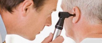
The procedure itself does not require special preparation. Before the examination, the doctor examines the ear canals, in other words, performs an otoscopy, using a backlight and a frontal reflector. The frontal reflector is worn to reflect the rays into the ear cavity. If necessary, the ear canals and sinks may be cleaned due to residual earwax. Then the eardrum is irrigated using isotonic sodium chloride solution.
Also, before coming for an ear diagnosis, you will be asked to remove your facial makeup and thoroughly cleanse your face. It is better not to apply makeup before the procedure.
If we talk about small children, the procedure can be performed under general anesthesia. But before that, you definitely need to discuss such issues in advance with your doctor.
Proper patient preparation
After a person has received an answer to the question: EMG, what is it, he needs to understand how to properly prepare for the procedure so that it is as safe and informative as possible. You can get similar recommendations from your doctor. It is important to note that no special preparation is required, however, patients should follow the following advice:
- In consultation with your doctor, avoid taking medications that can affect muscle or nerve tissue, as this may distort the results of the examination. For example, it is possible that amplitude contractions may appear with small stimuli, etc., which can be regarded as pathology.
- 24 hours before the procedure, it is necessary to avoid taking foods and drinks that increase the level of brain activity. These include coffee, strong tea, any types of chocolate, energy drinks, etc.
If you have diseases of the internal organs that require the use of drugs that reduce blood viscosity or its ability to form blood clots (Clopidogrel, Aspirin), you must consult a doctor before the procedure.
Methodology
The patient is invited to a special room, isolated from various sounds, and asked to lie on his side so that the ear that needs to be examined is on top. Then a special electrode is inserted using an otomicroscope, secured with clamps with a microphone under the strict guidance of the doctor. Another electrode is left in the area of the 7th cervical vertebra. It is intended for grounding. Another electrode with a negative charge is attached at the level of the mastoid process.
Most often, silver-plated cup electrodes filled with an electrically conductive mass are used.
When all the electrodes are attached, the patient will be asked to relax while listening to the doctor's electrical signals, such as clicking sounds. It is very important that the patient is relaxed for this test, as any stretching or muscle movement can slow down the process of transmission and evaluation of the results. Before the procedure, the patient should be informed that unusual sensations may occur when the electrode comes into contact with the eardrum. This test does not always require a response from the patient. While the patient listens to the click, the audiologist will measure the electrocochleographic response using a computer, which uses the necessary information in the form of potentials and amplitude graphs.
The audiologist will collect several responses from the ear and look for the presence of a large signal that contains two components: a summation potential (SP) and an action potential (AP). Both of these waveform components are a direct result of providing sound stimulation to the cochlea. The SP/PD ratio is calculated. The ratio of these two components is the key to deciphering the test results. An increased ratio may indicate excessive fluid pressure in the inner ear.
The study takes about one hour under normal conditions and patient behavior. The person can be free immediately after the examination, even if local anesthesia was used. Most doctors try not to use anesthesia to perform the test, since using anesthesia increases the risk of accidentally damaging the eardrum.
Simplified principle of xerography
| This section is missing references to information sources. Information must be verifiable, otherwise it may be questioned and deleted. You may edit this article to include links to authoritative sources. This mark was set on May 12, 2011 . |
Schematic representation of the electrographic process.
General scheme of electrography (xerography): 1) An electric charge is applied to the surface of the photodrum.
2) The light reflected from the document being copied selectively discharges areas of the drum. 3) Toner is applied to the drum; it lingers in areas that retain a charge. 4) Toner is transferred from the drum to paper, which has a greater negative charge. Before printing, the OPC is charged using a coronator (that is, it acquires a positive or negative potential), after which it is exposed using a lamp and a system of mirrors. The coating of the photodrum in places irradiated by light loses its dielectric properties, which leads to the flow of electric charge into the ground in these places (the photodrum is connected to it, as a rule, through its metal base). The next stage is called manifestation. The toner from the development roller is transferred to the charged areas of the photodrum due to its opposite charge. Then a sheet of paper (cardboard, transparent film, etc.) is rolled over the photodrum, on which to print. After this, the sheet enters the fusing unit (fuser), which melts and presses the toner into the sheet structure. After curing, due to the fact that, as a rule, not all the toner is transferred to the paper, the machine contains a cleaning module that removes the remaining toner on the drum.
A similar principle is used in laser printers, only in them the drum is discharged by a laser in accordance with the information received for printing.
In modern digital laser copiers and printers, the dark parts of the image are applied by a laser beam and the toner, due to the properties of the drums used in laser printing, “sticks” to its uncharged areas, and is repelled from the charged areas by the same electric charge. This principle allows you to increase the service life of the laser, since in most cases, dark areas during printing occupy a much smaller area.
Rehabilitation period
Best materials of the month
- Why you can't go on a diet on your own
- 21 tips on how to avoid buying stale food
- How to keep vegetables and fruits fresh: simple tricks
- How to curb your sweet cravings: 7 unexpected products
- Scientists say youth can be extended
Once this test is completed, your doctor may schedule a follow-up test in two weeks. At a subsequent visit, the attending physician will discuss the ECoG results and other aspects of the auditory test with the patient. When the doctor receives the report, he or she will compare it with the results of other tests to determine the next step in treatment. If endolymphatic hydrops or Meniere's disease is suspected, the doctor may ask for additional tests, such as vestibular evoked myogenic potential (VEMP), electroneurography (ENG), or magnetic resonance imaging (MRI), to confirm the diagnosis.
Visualization of the auditory nerve using MRI plays an important role. Every patient who receives a diagnosis of auditory neuropathy should have an MRI of the auditory nerve. This is why research shows that one in five patients actually have a cochlear nervous system deficiency, and sometimes no nerves at all. If there is no auditory brain response but normal otoacoustic emissions and normal summation potentials are detected, it is important for the patient to undergo magnetic resonance imaging of the auditory nerve to rule out cochlear deficits. If a patient does not have an auditory nerve, a cochlear implant is not the best option. It is known that such patients may also have intracranial problems. It is the detection of auditory neuropathy that often leads to valuable neurological information about the patient.
So, to summarize, only experience and good clinical knowledge are needed to correctly interpret an electrocochleographic study. When interpreting ECoG, it is important to consider the noise level, which is usually assessed by obtaining multiple trials. If they are all the same, then the standard deviation should be small and the result will probably be correct. If they vary widely, the reliability of the average SP/PD ratio may be questionable. Likewise, if several trials are performed and the ratio changes greatly, then confidence in the correct result should be lower than if the result was little different from the previous one.
Diagnostic methods
If a person has a large number of spots on his body, then he should visit a dermatologist. The doctor may prescribe the patient to undergo examination by a geneticist, ophthalmologist, orthopedist and neurologist. Diagnostic procedures may include the following:
- CT, MRI, ultrasound;
- X-ray;
- audiometry;
- Weber test;
- impedancemetry;
- electrocochleography.
The diagnosis of Recklinghausen's neurofibromatosis is based on the following:
- if there are more than 6 spots on the body, and they are more than one and a half centimeters,
- there are small freckles in places uncharacteristic for such a phenomenon,
- there are Lisch nodules - 2 or more,
- optic nerve glioma was discovered,
- a thin outer shell of the tubular bones was diagnosed,
- there is a hereditary predisposition.
Contraindications for diagnosing ear diseases
Typically, electrocochleography is an absolutely safe procedure with no complications and no contraindications. It is carried out regardless of age. Only when the patient has an allergic reaction to local anesthetics used during manipulation, only in this case their use is prohibited or the anesthetic substance is replaced with another.
During electrocochleography, the patient may feel some unpleasant discomfort in the sense organs being examined. But there is no need to worry, since these sensations disappear without a trace and without harm to health after the study.
The advantages of this diagnosis of ear diseases
An ear test is a valuable test that helps a doctor identify potential problems with a patient's inner ear. Electrocochleography is a painless, highly informative test for studying the hearing organs, both in the early and late stages. It helps the doctor make the correct diagnosis and decide on a further treatment plan.
More fresh and relevant information about health on our Telegram channel. Subscribe: https://t.me/foodandhealthru
We will be grateful if you use the buttons:
How is diagnosis done?
First of all, the patient lies on his side so that the ear that will be examined is on top. Then a small electrode and a special telephone device in the form of an earbud are inserted into it, which is securely fixed to the ear.
And finally, the doctor begins to give small electrical impulses (this is not painful and looks like a light mosquito bite). The patient does not feel any discomfort, but only feels a slight noise in the ears, which is created by pulses of different frequencies.
All results obtained during diagnosis are recorded and analyzed by the attending physician.
After the procedure, the patient must undergo a short period of rehabilitation. As a rule, it takes no more than 2 hours and almost immediately after the procedure a person can go about his business.
Article on the topic: Nebilet - instructions for use, indications and release form, composition, dosage and price