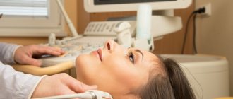
Cardiovascular diseases can sooner or later result in a significant deterioration in health and even death. Therefore, it is recommended to periodically check the blood vessels of the neck and other parts of the body for the purpose of prevention or as prescribed by a doctor. One of the most common ultrasound procedures is duplex scanning of blood vessels.
.
Indications
Duplex scanning of the arteries of the lower extremities is performed in patients who have the following symptoms:
- pain, heaviness in one or both legs after physical activity or prolonged walking;
- fatigue of the limbs, especially at the end of the day;
- development of edema in the ankle area;
- difficulties in determining the pulse at the site of the peripheral arteries;
- the appearance and spread of vascular networks on the skin;
- pigmentation, darkening, redness of the dermis;
- pain on palpation, presence of seals in the vein;
- the formation of trophic wounds that do not heal for a long time.
This type of diagnosis is recommended for patients with suspected pelvic vascular thrombosis.
Duplex scanning of the pelvic veins is recommended for patients with the following complaints:
- chronic and acute inflammatory complications of the genitourinary system;
- suspicion of the development of thrombosis in the pelvic plexus;
- pain in the lower body.
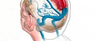
Transcranial ultrasound of the extracranial vessels of the neck is prescribed if a person complains of the following symptoms:
- ringing in the ears, visual impairment;
- dizziness, frequent headaches;
- high blood pressure;
- loss of consciousness;
- problems with the vestibular apparatus.
Angiography
Classic or X-ray angiography
Examination of the vascular system using fluoroscopy after injection of a contrast agent into the blood (visible in x-ray).
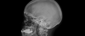
Angiography of cerebral vessels. On the left is the initial accumulation of contrast in the internal carotid artery, on the right is the distribution of contrast along its remaining branches. Click on photo to enlarge
X-ray angiography is divided into:
The higher the selectivity of contrasting the vascular tree, the lower the radiation dose during the study.
Feature of the method: requires hospitalization in a hospital.
Procedure technique:
- femoral artery puncture;
- installation and passage of a catheter to the study area;
- administration of a contrast agent and examination of vascular structures.
| Contraindications | Preparation | Time (min) |
| Intolerance to iodine-containing drugs Insufficiency of kidney and liver function Pregnancy and breastfeeding | Do not eat or drink for 12 hours before the test | 60–180 |
CT angiography
Obtaining a three-dimensional three-dimensional image of vascular structures by combining multiple images in different projections performed on a computed tomograph. Requires administration of a contrast agent.
Procedure technique:
| Contraindications | Preparation | Time (min) |
| The same as for X-ray angiography | If you are treating diabetes with Metformin, stop taking it 3 days before the test Have a blood test for creatinine within a week before the procedure | 20–30 |
MR angiography
A three-dimensional vascular pattern is obtained by recording the response of tissue structures to the action of the electromagnetic field of the tomograph. The main advantage of the method is the absence of radiation. In controversial cases, contrast enhancement is required.
Procedure technique: after fixing the area of interest in a special coil, a scan is carried out, during which you must lie still.
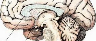
Results of MR angiography of cerebral vessels
| Contraindications | Preparation | Time (min) |
| Early pregnancy Threat of miscarriage Claustrophobia Presence of metal structures in the body (joint prostheses, fixed fractures) Mental illnesses during exacerbation | No | 40–60 |
Advantages and disadvantages
Duplex angioscanning of blood vessels involves the use of the properties of ultrasonic waves, so the procedure can be prescribed even to pregnant women, as it is absolutely safe and does not affect the condition of the fetus. Doppler ultrasound is considered an inexpensive technique, so all patients can afford it. There are other advantages:
- high information content;
- non-invasive and no discomfort;
- speed and simplicity;
- no age restrictions or side effects;
- no drugs are used.
This type of diagnosis has no disadvantages, but is still carried out only as prescribed by a doctor.
Encephalography
Also called EEG. Another routine activity for diagnosing cerebral vessels.
A special apparatus is used. It is capable of recording even the weakest electrical impulses of a few hertz. Maximum - 100 Hz.
The research is simple. A special corset equipped with sensors is put on the head. Then turn on and configure the device.
The device takes readings and displays them in the research protocol. Everything takes about 10-15 minutes. The patient does not experience any problems, discomfort or unpleasant sensations.
Attention:
The technique practically does not give direct answers. But you can suspect something is wrong by the intensity of the rhythm.
As a rule, standard encephalography is carried out in a system with other measures. EEG copes well with the functional picture of the disorder. That is, in order to determine how actively the brain itself works.
This is important, for example, after a stroke or long-term encephalopathy.
In addition to general indications, there are also specific ones for EEG:
- Epilepsy. Classic neurological diagnosis. As a rule, it has nothing to do with vessels. The brain produces too powerful signals. So the attack begins. Electroencephalography clearly sees such foci. Areas of increased activity.
- TBI. For example, concussions and others. EEG is used to exclude functional disorders.
- Stroke. Acute circulatory disorder in the central nervous system.
- Inflammatory processes. For example, meningitis or encephalitis. Characterized by high aggressiveness. If they are not treated promptly, the diagnoses lead to brain damage. Impairment of intelligence and other functions, features of higher nervous activity.
The technique is simple, cheap and accessible to most patients.
What should be the preparation?
Espumisan is recommended for getting rid of gas formation.
Duplex scanning of the vessels of the lower extremities does not require any preparatory measures or dietary restrictions. If an examination of the vessels located in the pelvis is prescribed, the patient will be advised to go on a fasting diet, in which animal protein, dairy products, bread, and foods that provoke fermentation are completely excluded from the diet. If you experience increased gas formation, you should take the medicine Espumisan. It is recommended to take a towel and napkins with you to the diagnostic room, because the procedure necessarily uses a gel that ensures tight contact of the ultrasound sensor with the skin. Thanks to this effect, the signal clarity will be maximum.
Electroencephalography
Recording electrical activity of the brain. Indirectly reflects changes in the blood supply to individual structures. In the study of blood vessels it is used only as an additional method.
Procedure technique:
Electroencephalographic study and its results
| Contraindications | Preparation | Time (min) |
| Mental disorders in the acute period Early childhood Psychotrauma during periods of emotional instability | Do not use stimulants within 12 hours before the test (nicotine, coffee, energy drinks) Wash your hair, do not use styling products Eating no later than 2 hours before the procedure | 45–120 |
How is diagnosis carried out?
Duplex scanning of veins is carried out in several stages. First, the groin area is to be examined, then the sensor is moved lower, where the deep femoral vein is carefully examined. The tibial vein is scanned under the knee joint. To assess the condition of the popliteal vessels in detail, the doctor asks the patient to roll over onto his stomach and also relax his limbs. To examine small choroid plexuses, low-frequency probes are used. The scanning procedure lasts on average 30-40 minutes, after which there are no restrictions, so a person can immediately return to normal life and do everyday activities.
Recently, color triplex scanning of the veins of the lower extremities (CDS), based on duplex, has gained popularity. The difference between these two methods is insignificant. The triplex study differs only in that it is supplemented with color coding, with the help of which the doctor sees the direction of blood circulation in color. CDS is often prescribed to patients to examine the vessels of the neck and brain, and for examination of the veins of the legs, an ultrasound scan will be sufficient.
Features of transcranial duplex scanning of the BCS
The bones of the skull are designed in such a way that the ultrasound with which we conduct research can penetrate inside the skull only in certain places - this is the area of the temporal bone and the foramen magnum. This is where the doctor will place the ultrasound sensor.
Unfortunately, in part of the adult population (about 10%), the structural features of the skull are such that it is very difficult or completely impossible to examine the vessels inside the skull.
In such cases, you may see the phrase “inadequate echo window” or “no echo window” in the study protocol narrative.
results
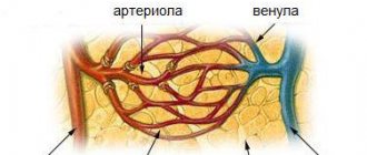
The vessel wall thickness parameter is used to assess the state of blood circulation.
The data obtained is decrypted immediately after the end of the study, which means that the patient does not have to wait long for the results, after 10-15 minutes. the document is handed out. To assess the state of blood flow, they are based on the following parameters:
- maximum systole velocity;
- minimal in diastole;
- wall resistance;
- ripple indexing;
- thickness of the wall under study.
Duplex scanning will help to diagnose disorders such as:
- varicose veins of superficial as well as deep vessels of the extremities;
- thrombosis;
- thrombophlebitis;
- thromboembolism;
- arterial atherosclerosis;
- obliterating endarteritis.
To make a final diagnosis, the doctor will refer the patient for a clinical and biochemical blood test. If the results of laboratory tests and duplex scanning show the same picture, and the diagnosis is confirmed, the doctor selects an individual treatment regimen or decides to undergo surgical treatment.
Ultrasound Dopplerography (USDG)
Doppler scanning of blood vessels is an accessible, simple way to study arterial and venous patency. Doppler ultrasound uses the Doppler effect, that is, the necessary information is obtained by recording changes in sound waves reflected by moving blood cells.
Ultrasound scanning is used when necessary:
- assess vascular patency;
- identify pathology of the venous valve;
- evaluate blood flow characteristics.
When a Doppler scan is performed, the vessels themselves remain invisible; only the patency and speed of blood flow are assessed based on the data of the recorded Doppler effect.
Dopplerography of the vessels of the head and neck, for example, can successfully diagnose the cause of headaches; in other cases, ultrasound scanning is useful in studying varicose veins and hypertension.
But if using this method it is quite easy to identify violations of vascular patency, then it is unlikely that it will be possible to establish the cause of this pathology. This is due to the fact that Doppler scanning cannot make visible the walls of blood vessels and their possible curvatures and other defects that directly affect the quality and speed of blood flow.
The very first ultrasound devices made it possible to obtain only thin flat projections of the studied areas of the body, but modern ones are already capable of producing a moving three-dimensional image, observed in real time.
You can read more about Doppler ultrasound here.
Video about Doppler scanning of blood vessels:
What are the contraindications?
A skin rash on the part of the body being examined is a temporary contraindication for diagnostics.
Duplex scanning has no direct restrictions. This method is safe; it does not in any way affect the course of the underlying pathology. There are temporary contraindications, which include:
- festering and bleeding wounds on the extremities to be examined;
- various skin pathologies in the form of rashes, pigmentation, ulcers;
- progression of a viral infectious disease, accompanied by fever and increased body temperature;
- exacerbation of bronchial asthma.
Rheoencephalography
Vascular structures are assessed by measuring the resistance of their walls when exposed to low-level electric current.
Currently, the method is used infrequently, due to the development of more accurate diagnostic methods.
Procedure technique:
- Electrodes (2–6) connected to a rheograph are fixed to the skin, the device passes an electric discharge through them and records changes in vascular structures;
- During the study, functional tests (medicinal, positional) are carried out.
Rheoencephalographic study
| Contraindications | Preparation | Time (min) |
| Early childhood | Do not smoke for 3 hours before the procedure | 30 |
MRI
Tomography is the most advanced imaging method. The device takes layer-by-layer images and then combines them. The result is a clear picture.
Attention:
Unlike CT, it does not create radiation exposure and is ideal for visualizing soft tissues, which include blood vessels and the brain itself.
If necessary, contrast-enhanced MRI is prescribed. A drug based on gadolinium is used. But for studying the vessels themselves, this is not necessary, since blood as such is a powerful natural contrast.
MRI is prescribed if other methods have failed. Or they are not informative enough.
There are special indications for this procedure:
- Suspicion of a tumor. Vascular disorders may be the result of compression by the formation. Gold standard for diagnosing neoplastic processes. Basically, the technique is prescribed for probable cancerous structures.
- A contrast study shows the size and shape of the tumor. Its exact location and degree of blood circulation. Conclusions can be given regarding the rate of cell division. Proliferative activity of the tumor. A clear picture is emerging.
Unfortunately, a biopsy must be performed to confirm the result. This is usually done directly during the operation. A tissue sample is collected and sent for histological evaluation. The laboratory gives the answer.
- Possible vascular formations. Two types: Malformations and aneurysms. Arterial wall protrusions. Both conditions are extremely dangerous and without treatment are fraught with complications. Up to massive bleeding and brain death. Accordingly, the death of the patient himself.
- Developmental defects. Congenital or acquired. In addition to malformations. For example, an open circle of Willis. And other anomalies. They are not always dangerous, but to establish this, one cannot do without research.
MRI is an indispensable method.
Source: cardiogid.com
Rheoencephalography
Similar research in nature. But there is a serious difference. If during an EEG, sensors take readings of the electrical activity of the brain itself, during rheoencephalography, doctors stimulate the work of the central nervous system. Using small current pulses.
It is safe and does not pose a health risk. The patient does not experience any discomfort. Based on the results of the examination, we can talk about something specific.
Rheoencephalography is completed within 10-15 minutes, like a standard EEG. It is impossible to say which method is more accurate and effective. Because they are used to register different values.
As in previous cases, there are additional indications for the procedure:
- Vascular diseases. Any. Be it aneurysms or malformations. Of course, doctors do not receive visual data, but based on the results of a functional study and the rhythms of the brain, a lot can be said. This is what doctors do.
- Evaluation of the effectiveness of the treatment. Usually conservative. Medication. In general, the same indication can be applied to other methods. But this one is the most effective in terms of results and accuracy.
- Atherosclerosis. As already mentioned, blockage of the arteries of cerebral structures with plaques. The greater the layering, the worse the situation is on rheoencephalography. The signal is poor. Therefore, the stimulation of cerebral structures is insufficient.
EEG and this technique are used in combination. This way doctors can describe the full picture of what is happening.