Head trauma and various brain diseases are the most dangerous. They can be identified by taking an X-ray of the brain. Although this formulation can hardly be considered correct.
It is important to understand that an X-ray of the head (in the classical sense) only includes an examination of the bones of the skull. X-ray examination of the brain is not carried out; there are other methods for this: computed tomography, magnetic resonance imaging.
Many will say that x-rays are harmful to the body, but thanks to it, more than one thousand people have been saved. The x-ray shows various injuries to the skull bones, pathologies, if any. The main thing is to diagnose deviations from the norm in time and prescribe the necessary treatment.
Photo title
The most common image of the head is called an x-ray. It is indicated in certain situations when abnormalities in brain function are suspected or there is damage to the skull. There are many indications for sending a person for an x-ray. In this case, the doctor has to choose which examination to take the picture with. This will determine what exactly can be learned through research.
Magnetic resonance imaging of the brain and neck is the most popular. This procedure allows you to find out a large amount of information and analyze the condition of the skull. Given the fact that photographs are taken from multiple angles, the specialist has the opportunity to examine all areas of interest.
They may be referred for Doppler ultrasound, which involves ultrasound diagnostics. It is designed to analyze the state of blood flow, as well as moving brain structures. At the same time, it is extremely rarely used for images of the brain itself, because with its help it is quite difficult to examine the internal structures of the brain.
There is also radiography, which is necessary to analyze the condition of the skull bones. Other structures cannot be seen with the help of research. Analysis is required so that the presence of a skull fracture, compactions, formations and shifts can be determined. As a rule, x-rays in the usual sense are allowed only once every six months, because the procedure is harmful.
It will be useful for each person to take a closer look at the situations in which x-rays and similar procedures are performed, as well as exactly what abnormalities can be seen in the images. Additionally, you should understand how you can prepare for the examination, as well as what negative consequences you may encounter.
Indications
There are many reasons why a head shot should be performed. In all situations, pathologies are suspected, or already identified diseases are monitored. If a person comes with a complaint about certain symptoms, the doctor cannot draw an accurate conclusion based on words alone. The specialist will definitely refer you for various examinations, the list of which depends on the specific situation. Thanks to the resulting images, it will be possible to more accurately judge the patient’s condition.
When is radiography prescribed:
- A person has constant pain in the head, which is difficult to relieve with conventional medications.
- Dizziness may be present, which is not dependent on external factors, such as hunger.
- There is bleeding from the nose that is not related to the injury.
- There may be pain when chewing.
- Hearing or vision has deteriorated. The violation can be either temporary or permanent.
Often a person is sent for an x-ray when he has had a head injury. It is also necessary to analyze the condition if fainting occurs or there are suspicions about oncological processes in the brain. Special X-rays will need to be used so that a number of specific pathologies can be seen.
The following symptoms are also additional reasons to take a head photograph:
- Speech impairment and coordination problems.
- Mood swings, in which a person suffers from increased irritability, can be aggressive and depressed.
- Numbness of the extremities is observed, mainly affecting one side.
- Nausea and vomiting not associated with poisoning or other usual causes.
- Mental disorders, hallucinations, delusions.
- Noise in ears.
As you can understand, a head examination is carried out in all cases when it is necessary to analyze the internal structures of the brain. When the symptoms described above appear, a person should not hesitate, because if there is a disease, it is important to identify it in time. If this is not done, then you will have to face various negative consequences. As you know, treatment is best carried out precisely when the disease is just beginning. Advanced forms are difficult to treat and also cause irreversible consequences.
What does a brain x-ray show?
X-ray of the head allows you to examine 3 groups of bone tissue of the skull: vault, base and facial skeleton. Spongy couplings and sutures are placed between the bony projections of the cranium. The only place where they are absent is the lower part of the skull. The base of the skull and jaw are attached to each other using joints.
An X-ray of the head allows you to assess the integrity and constitution of the jaw bone tissue.
Diagnostics shows disorders or congenital defects of the brain, if any:
- Destruction, decrease or increase in bone density, as well as when the sphenoid bone of the skull is deformed. These disorders appear due to physical pressure, which may indicate neoplasms in the pituitary gland.
- A large number of calcifications in the area of the inner part of the skull. The cause of their appearance may be toxoplasmosis, cysticercosis or chronic subdural hematoma.
- Deformations inside the bone tissue with pus inside. Osteomyelitis may be the culprit.
- The appearance of calcifications is characteristic of diseases such as oligodendrocytoma or arachnoidendothelioma. If the pineal gland is normal, it is located in the center and is clearly visible on the pictures. In case of displacement to the side, the cause is often a tumor on the opposite side.
- X-ray may show intrinsic hypertension. The latter occurs through compression of the brain and looks as if fingers were pressing on the plates of bone tissue.
Radiography
Head X-ray does not require special training from a person. A person just needs to show up for the procedure and then follow the doctor’s recommendations. Before entering the office, you will need to remove all jewelry and remove metal objects. You should also leave your mobile phone, tablet and similar devices. If the patient has dentures that cannot be removed, the doctor must be informed about this.
The person will be given an apron and then told how to position themselves to take the photo. After this, you will need to sit still and wait until the doctor from another office turns on the machine. The photo itself is taken instantly, and a series of photos may be required at once. Transcription takes more time, it takes about 15 minutes. The patient will be immediately given the image and sent to the doctor.
If we are talking about children, then they may also need to have an x-ray. Radiation negatively affects a fragile body, so the procedure is recommended as a last resort. For example, they may be referred to it if there was an accident, birth injury, or serious fall. In such a situation, equipment that emits less is used so as not to harm the minor.
On an x-ray you can see various pathologies, such as fractures, bone displacement, and various deviations from the norm. After the procedure, the doctor will be able to tell for sure whether everything is okay with the person. In some cases, additional research will be required if different information is needed.
What is a head x-ray called?
Head X-ray, even along with modern diagnostic methods, is relevant and has a number of advantages. This procedure is called radiography. It is the simplest and most cost-effective method of hardware research. In addition, it has no alternatives in the study of skull bones.
At the same time, radiography does not stand still, but is increasingly being improved. Thus, digital devices are now increasingly used, the advantage of which is lower radiation exposure, greater information content and high-quality digital images. The images are created and printed immediately, and after they are deciphered by the radiologist, the state of the brain becomes known.
Indications and benefits
Indications for an X-ray of the head are injuries, suspicion of cancer, fainting, asymmetric development of the facial bones, endocrine diseases or congenital pathologies. In addition, this radiation diagnostics is prescribed by a doctor for frequent headaches, dizziness, nosebleeds, trembling hands and a noticeable deterioration in vision or hearing.
Radiography allows not only to make the diagnosis as accurately as possible, but also to determine treatment and therapeutic measures, as well as monitor their effectiveness. In addition, following basic rules to prevent radiation overdose makes it a completely safe procedure. The advantage of x-rays also lies in the painlessness and absence of preparatory measures.
Taking pictures
Standard radiography is performed using two projections - frontal and lateral. However, these methods are not always acceptable. For example, for the temporal region of the skull it is optimal to use oblique projections, and for the mastoid process, facial and parietal regions - tangential ones.
Magnetic resonance imaging
As already mentioned, head images are obtained using various studies. One of them will be called MRI, it is used in cases where it is necessary to obtain detailed information about the internal structures of the skull. Indications for the study are various symptoms that indicate brain pathologies. The study itself is considered safe because it does not harm health.
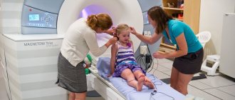
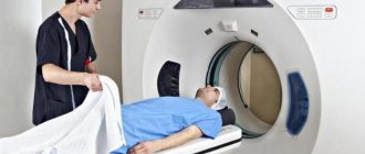
During the examination, the person will have to lie down on a special extendable table, after which the patient will be placed inside a special apparatus. After this, the head scan will begin, and images will be taken throughout the procedure. The brain can be viewed from different angles, so the pathology will not go unnoticed. The test is even used for children, but in this case anesthesia may be required because it is important to lie still during the procedure.
Based on the results of the MRI, it will be possible to understand whether there are any abnormalities, as well as what condition the brain is in. It is possible to identify hematomas, neoplasms, cysts, aneurysms and other disorders. Additionally, a contrast agent can be used, which allows a more accurate analysis of the state of the brain and blood vessels. It is extremely important to make sure that there are no contraindications so as not to cause harm to a person.
When MRI is not performed:
- During pregnancy in the first trimester.
- If a person weighs more than 120 kg, the patient simply will not fit into the device.
- If you have claustrophobia. The patient will have to be in a closed space, which will lead him to panic. They can only give you anesthesia if it is absolutely necessary to perform an MRI.
- If you are allergic to contrast media.
- If there are metal objects, prostheses and implants inside the body.
The procedure itself lasts from 30 minutes to 90 minutes, during which time the person must lie still. The doctor can communicate with the patient using a special microphone to give instructions. At the end of the study, it will be possible to obtain high-quality images that only a doctor can decipher.
First aid for bruise
The first thing to do is to examine the injured person.
If there are wounds or serious abrasions on the skin, they need to be treated and bandaged.
If there are bruises, apply something cold for 20 minutes.
Let the victim lie in bed for several hours. Do not give any medications.
If you have serious symptoms listed above, you should call a doctor. In this case, place the person in the most comfortable position for him and ensure peace.
It is important! Do not let the victim fall asleep until the doctor arrives. Continue to monitor his condition. In case of loss of consciousness, carefully shift the victim to his side, bend his knees closer to his chest, and place his hands under his head.
Doppler ultrasound
During the procedure, ultrasound waves are used to assess the condition of blood vessels and blood flow. It is possible to understand whether there are blood clots, whether the lumens are narrowed, and it will also be possible to assess the quality of blood flow. The examination is useful for situations where the heart rhythm is disturbed, there is hypertension, and atherosclerosis is suspected. The examination can be carried out even in the presence of vegetative-vascular dystonia, if it is necessary to monitor the person’s condition.
Before the procedure, you will need to give up alcohol, smoking and taking medications for blood vessels. During the diagnosis, the person will need to lie on his back, after which the doctor will apply a special gel to the desired areas. After this, the doctor uses an ultrasound sensor, with which you can assess the condition of the blood vessels. The procedure itself takes from 25 minutes to an hour. Everything will depend on how carefully the areas need to be analyzed.
During the procedure, you may need to perform certain actions, which the doctor will tell you about. For example, you will need to hold your breath in order to check the functioning of the blood flow during such a state. Based on the results of the examination, it will be possible to find out the speed of blood flow, the thickness of the vascular walls, the nature of blood circulation, as well as the resistive index. If there are deviations from the norm, then the doctor will draw specific conclusions.
All head scans are useful in their own way for diagnosing diseases. Only a specialist will be able to clearly say which examinations a person will need to attend. It is quite possible that you will have to undergo several procedures at once, based on the results of which a conclusion will be drawn.


