A relatively new diagnostic method, known as CT or computed x-ray tomography, is one of the most informative ways to detect changes in organs and tissues of the human body. This method has been widely used in medicine since the 80s of the last century, and makes it possible to identify even hidden pathologies at the initial stage.
Russian diagnostic doctors learned what CT is in the early 90s of the 20th century. At the same time, specialists were trained who were able to conduct examinations of organs using this method and interpret the results. However, until now, most patients do not understand the difference between MRI and computed tomography, although these types of diagnostics are based on the use of fundamentally different types of radiation and have different indications and contraindications.
The essence of the method, the differences between MRI and CT
In addition to computed tomography, another high-precision method is used today - MRI, or magnetic resonance imaging. In many ways, both methods are similar to each other. In both cases, the patient lies down on a special table, around which there is a contour of the device emitting rays. Under the influence of these rays, a three-dimensional image of internal organs and deep tissues appears, which is recorded on the monitor. The doctor can rotate the picture, studying it in detail from different angles. In this case, it is possible to examine sections of organs and tissues even 1 mm thick.
However, there are differences between CT and MRI. The main one is the use of waves of different origins. The tomograph used for CT scans uses X-rays. An MRI machine emits electromagnetic waves.
The different nature of the rays means that the indications and contraindications for CT and MRI are also not the same. For example, CT scans should not be performed on pregnant women. Magnetic resonance imaging is performed on expectant mothers; restrictions exist only for the first trimester of pregnancy.
Generally speaking, CT is better at detecting diseases associated with hard tissues of the body (bones, teeth) and bleeding. MRI most effectively examines soft tissues (vessels, cartilage, neoplasms).
Risks of examination
Despite the relative safety of MSCT, even here there are risks associated with its implementation, which later manifest themselves in the form of side effects, causing failure in various systems of the human body. Even if we consider that the risk of side effects after the procedure is minimal, you need to know about them in advance:
- The most difficult thing for young people and children to endure is even the small amount of radiation exposure given by modern devices. It is they who experience relapses of oncological processes, which has been confirmed by repeated studies that have shown the presence of a small percentage of such cases.
- Implanted electronic devices are a particular contraindication for the safer MRI procedure. But even with MSCT, although in rare cases, insulin pumps and neurostimulators can malfunction, causing a deterioration in the patient’s health.
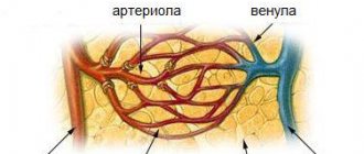
Due to the fact that the capabilities of MSCT are so wide, many hospitals and health centers prefer not to use conventional CT and ultrasound, preferring multilayer angiography. It makes it easy to examine the entire large circulatory system, consisting of the carotid artery, pulmonary vessels, liver, as well as the iliac and caval veins of the lower and upper types. The results are incredibly accurate and informative, helping not only to make an accurate diagnosis, but also to select effective treatment.
Types of computed tomography
There are several types of CT scans. To understand the differences, you need to understand what the device is on which the diagnostic procedure itself is performed (computed tomograph). It looks like a ring under which a table with the patient lying on it slides. Each type of CT scan works differently.
In spiral tomography, the tube rotates continuously and the table moves at the same time. This allows you to increase the speed of organ scanning and reduce the radiation dose.
Multislice tomography is similar to spiral tomography, but the radiation comes from two sources. This achieves an increase in the resolution of the resulting image, while reducing the procedure time. This type of CT is used to obtain images of small vessels and a detailed scan of a small area of tissue.
Multilayer tomography is distinguished by the fact that the speed of wave emission and rotation of devices is increased, and the sensors are arranged in several rows. This type of study provides a representation of the work of an organ in dynamics.
CT with contrast involves the introduction of a contrast agent, which enhances the contrast of the image of organs against the background of adjacent tissues. Thanks to this technique, the images in the photographs are clearer and easier to decipher for making an accurate diagnosis.
How is the procedure done?
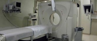
Vascular tomography is carried out in a medical institution (public or private) that has an equipped office and a properly functioning computed tomograph. The device consists of a large scanner ring, a movable couch and a computer to which the data is transferred.
- The patient is positioned on the couch, lying on his back, arms and legs extended along the body, head lying calmly, not thrown back.
- If the examination is carried out with contrast (and CT angiography always requires the administration of a contrast agent, more on this below), then before the procedure the nurse administers a special drug intravenously. It works immediately.
- The patient is then moved inside the ring and scanning begins.
- During a routine examination, a tomography of the blood vessels of the problem area of the body (or several areas) is prescribed. Separate angiography of the neck, brain, legs, and heart is distinguished. When examining a patient on a tomograph after an accident, falls, beatings and other serious large-scale injuries, the entire body is scanned.
- During a vascular CT scan, the scanner ring will rotate, making a characteristic sound. The patient should lie still for good image quality. If necessary, radio communication with medical personnel is possible.
- A vascular CT scan takes on average 30-40 minutes. With more modern devices, the procedure takes a little longer, since a more accurate result is obtained.
- A tomograph uses X-rays to scan blood vessels, transmitting data to a computer. At the end of the study, the program will create a 3D picture of the vessels of the examined organs. The results are prepared within 30-60 minutes and are provided on paper and electronic media.
When is contrast needed?
When performing vascular tomography, it is impossible to do without the use of a contrast agent. The contrast will “highlight” the vascular structure of the organ, highlighting the smallest branches. This will provide more accurate and complete images. Iodine is used as a contrast agent. A drug based on it is administered to the patient before the procedure begins. And this is one of the reasons why vascular CT may be contraindicated.
What diseases can a CT scan help detect?
CT is used in almost all medical fields, as it can detect many pathologies in different parts of the body and internal organs:
- enlarged lymph nodes,
- the presence of inflammatory processes in tissues,
- various types of neoplasms (cysts, oncological tumors, etc.),
- types of liver dystrophy,
- tuberculosis, sarcoidosis,
- in some cases - pneumonia,
- heart and vascular diseases,
- disorders in the thoracic, cervical, lumbar spine,
- kidney stones, gall bladder,
- threat of stroke, hardening of brain tissue, aneurysm,
- hernias of various localizations,
- congenital anomalies of various internal organs,
- intestinal polyps,
- fractures, bone diseases,
- sources and causes of internal bleeding.
Indications for CT scanning
An examination is prescribed to clarify diseases in many cases. This may be an initial examination or a test to definitively confirm the diagnosis. The reason for performing a CT scan is usually:
- Suspicion of tumors in various organs.
- Chronic pain, fainting.
- Emergency diagnostics for injuries, strokes, bleeding, convulsions.
- To clarify the diagnosis with a biopsy.
- To record the exact location of organs and vessels (for example, before operations).
Who is it shown to?
It’s clear how MSCT is done, but who should undergo it? The procedure is prescribed by the attending physician, who can be either a therapist or a neurologist. It is indicated for:
- identifying the cause of pain in the middle ear area;
- if there are signs of subatrophy;
- to find an aneurysm or vascular malformation;
- to understand why the patient’s consciousness is confused or their limbs are numb, they are struck by paralysis, they suffer from migraines, visual system failures or vertigo, which may indicate a tumor, hemorrhage in the brain or be a consequence of injury or rupture of an aneurysm;
- determining the size of the brain cavities, which indicate the presence or absence of hydrocephalus or intracranial hypertension;
- in the preoperative period;
- monitoring the effectiveness of therapy after identifying a problem;
- visual determination of damage to bone and soft tissues of the organ;
- determining the causes of fainting;
- brain biopsies to monitor the process at each stage;
- for diagnosing skull fractures or soft tissue injuries after injuries and accidents;
- to find tumors;
- in case of stroke to detect bleeding or blood clot;
- to check the integrity of the temporal bone, such an injury can cause hearing loss, both partial and complete;
- diagnostic measures to identify meningitis and its various complications.
In what cases is CT scan contraindicated, is there a danger to the body?
Despite all its advantages, computed tomography has contraindications. This is due to the use of x-rays. CT scans are not performed:
- women during pregnancy, regardless of stage,
- patients with renal failure,
- with diabetes mellitus in advanced stages,
- CT with contrast is prohibited for people with an allergy to iodine, as well as for patients with hyperthyroidism,
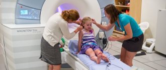
- for young children and people with behavioral problems who cannot remain in a still position, when the child is being examined, the presence of parents in protective aprons is allowed nearby,
- There is a restriction for nursing mothers: after the CT procedure, you will have to take a break for a day in feeding the baby with breast milk.
CT angiography of the vessels of the brain and neck: indications, contraindications
Any radiation diagnostic method is carried out after a doctor’s prescription. Determining the benefit and harm to the patient is the basis for choosing a study.
Classic CT angiography is prescribed for:
- Confusion;
- Severe skull injuries;
- Progressive loss of hearing and vision;
- Chronic incurable headaches;
- Ear noise;
- Increased intracranial pressure;
- Dynamic control of the treatment of brain pathology.
An alternative to classical tomography is a multislice version (MSCT of the brain). Modern equipment allows scanning to be performed faster, which reduces the time a person remains stationary. The capabilities are achieved by installing several source-receiver blocks.
Classic step-by-step CT angiography of the vessels of the brain and neck is equipped with one X-ray tube and a signal receiver. When scanning, the block moves from one position to another at a specified interval.
The search for oncology requires the production of tomograms at a minimum interval - usually 1 mm. Modern multi-slice MSCT makes slices every 0.5 mm, which makes it possible to determine the tumor “in situ”, when it is still possible to perform a radical operation and save a life.
Spiral CT angiography is faster, the resolution is higher, and the specificity and reliability of the analysis is not lost. The method is x-ray. Theoretical harm to human health cannot be ruled out.
Contraindications to contrast CT scan of the brain:
- Bearing a fetus;
- Lactation. Refusal of lactation will be required;
- Weight over 150 kilograms is the weight limit of the equipment;
- Hypersensitivity to iodine and other contrast components.
Individual restrictions are determined by the attending physician. To analyze the factors, you need to bring the results of previous diagnostics, an outpatient card, and other health information.
MSCT vascular angiography does not differ in technical features from the classical method. The contrast agent is injected into the brachial artery. In a few seconds the substance reaches the cerebral arteries. It is excreted by the kidneys after tens of minutes, so renal failure is a contraindication to angiographic examination.
Preparing for CT and performing the procedure
Examination of the abdominal organs is performed on an empty stomach. It is recommended to refrain from eating the evening before the procedure. In other cases, no special preparation is required for CT scanning.
It is necessary to warn the doctor if the patient is taking medications for diabetes or has a pacemaker. This is especially important with contrast tomography. Some medications must be temporarily avoided. If you have cardiac devices, the doctor will adjust the radiation dose.
In addition, you need to tell doctors if there may be traces of bismuth or barium in your body. These substances are usually used to examine and treat the gastrointestinal tract.
The patient needs to be prepared to lie still for 15-30 minutes during the CT scan.
How is a CT scan done?
The patient lies down on a table, which slides into a semicircular tunnel bordered by a ring-shaped tomograph. A monitor connected to the tomograph captures a three-dimensional image of the internal organs. The table moves through the ring to improve visibility of the area being examined. Some parts of the body are secured with straps to ensure complete immobility. You can talk to the doctor through a special device.
Before the procedure begins, the patient will be asked to remove all metal jewelry, remain still, and follow medical commands. Movement during a CT scan will disrupt the clarity of the image on the tomogram. This will prevent the doctor from studying the results in detail and correctly interpreting the data obtained.
What can be seen in the resulting photographs?
The doctor will report some examination results immediately after the procedure. However, the full transcript will be ready in 1-3 days.
In the images you can see the silhouettes and size of the internal organs. If the size of the organs is larger or smaller than normal, this indicates a disease. The same applies to changing their outlines.
Also visible will be a change in tissue density, the presence of cavities, fluid filling, the presence of foreign bodies, areas of vascular obstruction, enlarged lymph nodes, tissue proliferation, and the heterogeneous nature of the structure in the tissues.
Free consultation with a urologist
What is computed tomography (CT)?
Computed tomography (CT) is a non-invasive diagnostic method that helps doctors make the correct diagnosis and prescribe the necessary treatment.
Computed tomography (CT) is a combination of special X-ray equipment and a computer station to produce images of internal organs. Images of cross-sections of the studied area of the body, tomograms, are displayed on a computer monitor and can be printed.
CT scans of internal organs, bones, soft tissues and blood vessels provide greater clarity and detail than conventional x-rays.
Using computed tomography (CT), various tumors (kidney cancer, prostate cancer, bladder cancer), cardiovascular diseases, infectious diseases, trauma and diseases of the musculoskeletal system can be diagnosed.
Indications for CT examination
Computed tomography (CT) is
- one of the best and fastest research methods for studying the chest, abdomen and pelvis, as it provides detailed, cross-sectional images of all types of tissue.
- one of the best methods for diagnosing various tumors, including lung cancer, liver and pancreatic cancer, allows you to confirm the presence of a tumor, measure it, determine the exact location and extent of damage to surrounding tissues.
- a study that plays an essential role in the detection, diagnosis and treatment of vascular diseases that can lead to acute renal failure or death. CT scans are commonly used to diagnose pulmonary embolism (blood clot in the blood vessels of the lungs) and also to diagnose abdominal aortic aneurysms.
- An invaluable method for diagnosing and treating spinal problems, injuries to the arms, legs and other skeletal structures, since even very small bones can be clearly seen with a CT scan, as well as surrounding tissues such as muscles and blood vessels.
Doctors use computed tomography (CT) scans to:
- rapid detection of injuries to the lungs, heart and blood vessels, liver, spleen, kidneys, intestines or other internal organs in case of injury;
- carrying out kidney biopsy, prostate biopsy and other therapeutic and diagnostic procedures - brachytherapy, HIFU, drainage of kidney abscess, prostate and minimally invasive methods of treating tumors under CT control;
- monitoring the results of surgical treatment, such as organ transplantation or restoration of urinary tract patency;
- determining the stage, plan and need for radiation therapy to treat tumors, as well as to monitor the tumor response to chemotherapy;
- measuring bone mineral density to detect osteoporosis.
How to prepare for a computed tomography (CT) scan?
You must come to the examination in comfortable, loose clothing. You may be asked to change into hospital gowns during the examination.
Metal objects, including jewelry, glasses, dentures, and hairpins, can interfere with CT scans and should be removed before the test or left at home. You may be asked to remove your hearing aids or dentures.
You will need to avoid eating and drinking for several hours before your CT scan, especially if contrast material is planned. You should tell your doctor about any medications you are taking or if you have any allergies. If you are allergic to contrast material, or “dye,” your doctor will prescribe medications that will reduce the risk of an allergic reaction.
Also tell your doctor about any diseases or other medical conditions you have, such as heart disease, asthma, diabetes, kidney disease, or thyroid disease. Any of these conditions may increase the risk of adverse effects.
Women should always tell their doctor before a CT scan that they may be pregnant.
What does a computed tomography (CT) machine look like?
A computed tomography (CT) machine is a large, square machine with a short tunnel in the center. You will be placed on a movable examination table that moves through the tunnel. CT images are produced using a narrow, rotating beam of X-rays and a system of sensors arranged in a circle called a gantry. The computer station that processes the images is located in a separate room, where the technologist controls the scanner and monitors the progress of the study.
How does the computed tomography (CT) procedure work?
A CT scan uses X-rays to produce images. X-rays are a form of radiation, like light or radio waves. Different parts of the body absorb X-rays differently.
In conventional radiography, a small pulse of x-ray radiation passes through the body, creating an image on film or a special recording plate. On an x-ray, the bones appear white; soft tissues are in shades of gray, and air is depicted in black.
In a CT scan, a narrow beam of X-rays rotates around you and a series of electronic sensors that measure the amount of radiation absorbed by your body. At the same time, the table moves through the scanner so that the X-ray beam moves in a spiral. A special computer program processes large amounts of data to create two-dimensional cross-sectional images of your body, which are then displayed on a monitor. This imaging technique is called helical CT. Spiral CT can produce detailed two- and three-dimensional images of internal organs.
The advent of improved sensors allows new CT machines to obtain a large number of “slices” during a single rotation. These machines, called multidetector helical CT scanners, produce thinner “slices” in a shorter period of time, resulting in a more accurate diagnosis.
Modern CT scanners are so fast that studying one anatomical area of the body takes several minutes. This speed of examination is an advantage for all patients, but especially for children, elderly patients and critically ill patients.
Some CT scans use contrast material to produce more detailed images of the areas of the body being studied.
How is a CT scan performed?
The radiologist will place you on a moving table, lying on your back, side, or stomach. Straps and cushions may be used to help you maintain and maintain correct position during a CT scan.
If contrast material is used, it will be given intravenously or by mouth or by enema into the rectum. The method of administering contrast material depends on the type of CT scan you need.
The table will move quickly through the scanner to determine the correct starting position for the examination. Then, as the table begins to move slowly through the scanner, a CT scan is performed.
You may be asked to hold your breath during the CT scan. Any movement - breathing or body movements - can lead to defects on a CT scan. These defects are similar to the blurry photograph you get when shooting a moving subject.
When the CT scan is completed, you will be asked to wait while the radiologist checks the quality of the images.
A full body CT scan is usually completed within 30 minutes.
What will I experience during and after the procedure?
CT is a painless, quick and simple diagnostic method. Spiral CT reduces the amount of time the patient must lie still.
Although the CT scan is not painful, you may experience some discomfort from having to remain still for several minutes. If it is difficult for you to lie still, or you suffer from claustrophobia or chronic pain, then a CT scan will be a difficult test for you. The technologist or nurse, under the direction of your doctor, may give you a mild sedative to help you tolerate the CT scan.
If intravenous contrast material is used, you will feel a small prick where the needle is inserted into the vein. You may experience a feeling of warmth, redness where the contrast was injected, or a metallic taste in your mouth that goes away after a few minutes. Some patients feel the urge to urinate, which quickly passes.
Sometimes, the patient is bothered by itching and urticaria, which decrease with treatment. If you feel dizzy or find it difficult to breathe, you should tell your doctor or nurse right away. A radiologist or other doctor will provide you with the emergency care you need.
If contrast material must be swallowed, you may experience an unpleasant taste; however, most patients tolerate it easily. You may feel the urge to have a bowel movement if contrast material is given through an enema. In this case, be patient as the mild discomfort will not last long.
While you are in the CT scanner, a special light may be used to make sure you are positioned correctly. While the CT machine is operating, you will hear a slight buzzing or other sounds.
You will be alone in the room during the CT scan. However, the technologist or radiologist will see, hear, and talk to you throughout the test.
During CT scans of children, parents may be allowed, wearing a special lead apron, to be present in the room where the examination is being performed.
After the CT scan, you can return to your normal lifestyle. If you have been given contrast material, you will be given special recommendations.
Who interprets computed tomography (CT) results? How will I receive them?
A radiologist who is trained in performing and interpreting radiological tests will analyze the images and send the results to your doctor. Your doctor will tell you the results.
Benefits and risks of computed tomography (CT)
Benefits of CT Scan
- CT examination is painless, non-invasive and accurate.
- The main advantage of CT is its ability to simultaneously image bone, soft tissue and blood vessels.
- Unlike conventional X-rays, CT scans provide very clear images of many types of tissue, such as the lungs, bones, and blood vessels.
- CT examinations are fast and simple; in emergencies, they help quickly identify internal injuries and bleeding to help save lives.
- CT is a relatively inexpensive tool for diagnosing a wide range of clinical problems.
- CT is less sensitive to patient movement than MRI.
- A CT scan can be performed if you have any type of implanted medical device in your body, unlike an MRI.
- CT scanning provides real-time images, making CT a good tool for minimally invasive procedures such as fine-needle biopsies of many areas of the body, especially the lungs, abdomen, pelvis and bones.
- The diagnosis determined by CT examination may eliminate the need for exploratory surgery and surgical biopsy.
- There is no radiation left in the patient's body after a CT scan.
- The X-rays used in CT scans usually have no side effects.
Risks of CT Scan
- There is always a small risk of developing cancer from overexposure. However, the possibility of an accurate diagnosis outweighs this minimal risk.
- The effective radiation dose from CT is 2 to 10 mSv, which is the same as what the average person would receive from background radiation after 3 to 5 years. Women should always tell their doctor or radiologist if there is any possibility that they are pregnant. CT scans are generally not recommended for pregnant women due to the potential risk to the baby.
- Breastfeeding mothers should take a break from breastfeeding for 24 hours after a contrast injection.
- The risk of serious allergic reactions to contrast materials containing iodine is extremely rare. But radiology departments are well equipped to deal with them.
- Since children are more sensitive to radiation, CT scans in children should only be ordered when absolutely necessary.
What are the limitations of whole body computed tomography (CT) scans?
MRI provides clearer images of soft tissue details in areas such as the brain, pelvic organs, knee, or shoulder than CT scans. The study is not conducted in pregnant women at all.
A person with a large body mass may not fit into the opening of a regular CT scanner or may exceed the weight allowed by the moving table.
| Three-dimensional reconstructed CT image of the kidneys and ureters, which connect the kidneys to the bladder. This image shows the lower ribs, spine, and pelvis. | |
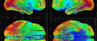
| Part of a CT scan, a transverse "slice" through the middle of the abdominal cavity, showing the abdominal organs. |
The article is for informational purposes only. For any health problems, do not self-diagnose and consult a doctor!
Author:
V.A. Shaderkina is a urologist, oncologist, scientific editor of Uroweb.ru. Chairman of the Association of Medical Journalists.
‹ Examination of the prostate gland Up Computed tomography of the kidneys ›
Pathologies of which areas of the spine are determined by CT
The structure of the spine is very complex. It consists of bone, muscle, nervous, cartilage, vascular tissue, and they are all collected in a relatively small area of the body. Scanning the spinal column for diagnostic purposes is a painstaking process. But the problem can be completely solved with the help of computed tomography.
Typically, the vertebral region is examined using spiral and multi-slice (multi-slice) CT.
The obtained visual information helps to identify a number of pathologies:
- spondylosis, most often of the cervical spine (proliferation of ligaments in the form of spines),
- osteochondrosis in any part of the spine (changes in cartilage in intervertebral discs, bone tissue),
- osteoarthritis (loss of elasticity of cartilage tissue with pain and impaired mobility),
- lumbar stenosis (overgrowth of the spinal canal with bone tissue, leading to paralysis).
Why is a CT scan with contrast performed?
A contrast-enhanced CT scan is performed to improve visualization of organs. It comes in two types:
- Angiography using a CT machine to examine the body’s circulatory system or its individual sections.
- Perfusion to examine the blood supply to the brain.
Before the procedure, the patient is given a contrast agent intravenously. When examining the stomach, the drug can be administered orally. In X-rays, the areas of interest appear in contrast to other tissues.
When the procedure is impossible
Doctors have not identified any absolute reasons why a head examination cannot be performed using MSCT. But each attending physician, at his own discretion, can determine whether the procedure will be safe. Most often, it is not performed on pregnant women and young children, replacing it with MRI. The doctor must accurately determine the line between the necessary harm and the patient’s health. And this is provided that the load of radiation on the body is minimal. The list of relative contraindications includes patients:
- pregnant women;
- asthmatics;
- with kidney disease and heart failure;
- with any form of diabetes;
- having multiple lesions of the body with myeloma spots;
- with a negative reaction to the contrast agent gadolinium.