EEG video monitoring is a research method in neurology that provides good recording of the oscillatory electrical process occurring in the human brain, followed by decoding. Activity is recorded by special equipment with electrodes attached to the patient's skull. Observation continues for a long time - for several hours or days. During the study, it is important to assess brain activity in two phases - wakefulness and sleep.
The main task of daily EEG video monitoring is to diagnose epileptic and non-epileptic disorders. Long-term observation makes it possible to evaluate brain activity in several states using an electroencephalogram (wakefulness, including during exercise/tests and at rest, sleep). The method differs from routine EEG in complexity, labor-intensiveness and higher information content.
What is video monitoring of brain BEA?
An electroencephalogram is a method of examining the central nervous system using a device that records indicators of its electrical activity.
Sensors are connected to it, which are placed on the patient’s head in the form of a mesh cap. They transmit signals (waves) from the brain. Data is recorded on paper or electronically. Why is an encephalogram done:
- Examination for obtaining a license to drive vehicles, passing a commission at work, at the military registration and enlistment office.
- Chronic headaches.
- Migraine.
- Episyndrome.
- Vegetative-vascular dystonia, hypo- and hypertension.
- Diencephalic (hypothalamic) syndrome.
- Previous strokes.
- Central nervous system tumors, injuries, cysts, operations.
- Mental disorders, attention deficit disorder, tics, stuttering, neuroses.
- Cerebral palsy.
- Meningitis, encephalitis.
The central nervous system contains pacemaker cells that set the rhythm of electrical activity when generating wave impulses of a particular frequency and length. As conditions change, their intensity may weaken or intensify.
Some diseases of the central nervous system are characterized by changes in the intensity of certain waves. With epilepsy, ectopic foci appear that generate their waves, disrupting the normal activity of the nervous system and leading to convulsive seizures.
EEG is a way to assess the state of the brain, which is based on recording the electrical activity of the organ. Diagnosis is carried out using a special device, to which electrodes are connected, which are then attached to the head of the subject. They read all, even the least pronounced, impulses, which are subsequently processed and analyzed by a computer program. This way you can determine whether there are abnormalities in brain activity.
By studying the results of an electroencephalogram of the brain, a specialist is able to identify all changes - both reversible and structural, as well as problems in the functioning of the central nervous system. This can be achieved due to the fact that the so-called EEG rhythms reflect the synchronous activity of all organ structures.
EEG video monitoring is a long-term recording of brain activity. The exact time for this technique has not been established, so it can last for hours. How much time it will take to study a particular pathology is decided by the attending physician and a specialist in the diagnostic room, guided by the characteristics of each specific case. Sometimes even daily observation is prescribed, which can show all possible deviations of neural activity.
EEG video monitoring is carried out if necessary:
- track the patient’s troubling conditions and find out their nature;
- do a long-term EEG if the standard recording shows no changes;
- obtain a full-scale electroencephalogram - while awake and asleep.
Long-term monitoring significantly increases the likelihood of detecting pathological signs compared to standard EEG. Not only the time factor plays a role here, but also the opportunity for the patient to be in familiar conditions, as well as perform his everyday actions. Communication with relatives, reading, watching TV, sleep and other reasons can lead to attacks and are recorded on camera, allowing the doctor to study them thoroughly.
In simpler and more understandable cases, a short EEG recording performed while awake is sufficient. For example, if the child’s close relatives are worried about his specific states during wakefulness (tics) and it was these that were able to be recorded during video monitoring, then the study does not have to be carried out during sleep. On the other hand, during sleep you can discover the baby’s conditions, which his family has no idea about yet.
Therefore, a qualified neurologist must highlight these subtleties in advance to patients, relatives, and diagnostic room staff. To the question of which video monitoring is “better,” the clear answer is nighttime. Since it, as usual, is longer and contains a recording of sleep. Not everyone can fall asleep during the day - not only adults, but also children, and the duration of daytime diagnostics, depending on the laboratory, is 2 - 4 - 6 hours.
You can read more about conducting an EEG for a child in this article. Of course, when choosing the type of EEG monitoring, the doctor will rely on symptoms. If certain events bother you during the day, the procedure will be scheduled for the day, and if at night, then, accordingly, at night. Do not forget that a long-term examination in an isolated room is quite tiring, especially for young patients.
In addition, night video monitoring is a more expensive procedure, and in government institutions where it is done free of charge, there are huge queues. Therefore, if the main issues can be resolved upon receiving the recording and photos taken during the day, then you should not subject patients to unnecessary expenses and painful manipulations.
In certain forms of epilepsy, pathologies on the EEG are determined only during night-time studies. Monitoring during sleep makes it possible to detect epileptic activity even in those patients in whom functional tests could not detect it during the daytime.
This terminology is used to describe one of the methods for diagnosing the state of the brain, which is based on the process of recording its electrical activity.
Based on what the electroencephalogram of the brain shows, doctors can identify various pathologies in the blood vessels, the development of inflammatory diseases, as well as signs of tumors and epilepsy. It is worth noting the important fact that EEG is the only outpatient research method that makes it possible to diagnose an unconscious person. Moreover, this technique does not pose a danger to patients of any age category, including children.
Using what electroencephalography of the brain shows, doctors can record the effects of various drugs on patients, assess the dynamics of the course of the disease, and also make adjustments to treatment methods. Continuing to consider the features of EEG, it should be noted that this type of study is used to track all changes in the brain - from reversible to structural. This is one of the main differences between this technique and other methods of examining a patient.
Who should be examined using electroencephalography?
Electroencephalography (EEG) can help clarify many diagnoses related to infections, injuries, and brain disorders.
The doctor may refer you for examination if:
- There is a possibility of epilepsy. Brain waves in this case show a special epileptiform activity, which is expressed in a modified form of graphs.
- It is necessary to establish the exact location of the injured area of the brain or tumor.
- There are some genetic diseases.
- There are serious disturbances in sleep and wakefulness.
- The functioning of cerebral vessels is disrupted.
- An assessment of the effectiveness of the treatment is needed.
The electroencephalography method is applicable to both adults and children; it is non-traumatic and painless. A clear picture of the work of brain neurons in different parts of the brain makes it possible to clarify the nature and causes of neurological disorders.
Comparison of EEG with other types of diagnostics
Despite the fact that any orders of tests and studies should be carried out only by a professional doctor, many people have a question: is it possible to replace an encephalogram with an MRI of the brain, ultrasound of cerebral vessels, or another study.
Of course, there is a difference between these research methods, so the question of which is better: an encephalogram or an MRI is incorrect. Often MRI and EEG are prescribed together as part of a comprehensive examination of the patient. The result of magnetic resonance imaging is a detailed three-dimensional image of tissues (including soft ones) with highlighted areas of compactions, tumors and cavities.
Several EEG research methods are used:
- routine;
- with deprivation;
- long;
- night.
Depending on the duration and purpose, computer encephalography is divided into types:
- Electroencephalogram of the brain - used in the initial stages of the examination. Both background activity and stress tests (hyperventilation, sharp sounds, flashes of light) are recorded.
- EEG monitoring is a long-term recording of brain activity. It is used when it is necessary to cover all possible physiological states of the central nervous system (sleep, wakefulness, mental work, emotions).
- Rheoencephalography is a study of cerebral vessels. Diagnostics is based on recording the changing value of electrical resistance of tissues when a weak high-frequency current is passed through them. Provides information about the tone and elasticity of the vascular wall, the amount of pulse blood supply.
Routine method
The routine method consists of short-term (approximately 15 minutes) recording of brain biopotentials. This is necessary to examine and evaluate the dominant rhythms, the presence of pathological potentials and paroxysmal activity.
Functional tests are also carried out, during which the reaction to:
- opening - closing eyes;
- clenching a fist;
- hyperventilation - forced breathing;
- photostimulation - blinking LEDs with eyes closed;
- sharp sounds.
The video shows an EEG with functional tests. Filmed by the channel “Clinic Doctor SAN”.
Deprivation encephalography is performed with complete or partial sleep deprivation. Determines epileptic activity in situations that did not arise during provocative tests.
The patient either does not sleep all night or wakes up 2–3 hours earlier than usual. A routine EEG will be performed no earlier than 24 hours after the initial awakening.
Long-term recording of parameters during sleep is often carried out after deprivation EEG, since sleep is a powerful activator for the detection of epiactivity.
Only by performing a sleep EEG can a differential diagnosis of epilepsy with cognitive impairment be made. Therefore, this type of examination is prescribed if the doctor suspects that changes in the brain occur while the patient is sleeping.
Night EEG
Night EEG recording occurs in a hospital setting as follows:
- begins a few hours before bedtime;
- covers the period of falling asleep and the entire night's sleep;
- ends after natural awakening.
If necessary, additionally:
- video monitoring;
- electrooculography (EOG);
- recording of a cardiogram (ECG);
- electromyograms (EMG);
- spirography.
The bioelectric potential of brain activity is studied using several electrocyfogram methods:
- A routine technique with stress recording tests lasts no more than 15-20 minutes, reveals the cause of paroxysmal conditions and hidden brain disorders using hyperventilation, photostimulation or electrophonogram described above.
- Encephalography with deprivation - partial or complete deprivation of night sleep or awakening earlier than the appointed time of the biological alarm clock.
- Polysomnography is a recording of brain activity in a sleepy state (dream).
How long (procedure duration) the EEG lasts depends on the chosen method.
Advantages and disadvantages of the method
Like any diagnostic procedure, the described method has its pros and cons. They determine the indications for the procedure and also limit its implementation in some patients. The main advantages are as follows:
- high sensitivity to changes in the bioelectrical activity of the brain;
- does not require special and complex preparation of the patient;
- can be performed in an unconscious state of the patient, including in a coma and in severe illnesses;
- available in all medical institutions, which makes it possible to conduct research on a large number of people;
- makes it possible to identify the disease at the initial stage before the development of pronounced organic changes in the brain.
- The patient should remain motionless during the procedure and not be nervous. This is especially important when examining children;
- with psycho-emotional stress, tremor and minor movements, changes occur in the electroencephalogram that can be regarded as pathology;
- The method is difficult to use in children while they are awake.
These disadvantages do not have a major impact on the use of EEG in clinical practice.
History of EEG development
Encephalogram of the brain as a high-precision study has been performed by doctors for almost 90 years - the first diagnosis was carried out in 1928. This was a real breakthrough in the field of studying brain diseases, which made it possible to discover new types of diseases and methods of treating them.
Since then, medicine has made great progress, and the encephalograph (examination device) has been improved many times. With the rapid development of information technology, it began to be used in conjunction with computers and other computing equipment. This significantly improved the quality of the research being conducted.
Today, the encephalograph, like many years ago, consists of sensors, which are electrodes. They are attached to a special mesh made in the shape of a cap. Electrodes detect electromagnetic bursts and transmit readings to a device for recording and recording. Subsequently, these bursts are processed and analyzed.
Today, an encephalogram of the brain has become so popular and accessible that it can be performed in almost any clinic or neurological hospital.
How to decipher an EEG
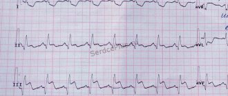
The graph produced by the EEG machine shows several types of waves. Each of them has a specific interpretation and reflects the type of activity that can complement the clinical picture and confirm the presence of the disease. Only a specialist can decipher EEG data and issue the patient a conclusion explaining the indicators after the procedure.
Types of human brain activity recorded during diagnosis
There are four types of waves that record human brain activity.
- Alpha wave. Symbolizes the activity of the brain during wakefulness, but without active brain work and stress. Determines the presence of anxiety, fear, depression.
- Beta wave. Active brain activity, wakefulness, has weakly expressed indicators during the rest period.
- Gamma wave. Occur when performing activities that require concentration.
- Kappa wave. Another rhythm characterizing cognitive processes.
- Lambda wave. Oscillations occur when it is necessary to make visual decisions.
- Mu wave. Starts during the dormant period.
- Theta wave. Observed during sleep. Strengthening is observed after prolonged wakefulness and exercise, as well as with neurological diseases.
- Delta wave. Determines the phase of natural deep sleep; when activated during wakefulness, it indicates certain disorders.
- Sigma wave. It starts in the initial phase of sleep.
The process of studying the results and deciphering the values at different age intervals
When analyzing EEG indicators using computer processing, age indicators, the presence of serious diseases, poor vision and uncontrolled tremors in the limbs must be taken into account. In addition, the time of the last attack and heart rate are taken into account. To get a complete, detailed picture, it is necessary to apply additional examinations.
There are age-related differences in the normal electroencephalogram. Thus, in children under 15 years of age, the alpha rhythm predominates; by adulthood, a balance of rhythms characteristic of an adult is achieved. From the age of 50, a restructuring of rhythm begins - alpha oscillations decrease, beta and delta rhythms increase. After 60 years, the frequency fades, delta and theta oscillations are noted on the EEG.
Possible decryption problems
In the event that a patient undergoes electroencephalography after a stroke, or he has an epileptic predisposition, it is much easier to see the norm and abnormalities than when a patient presents with vague headaches. In the case of a stroke, even a short recording will confirm the presence of a lesion. In other situations, for example, with a headache or panic attack, it is more difficult for a neurologist to determine an accurate diagnosis. Perhaps in the conclusion the entry “dysfunction of the middle structures” will appear.
For the patient, this conclusion is not a final diagnosis, so it is necessary to consult a doctor to prescribe an additional examination and undergo the necessary tests. EEG monitoring allows you to obtain more information than the “classical” routine encephalogram.
Features of the EEG procedure for children
There is no fundamental or functional difference in diagnosing brain pathology in a child. Despite the safety and simplicity of the procedures, performing an EEG on a child is not so simple. Often a child is frightened by a helmet with all kinds of wires and sensors, and for active children, sitting still calmly is a very difficult task.
The main task of parents and doctors is to explain to the little patient how an EEG is performed, to tell him in a language he understands how the procedure will take place. It is good if the doctor and parents manage to turn the study into a quiet game. For the study to proceed correctly, the child must be completely calm and in the right mood.
Adults, having received an order to examine a child’s EEG, begin to doubt its safety. There is nothing wrong with this procedure, but the process itself can be difficult. Children under 1 year of age undergo the procedure in sleeping mode. The baby's hair is washed, fed and the EEG is adjusted for sleep.
While it is not a problem to have a brain encephalogram performed on a child under 1 year of age, it is more difficult for children under 3 years of age. They need to be persuaded. If children are contact children, then the procedure can be carried out while awake, but in other situations, preference is given to sleeping mode. At this age, it is difficult to persuade a child to sit quietly for even 15 minutes.
For children under one year of age, an EEG is done during sleep: the duration of the procedure is adjusted according to the schedule.
Before the study:
- wash your hair with shampoo;
- fed;
- go to bed on schedule.
After one year, the child can be examined while awake. The parents’ task is to psychologically prepare the baby, talk about the procedure and its importance. You can come up with a game of astronauts or superheroes so that the child adapts faster.
EEG is performed for children without stress tests.
In order for the diagnostic procedure to be extremely effective, you need to wake up the child 2 hours earlier than his usual awakening. After this, during the day you need to make sure that the baby does not fall asleep. To do this, you will have to spend time with him quite actively.
Preparation for the examination can begin after 18-00. Its essence comes down to limiting the consumption of sweets and liquids, as well as salty and spicy foods. At the same time, you should avoid any factors that could lead to overstimulation of the child.
– when there is suspicion of ischemic damage to the central nervous system;
– to assess the severity of the disease;
– for febrile convulsions, epileptic seizures and pseudo-seizures;
– to assess the correct development and formation of bioelectrical activity in children at the earliest stage of their life (infants);
– in the case when there is a need to predict the development of brain damage and assess its dynamics.
The procedure itself is carried out between 8 and 9 pm at home. This allows you to record the activity of the child's brain before bed and after falling asleep. Otherwise, the algorithm of actions is the same as when working with adults.
It is worth noting that even for infants such a research method as EEG of the brain is absolutely safe. There will always be important diagnostics, regardless of the technique, but this type of assessment of the state of the brain will remain one of the most relevant for a long time.
Decoding EEG in an adult
- Alpha rhythm. Normally it is 8-14 Hz, and its amplitude in a healthy person between the hemispheres is within 100 μV. Pathological signs of the alpha rhythm: unstable frequency, asymmetry between the hemispheres is more than 30%, the index is less than 50%, the amplitude is more than 90 or less than 20 μV.
- Beta rhythm. Mainly expressed in the brain's frontal lobes. In both hemispheres it has a symmetrical amplitude from 3 to 5 µV. Pathological signs: asymmetry between the hemispheres of more than 50%, amplitude from 7 μV, sinusoidal rhythm, paroxysmal discharges.
- Delta rhythm and theta rhythm. The device records them, but only during sleep. If they appear while awake, this indicates the presence of dystrophic pathologies in the brain tissue.
- BEA (bioelectric activity). Normally, the indicator should be rhythmic, without paroxysms, synchronous. Changes in BEA are detected in depression, a tendency to seizures, and the presence of epilepsy in early childhood.
Preparation for diagnosis and methodology for its implementation
Before doing an encephalogram of the brain, you need to pay attention to the rules, compliance with which will make the study as informative as possible.
- It is important to notify the doctor about the medications the patient is taking. Anticonvulsants, tranquilizers and some others affect brain activity and contribute to distorted results. That is why the specialist will most likely advise the patient to stop taking them a few days before the test;
- For the same indication, the subject needs to stop drinking alcohol, as well as foods and drinks containing caffeine (black tea, coffee, cola, chocolate, energy drinks);
- In the morning before the study, it is recommended to wash your hair with shampoo - the electrodes are well attached to clean skin, and this improves the quality of the signal. Before the procedure, you should not use hair styling products, gels, conditioners, or masks;
Functional diagnostics doctor Vladislav Vitalievich Lebedev tells how to prepare a child for the diagnostic procedure:
- A few hours before the head encephalogram, the patient should eat a full meal: a decrease in blood sugar levels caused by fasting will distort the results of the study;
- If the patient has been prescribed a type of study such as a sleep EEG, then he should not sleep the night before it is performed. This is necessary so that he falls asleep faster during the procedure after taking a sedative;
- Before the actual procedure, it is necessary to remove all metal jewelry;
- Those with long hair are advised to take a towel with them - it can be used to get rid of the medical gel that is applied to better fix the electrodes.
For adults, the preparation and the procedure itself go without any particular difficulties, but persuading children to undergo the examination is not easy. If an EEG is performed not on an infant, but on a child at an age at which he is already aware of what is happening around him, preparation should be carried out with him. It is important to convince the child that a painless procedure awaits him, to show in a playful way that a special cap will be put on his head.
For each person, the doctor selects an individual load to conduct a quality examination:
- oculogram - at the request of the doctor, the patient blinks, opens and closes his eyes at the required interval in order to examine the cerebral cortex in different states;
- electrophonogram - the reaction to external stimuli (sound, finger clicks, etc.) is checked;
- photostimulation is a test of the reaction to a bright light or flash, which allows you to assess the psychomotor and speech state of a child or adult, as well as identify epilepsy;
- hyperventilation - rhythmic breathing at a certain interval, quite clearly reveals the presence of diseases in the brain cortex;
- Additionally, during the procedure, the patient may be asked to perform some actions, for example, to fall asleep without the use of sleeping pills.
The average duration of a functional EEG study is from 30 to 45 minutes. Sometimes it takes longer. Using electroencephalography data, it has become possible to identify various vascular pathologies, the development of inflammatory processes, signs of a cyst, tumor, neoplasm or cancer.
The disease is usually accompanied by a violation of the motor reflex, mental-vegetative, sensory functions, loss or change in consciousness.
The study of the EEG - electrocelogram determines the focus where epiactivity is expressed or indicates the onset of an epileptic attack.
Patient preparation
Computer electroencephalography does not require special preparation of the patient. The procedure is performed both in the hospital and on an outpatient basis. All patients are advised to abstain from alcoholic beverages and caffeine-containing products, including cocoa and chocolate, 24 hours before the study. They stimulate the central nervous system and can lead to false results. Smoking is prohibited for 3-4 hours before the procedure. Nicotine affects the blood vessels in the brain and may cause abnormal results.
What is an EEG procedure?
The EEG study is carried out in a room completely isolated from light and sound. The patient is advised to close his eyes and relax as much as possible - this state is called passive wakefulness.
The technique of the procedure depends on the purpose for which it is being carried out. Usually the diagnosis is carried out in the morning or daytime, but sometimes an EEG is required during sleep. The duration of the procedure usually does not exceed 30 minutes, but sometimes lasts up to 2 hours.
The patient sits or lies down on the couch. Many electrodes are installed on the surface of the head, connected to each other by wires. All of them are connected to an electroencephalograph. The pulses received from the sensors are amplified by the device and output to electronic or paper media in the form of multiple broken lines.
In order to assess technical errors from blinking, the subject is asked to close and open his eyes several times. After this, the patient should close his eyes and remain motionless.
When examining an adult or child, additional stress tests can be performed - this allows you to assess the brain’s reaction to situations that are stressful for it. In particular, the tests involve the patient breathing deeply for 3 minutes or using a light source flashing at a certain frequency.
There are clinical cases in which fixation of brain activity during an epileptic attack is required. In this case, EEG monitoring is used. The bottom line is that the patient is admitted to the hospital for several days, and if necessary, weeks, and daily EEG video monitoring is carried out along with an audio recording. In this case, an overnight EEG is performed.
To provoke an epileptic attack for diagnostic purposes, anticonvulsant medications are discontinued. EEG monitoring provides a much more complete picture of epilepsy than a standard EEG procedure. This is due to the fact that EEG video monitoring is longer and usually contains a recording of the patient’s sleep.
| Name | price, rub. |
| EEG with tests | 1700 |
| EEG with sleep deprivation | 3800 |
| Prices are relevant for three regions: Moscow, Chelyabinsk, Krasnodar. | |
There are a number of nuances that may seem unusual to those who have never attended an EEG. An electroencephalogram of the brain is carried out using a specific device that looks like a cap, which is placed on the patient’s head. This device is necessary for high-quality installation of electrodes.
The process does not end there: the electrodes are filled with a substance that allows the rapid transmission of electrical impulses. Next, the device, fixed on the patient’s head, is connected via wiring to an electroencephalograph, which initially amplifies the received signal and then transmits it to a computer for subsequent processing.
Already in the computer, the signal takes on a wave-like form, allowing doctors to assess the state of the brain in general and the activity of cells in particular.
Who is it for?
There are strict indications and contraindications for electroencephalography, which are assessed by the doctor during the preliminary examination. The indications are as follows:
- prolonged presence of dizziness or fainting that occurs without an established cause;
- frequent night awakenings and insomnia;
- severe headache;
- convulsive seizures, including absence seizures in children;
- poisoning with lead, mercury, carbon monoxide and other neurotoxic substances;
- meningitis and encephalitis of a bacterial, viral or fungal nature;
- neurological symptoms of a benign or malignant brain tumor;
- coma;
- traumatic brain injury;
- strokes of any nature and location;
- persistent retardation of the child in psychomotor development;
- monitoring the effectiveness of drug treatment for epilepsy.
There are no absolute contraindications. Conditions are identified in which the study should be postponed. These include recent operations, open head injuries, and acute infectious diseases.
Electroencephalography can be performed on patients with mental illness. In this case, it is not recommended to use stress tests with flashing lights or loud sounds. If the examination is necessary, the patient may be sedated with medication.
Indications and contraindications
There are no strict contraindications for performing this examination. The small current strength in the electrodes is not capable of causing any harm to human health. In the case of previously diagnosed epilepsy attacks or other life-threatening conditions for the patient, a doctor who is a specialist in the field of the problem at hand is invited for the period of the study.
Before proceeding with the diagnosis, it is necessary to check the well-being and condition of the patient. The examination is carried out under strict control of the condition of the person being diagnosed; If there is a deterioration in his health, the diagnosis is stopped.
An encephalogram is performed in the following cases:
- complaints of insomnia, problems falling asleep, waking up at night;
- frequent dizziness, fainting;
- severe causeless headaches;
- epileptic seizures;
- psychopathy, psychosis, nervous breakdowns;
- poisoning with neurotoxic substances (lead, mercury, manganese, pesticides, carbon monoxide and others);
- infectious and viral diseases affecting the brain (encephalitis, meningitis);
- suspicion of a tumor;
- comatose state of the patient;
- delay in speech or mental development in children;
- head and neck injuries;
- all types of strokes;
- diseases of the endocrine system;
- study of the sleep-wake cycle;
- before, after and during brain surgery.
There are no absolute contraindications for EEG of the brain, however, you can undergo the procedure on another day if you have:
- open head injuries;
- postoperative wounds;
- colds or ARVI, flu.
Caution should be exercised in studies in patients with acute mental disorders, as well as violent patients. Exercise tests (sounds, flashing lights), and even the very sight of a cap with electrodes can trigger an attack. If the benefit of the study outweighs the possible risk, then EEG is performed on such patients with preliminary drug sedation in the presence of an anesthesiologist.
Currently, EEG is often used to obtain a driver’s license or a certificate for the right to carry a weapon, which has a limited validity period. Why is electroencephalography prescribed for therapeutic purposes:
- when necrosis is detected after surgery;
- to confirm or refute the presence of schizophrenia, cerebral palsy, SVD, epilepsy, Alzheimer’s, tumors, cysts, as well as many other diseases;
- if you complain of constant headache, dizziness, fainting, seizures or numbness of the limbs;
- if a person suffers from prolonged insomnia and lack of sleep;
- for vegetative-vascular diseases, especially hypertension;
- in the presence of depression, thalamic syndrome, panic phobia, nervous overexcitation, breakdowns;
- slow mental development in children, signs of an oculogram (eye movements, frequent blinking), autism, hearing impairment, vision impairment and other abnormalities;
- serious condition after injury or poisoning;
- to evaluate drug treatment;
- coma.
Diffuse changes in the bioelectrical activity of the brain are common. Such anomalies will not cause harm if detected in time.
When the harmony of the system is already disturbed, then it is dangerous to wait. Mild changes can quickly develop into serious illness.
If necessary, additional EEG, MRI, RVG of the lower extremities, echoencephaloscopy, electro-oscillogram and other procedures are prescribed.
There are practically no contraindications. The following factors are exceptions:
- excited nervous state;
- severe mental disorder;
- ischemic disease - an ECG procedure (cardiogram of the brain) is recommended after an EEG of the heart;
- the presence of a virus, for example, influenza or ARVI.
It is important not only to know the essence of the definition of “electroencephalogram of the brain”, what this study shows and what characteristics it has, but also to understand for whom this type of diagnosis is relevant.
Initially, it is necessary to clarify the fact that no one will do an EEG without a referral. And although this procedure is not capable of causing harm to patients in any condition, doctors, before using this diagnostic resource, collect a picture of the disease of a particular person. And only if classical methods do not allow one to accurately determine the essence of the disease, an EEG is prescribed.
– presence of epileptic seizures;
– if there is a suspicion of a tumor;
– when the patient is not able to objectively assess his own feelings or is too young for this (children);
– if the patient has had a disturbed sleep pattern for a long time or suffered from insomnia;
– in case of psychopathy, nervous breakdowns and psychoses;
– if a brain lesion was recorded that developed from a nosological form;
– when the patient has vascular diseases;
– development of necrosis during surgery;
– if the patient is in serious condition resulting from poisoning or injury;
– when the patient is in a comatose state.
With such difficulties, an electroencephalogram of the brain is extremely important, which shows the relevance of this technique in working with patients of various groups.
general information
EEG is a method for assessing the bioelectrical activity of the brain, including its individual areas. The procedure is carried out using an electroencephalograph and a computer that processes the received data. As a result, the doctor receives an electroencephalogram - a graphic display of the activity of clusters of neurons.
It is important to know that the EEG also shows areas of increased seizure activity. Similar changes occur in epilepsy, against the background of tumor formations and as a result of strokes. Organic diseases of the brain affect its bioelectrical activity.
In epilepsy, electroencephalography is used to monitor the effectiveness of drug therapy. It is carried out dynamically, assessing the number and severity of lesions with high convulsive readiness.
How long are the research results valid?
Interpretation of the results obtained
Typically, computerized electroencephalography involves recording the results on a computer to create a database of each patient. As a result of recording the received data, rhythmic oscillations of two types are formed. Conventionally, they are called alpha and beta waves.
The first ones are usually fixed at rest. They are characterized by a voltage of 50 µV and a certain rhythm - up to 10 per second.
Sleep electroencephalography is based on the detection of beta waves. Unlike alpha waves, they are smaller in size and occur in the waking state. Their frequency is about 30 per second, and the voltage is around 15-20 μV. These waves usually indicate normal brain activity while awake.
Clinical electroencephalography is based precisely on recording these waves. Any deviation (for example, the appearance of alpha waves in a state of wakefulness) indicates the presence of some pathological process. In addition, pathological waves may appear on the encephalogram - theta waves, peak waves - or a change in their nature - the appearance of pointed complexes.
Research principle
The brain contains more than 6 billion neurocytes. These are cells of the nervous system that have a body and many fibers (processes) connecting to each other (nerves consisting of motor axons and sensory dendrites). The functional activity of neurocytes is ensured by the production of nerve impulses in them, which represent an electric current of very low strength. All processes in the brain - thinking, emotions, feelings - are ensured by the flow of electrical processes in neurocytes. Moreover, any somatic or infectious pathology leads to a change in electrical activity. Electroencephalography (EEG) is carried out using electrodes that are attached to the scalp in a certain order and record electrical potentials arising on the skin. Then, in a special diagnostic apparatus (electroencephalograph), these potentials are amplified and fed to recorders, which draw a curved line on a moving paper tape. The result obtained is called an electroencephalogram of the brain. Based on the changes in the resulting curve, a conclusion is made about the absence or presence of disturbances in the functioning of the brain, their nature and severity. Modern electroencephalographs digitize the received data and record it in a file. If necessary, the result can be printed, sent by email or saved in a medical database.
The first EEG was performed by the German psychiatrist Hans Berger in 1928. He also coined the term electroencephalography.


