Author of the article: Galina Nikolaevna Chudinskaya, neurologist, member of the Association of Interdisciplinary Medicine
A condition in which the size of the spinal canal or intervertebral foramina decreases is intervertebral canal stenosis. As a result of this process, the roots of the canal and the spinal cord are severely compressed. This disease is most often diagnosed in the lower lumbar vertebrae. But cases when stenosis of the intervertebral canal makes itself felt in the thoracic and cervical spine are no exception.
The spinal canal also has a second name – the spinal canal. It represents a certain space that is located inside the spinal column. It forms:
- in front – vertebral bodies;
- intervertebral discs;
- behind and on both sides - the vertebral arches (they are connected to each other by a special biological yellow ligament).
Looking at it in cross section, you can see that it can be either round or oval.
The spinal canal includes:
- the spinal cord with paired nerve roots that depart from it and extend beyond this canal through their specific openings (they are surrounded by the dura mater);
- loose and fatty connective tissue, which consists of nerves, arteries and veins.
The spinal cord, in turn, performs two main functions:
- conductive (sends and transmits a nerve impulse from the center of its location to the periphery, and then returns this signal back);
- reflex (forms a response to all kinds of irritations from the nervous system).
Spinal stenosis of the lumbar, as well as thoracic and cervical, can be either congenital or acquired.
General information
Spinal canal stenosis - what is it?
Spinal canal stenosis is a chronic pathological process that leads to a reduction (narrowing) of its anatomical dimensions and cross-sectional deformation, causing compression of the vascular/neural structures of the spinal cord (roots, spinal vessels and nerves) at any level of the spinal column. In most cases, the pathological process develops as a result of various diseases of the spine, accompanied by degenerative-dystrophic processes ( degenerative spondylolisthesis , spondylosis , arthrosis , spondylolytic spondylolisthesis , disc protrusion ), compression of altered soft tissues (hypertrophy of the yellow ligaments of the spine), post-traumatic conditions and metabolic disorders.
Anatomically, the spinal canal is formed by the vertebral bodies/intervertebral discs in the front, the vertebral arches/the lateral part of the ligamentum flavum in the back, and the joints/ligaments on the sides. The spinal canal contains the membranes of the spinal cord, the spinal cord and its roots, and the space located between the walls of the spinal canal and the spinal cord is filled with cerebrospinal fluid and loose fatty tissue.
It is this space that provides compensation for minor narrowing of the spinal canal, preventing the development of neurological complications. However, as the stenosis progresses, compression of the nerve roots/spinal cord inevitably develops, which is accompanied by the manifestation of neurological symptoms.
An objective indicator for diagnosing a stenotic process is the size of the intervertebral canal in direct/lateral projections (according to a standard spondylogram) - sagittal and frontal dimensions. Normally, the size of the canal and its area vary depending on gender and the level of the spinal column and range from 16 to 27 mm (sagittal size) and from 21 to 37 mm (frontal size), and the area of the spinal canal varies depending on the level of the spinal column from 162 mm2 to 205 mm2. Below are the sagittal frontal dimensions of the normal spinal canal.
Depending on the degree of narrowing of the spinal canal, relative spinal canal stenosis and absolute spinal canal stenosis are distinguished:
- relative stenosis is a pathology in which the sagittal size is up to 12 mm, and the frontal size is from 16-20 mm or the area of the spinal canal is equal to 100 mm2;
- absolute stenosis is a pathology in which the sagittal size is less than 10 mm and the frontal size is less than 16 mm or the area of the spinal canal is 75 mm2 or less.
Thus, the narrowing of the canal leads to a discrepancy between the capacity of the osteo-fibrous sheath of the spinal column and the volume of the neurovascular formations located in it.
It has been established that neurological symptoms with slowly developing compression appear in most patients when the area of the spinal canal decreases to 45-52%, while with acutely developing compression this figure is significantly less.
The incidence of spinal stenosis varies depending on its location. The largest share falls on spinal canal stenosis at the lumbar level, the incidence of which is about 11.5 cases per year per 100,000 population. Also, in almost 20% of senile/elderly people, there is an asymptomatic narrowing of the spinal column, but the diagnosis of “spinal stenosis” is not made in such cases, since narrowing of the spinal canal without the presence of characteristic clinical signs is not a disease.
As a rule, the cause of narrowing of the canal at the L4-L5 level is structural wear of the structures of the spinal column, caused by prolonged loads on the spine, so the lumbar region most often suffers, since it bears the main load, and the cervical and thoracic regions suffer much less often.
Spinal stenosis most often develops in any part of the spine, but in 5-20% of cases there is combined (tandem, combined, parallel, simultaneous) stenosis in different parts of the spine, most often in the cervical and lumbar regions. At the same time, the symptoms of compression can dominate only in one of the sections of the spine.
Stenotic processes of the spinal canal in neurosurgical practice are considered serious conditions, since they are accompanied by a high degree of disability, which makes it possible to classify them as a socially significant problem.
Complications
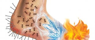
The narrower the spinal canal becomes, the worse the condition of the vessels and nerves passing through it. Vessels and nerves are pinched - tissue nutrition and innervation deteriorate. The inflammatory process begins. Due to a lack of nutrient supply, there is a risk of ischemic spinal cord stroke, which often leads to lifelong disability. In this case, nerve cells can begin to die en masse, and the person loses the ability to move normally and feel the limbs.
- Recommended reading: spinal lateral stenosis
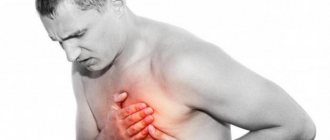
The spinal cord is the organ that feels the lack of oxygen and blood more than any other. If you block their access for only a short time, the tissues begin to die almost immediately. The result is paralysis of the lower extremities, or, if the stenosis is localized in the thoracic region and higher, death due to the inability to breathe.
Pathogenesis
The narrowing of the spinal canal is based on changes of various types: thickening of the spinal ligaments, arthrosis of the facet joints , thickening of the bone, slipping of the vertebrae, etc. The pathophysiological mechanisms of neurological symptoms are caused by a combination of three groups of factors: increased pressure in the epidural space, ischemia and aseptic inflammation , each of which develops under the influence of chronic compression of the vascular/nervous structures of the spinal canal.
In the presence of prolonged compression, the blood supply to the nerve structures suffers, since the amount of blood flow does not correspond to current needs. Developing against the background of a decrease in the amount of incoming blood, ischemia of the nerve root contributes to the development of demyelination , the formation of adhesions between the paranoid and soft membranes of the spinal cord, as well as the development of interstitial fibrosis / cicatricial adhesive epiduritis .
The specificity of the pathogenesis of lumbar spinal stenosis is the pronounced dependence of the volume of the spinal canal on the position of the body: thus, at the time of squatting, the lumbar lordosis becomes kyphotic/straightened, and the articular processes diverge and the lumen of the intervertebral foramen increases, thereby freeing the blood vessels that are under compression, which contributes to normalization blood flow and nutrition of ischemic neural roots. When the spine is flexed, the height of the intervertebral foramen increases by 12%, while during extension it decreases by 15%, which explains the regression of pain when sitting down/bending the body.
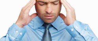
Symptoms of cervical spinal stenosis
Cervical spinal stenosis develops very slowly and is perceived by patients as a chronic problem. When the compression level reaches a critical level, surgery is the only option. The pathology has the following symptoms:
- Pain in the neck, back of the head and shoulder girdle. It can be dull, aching or sharp, expressed in the form of “lumbago”.
- Weakness of the arms and legs, loss of sensitivity, “woolly” condition, tingling in the fingers. Physical exercise, walking or simple work become inaccessible to the patient.
- Breathing problems or complete cessation.
Cervical spinal stenosis is dangerous because a person loses sensation throughout the body below the affected area. Arms, legs, genitourinary area (difficulty urinating or incontinence), and genitals suffer. The consequences of advanced pathology can be life-threatening.
Classification
The classification of spinal stenosis is based on a number of signs, according to which the following are distinguished:
- Primary and secondary stenosis of the spinal canal. Primary stenosis is caused by congenital pathology (underdevelopment) of the vertebral foramen, manifested by shortening of the vertebral arch, fibrous/cartilaginous diastematomyelia and achondroplasia (decreased vertebral body height/increased arch thickness/shortened pedicle), while secondary spinal stenosis (acquired) develops as a result of various diseases spinal column ( deforming spondyloarthrosis , occurring with the formation of marginal osteophytes/hypertrophy of intervertebral joints, ossification / hypertrophy of the ligamentum flavum , degenerative spondylolisthesis , ankylosing , ossified herniated intervertebral discs, Forestier disease , postoperative scars/subarachnoid adhesions, etc.).
- According to anatomical criteria, central stenosis , manifested by a decrease in the anteroposterior size of the spinal canal, which occurs due to the development of pathological processes in the anatomical processes that directly form the spinal canal (intervertebral joints/discs, posterior longitudinal and yellow ligaments) and lateral stenosis - narrowing of the intervertebral foramen, developing as a result of hypertrophy of the superior articular process, peculiarities of the formation of the facet joint, the development of osteophytes localized at the site of attachment of the yellow ligament (foraminal stenosis). In turn, lateral stenosis is divided into “middle zone stenosis”, “entry zone stenosis” and “exit zone stenosis” of the nerve root from the intervertebral foramen.
- Relative and absolute spinal stenosis.
Etiology
The main provoking factors for the formation of spinal canal stenosis include the following:
- displacement of the vertebral discs due to injury;
- chronic stage of osteochondrosis;
- dissection of a section of the vertebral artery;
- ailments in the neck or spine;
- increased physical activity due to work or professional sports;
- obstruction of patency in the arteries.
Most often, spinal stenosis can form due to injury or improper distribution of loads on the spinal column.
Causes
The development of spinal stenosis can be based on a variety of reasons:
- Congenital pathology of the spinal canal - achondroplasia (congenital chondrodystrophy), diastematomyelia .
- Injuries of the spinal column with displacement of vertebrae/fragments, intracanal hematomas.
- Facet arthropathy (changes in intervertebral joints of degenerative-dystrophic origin in the form of bone growths inside the spinal canal).
- Discopathies with prolapse/ossification/sequestration of intervertebral hernia
- Spondylolisthesis (anterior displacement of the vertebra) due to an anatomical defect of the vertebral arch.
- Thickening of the capsule of the intervertebral joints as a result of their inflammation ( Bechterew's disease ).
- Calcification/thickening of the yellow ligaments of the spine as a result of an inflammatory process or their degeneration.
- Forestier's disease (hardening of the longitudinal anterior ligament).
- Cysts/tumors and scar changes inside the spinal canal.
- Stagnation of venous blood inside the spinal canal.
Treatment of stenosis: basic methods
Treatment of spinal pathology is possible using two methods: conservative and surgical (operative). The second is resorted to when the first does not give the desired result.
Conservative treatment can be used for minor (relative) stenosis, when neurotic disorders are practically absent, and the symptoms of the pathology are limited to pain in the legs and lower back. Treatment without surgery involves taking medications, special exercises selected by a doctor, physiotherapy and manual techniques. A positive result is possible only with an integrated approach. Drug treatment includes taking the following medications:
- non-steroidal anti-inflammatory drugs - to eliminate pain and relieve inflammation (Ibuprofen, Diclofenac, Xefocam);
- muscle relaxants – relieve muscle tension (Mydocalm);
- B vitamins – have a positive effect on the nervous system, relieve pain (Milgamma, Neurovitan);
- vascular agents - to normalize venous and outflow (Eskuzan, Cavinton).
For severe pain, medical blockades based on anesthetics and hormonal agents are used. Successful treatment of stenosis necessarily includes physical exercises and physiotherapy. The latter include therapy with electric current and magnetic field, electrophoresis, exposure to healing mud, and water procedures. The choice of method is determined by the severity of the patient’s disease and individual contraindications.
To improve local blood flow and stimulate metabolism, massage courses are recommended for patients with stenosis. It is also important to engage in physical exercise that improves overall well-being and relieves pain. If the stenosis is absolute, i.e. has entered an advanced stage, the only option for the patient is surgery. Its goal is to free the nerve roots from compression. Today, different types of surgical intervention are practiced: open and endoscopic. The most popular methods are:
- Decompression laminectomy is the removal of intervertebral joints or part of the arch, thereby achieving expansion of the spinal canal. This is the most traumatic method of surgical therapy.
- Stabilizing effect - it usually complements laminectomy. To improve the supporting functions of the spine, metal staples are implanted into it.
- Microsurgical decompression with the establishment of dynamic fixation is the most physiological method that allows you to preserve the ability to flex and extend the spine.
The surgical technique is selected individually, taking into account the characteristics of a particular case. In most situations, such therapy ensures recovery. The patient’s behavior at the rehabilitation stage plays a significant role. He needs to follow a gentle regimen, perform special exercises, and eat right.
Spinal stenosis is a dangerous pathology that can lead to complete disability. Absolute stenosis can only be treated surgically. To prevent the development of the disease, it is important to monitor your diet and daily routine, get rid of bad habits and add physical exercise to your life.
Symptoms
Clinical manifestations of spinal canal stenosis are determined by a number of factors - the localization of the narrowing, the reasons for the development of the process and its stage.
Spinal stenosis at the level of the cervical spine
Stenosis in this area of the spine is a serious danger, since chronic compression of the spinal cord often leads to chronic vascular disorders, leading to changes directly in the spinal cord ( myelopathy ), with the development of disturbances in the conduction of nerve fibers in the top-down direction and the development of neurological symptoms with impaired sensitivity and motor disorders in the upper/lower extremities.
Specific manifestations of such stenosis are:
- stiffness of the neck muscles;
- numbness of hands;
- changes in vision/hearing;
- disorders of the vestibular apparatus;
- sudden short-term loss of consciousness;
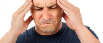
- periodic attacks of migraine localized in the temples/eyebrow area;
- breathing problems.
Spinal stenosis at the level of the thoracic spine
Relatively rare. Manifested by the patient's complaints of a burning sensation on one side of the chest; paresthesia of the epigastric region; pain that radiates along the pinched nerves; complete/partial loss of motor activity below in the area below the level of the lesion.
Lumbar spinal stenosis
Spinal narrowing in the lumbar region is most common. In the initial stage, slight discomfort appears in the lower back, but mild pain quickly gives way to relative well-being. More often, pain occurs due to inadequate load or awkward movement, but over time the duration of the pain syndrome increases and can move from the lower back to the lower extremities. When bending and squatting, the patient feels relief, but when returning to the previous position, the pain returns and intensifies.
Compression of the neurovascular structures in the vertebrae of the lumbar region contributes to the development of their inflammation and manifests itself with the following symptoms:
- lower back pain that gets worse while walking;
- pain radiating to one/two lower limbs;
- lameness;
- numbness in the perineal area;
- paresthesia of the legs;
- bowel/bladder disorders;
- decreased/absent tendon reflexes;
- erectile dysfunction;
- decreased sensitivity of the toes;
- partial paralysis of the legs.
Symptoms usually develop slowly, but the process inevitably progresses. Neurological manifestations are determined by the stage of development of stenosis. In the initial stage, subjective symptoms predominate in the form of pain, mild paresthesia, and transient motor disturbances. Signs of damage to the nervous system are often absent or mild. Their manifestation occurs in the late stage of the disease, and they are caused by the development of ischemic-compressive radiculomyelopathy / less often - spinal cord lesion syndrome .
by the syndrome of neurogenic intermittent claudication observed in the vast majority of patients . The pathogenetic mechanism of intermittent claudication syndrome, in addition to purely mechanical compression and reduction in the volume of the spinal canal, is transient ischemia of the spinal cord /its roots, which develops as a result of venous/cerebrospinal fluid hypertension in the radicular/spinal canal and vasospasm .
Ischemia increases when standing/walking, that is, when the patient is in an upright position. The reason for the exacerbation of symptoms during movement is segmental rotation that occurs when walking, leading to an even greater narrowing of the stenotic canal and, accordingly, a deterioration in the blood supply to the spinal cord.
The main complaint of patients with lumbar stenosis is the presence of constant pain in the lower back, which radiates to one/both lower extremities.
Lumboischialgia is characterized by a progressive, remitting course. The pain syndrome is accompanied by a feeling of cold/heat in the extremities and dysesthesia . Less commonly, patients experience shooting pain in the lower extremities and short-term weakness in the legs. Later, the pain syndrome is joined by the syndrome of unilateral/bilateral neurogenic intermittent claudication. Typically, unilateral intermittent claudication (pain in one leg when walking) initially develops. In the advanced stage, a symptom of bilateral intermittent claudication appears, sometimes they are asymmetrical.
Gradually, the severity/duration of the attack increases, postural weakness in the legs is noted, in some cases patients cannot stand upright (postural/orthostatic variant of the manifestation of “intermittent claudication of the cauda equina”). Less commonly, intense pain forces the patient not only to stop, but also to lie down or take a specific position with slight flexion of the lower limbs at the hip joints and tilt of the torso forward.
Constant motor, sensory or reflex disorders are evidence of the development of cauda equina compression syndrome, manifested by hyporeflexia , asymmetric muscle wasting, hypoesthesia . At a later stage, disorders of the pelvic organs occur - urinary retention/urinary incontinence ( neuropathic bladder ) and fecal incontinence, which is especially pronounced after physical activity, prolonged standing or walking.
The appearance of neuropathic bladder symptoms directly correlates with a decrease in the anteroposterior size of the spinal canal: the incidence of urological disorders is greater as its size decreases.
Lateral degenerative spinal canal stenosis is manifested by pain in the radicular syndrome, which has a clear localization, and the pain is limited to the zone of innervation of the root. Later, sensory disorders also appear along the radicular type. Sometimes pain/impaired sensitivity is combined with decreased/loss of reflexes or paresis of certain muscle groups.
With combined spinal/radicular canal stenosis, symptoms may be dominated by both intermittent claudication and radicular pain syndrome . In cases of multiple stenosis of the root canals, the syndrome of intermittent claudication naturally develops, combined with painful cramps (spasms) in the calf and other groups of large muscles (fascicular twitching mainly after physical activity).
General symptoms
First of all, it is worth considering that in addition to the general symptoms, each form of this pathological process has additional signs. Common symptoms of spinal stenosis are:
- pain in the lumbar region;
- decreased sensitivity in the perineum and lower extremities;
- fatigue when walking;
- amyotrophy;
- erectile dysfunction (in men);
- paralysis;
- dysfunction of the bladder.
It is such a symptom as pain in the lumbar region, which radiates to the left side, that indicates the progression of pathology in the spinal column. Therefore, if you have this symptom, you should immediately contact a surgeon.
Tests and diagnostics
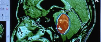
The diagnosis is made on the basis of clinical complaints, the results of a physical examination (numbness, weakness, palpation of areas where pain is present, examination of skin sensitivity, tendon reflexes, muscle strength), as well as data from instrumental research methods that allow visualization of the narrowing of the lumen of the spinal canal (spondylography , MRI and CT of the spine).
X-ray (survey and functional) allows you to visualize bone formations (osteophytes), a decrease in the height of the intervertebral space, tumors and vertebral fractures, instability of the spinal motion segment, hypertrophy of the facet joints.
CT (MRI) is the main method for diagnosing narrowing of the spinal canal. At the same time, CT is more preferable in identifying the degree of destructive-degenerative changes in the facet processes/vertebral bodies, herniated intervertebral discs and ossification of the ligamentous apparatus . MRI is preferable for determining pathological changes in the structures of the soft tissue component (determining the structure of the spinal cord, nerve roots), as well as hemorrhage, abnormalities of the ligamentous apparatus, swelling of the nerve root, the presence of cysts , epidural/perineural fibrosis , inflammatory changes, etc.
If necessary, myelography (electroneuromyography) and scintigraphy .
Forms of the disease
There are only two forms of this disease - congenital or acquired (secondary).
Primary (idiopathic) spinal stenosis is quite rare. Treating this form of the disease is very problematic, since the pathological process progresses due to genetic predisposition.
Acquired spinal stenosis
This disease develops in people with age. The main reason for its progression is degenerative changes in the spine or injury.
It is based on dystrophic changes in cartilage tissue. Due to certain circumstances, smooth cartilage tissue becomes rough. The cartilage begins to wear out quickly, as a result of which the joints cease to be protected and rub against each other. As a result, bone spurs are formed. When such formations begin to form in the facet region of the spine, a narrowing occurs in the spinal canal.
Most often, such pathological deformities occur in the lumbar region.
Prevention
There are no specific measures for spinal stenosis. Among the general activities we can recommend:
- A lifestyle with systematic sufficient and adequate physical activity, including morning exercises, walking, swimming, and sports games.
- Avoid dynamic/static overloads of the spinal column.
- Maintaining hygiene/proper organization of work and sleeping space.
- Timely diagnosis of the condition of the spinal column and, if necessary, correction/treatment of changes (weight loss, wearing a corset, exercise therapy, wearing a corset).
Forecast
The prognosis for the course of spinal stenosis is determined by its causes, duration of the disease, and the characteristics of clinical symptoms. With a timely diagnosis of the disease (in the early stages) with adequate conservative/surgical treatment, the prognosis is favorable. Depending on its cause, you can always choose a treatment method for the patient. Delayed diagnosis (in advanced cases), postoperative complications and rough manual interventions make the prognosis unfavorable and can lead the patient to permanent disability.
Therapeutic measures
Spinal stenosis requires mandatory treatment. An advanced condition or attempts to independently get rid of a pathological condition in the vast majority of cases lead to disability. An integrated approach taking into account the stage of narrowing will help to achieve a lasting positive result from treatment. It should be borne in mind that canning methods are only effective in case of minor deformation.
Conservative therapy
Medicines for symptomatic treatment, which are prescribed and prescribed by the attending doctor:
- Non-steroidal anti-inflammatory drugs (Indomethacin, Ketorolac, Diclofenac) to relieve pain and inflammatory response.
- Local blockades with an analgesic (Novocaine) or glucocorticosteroid (Hydrocortisone).
- Muscle relaxants (Tizanidine, Baclofen) to relieve spasms from muscle fibers.
- For local analgesic and anti-inflammatory effects, it is recommended to use ointments (Voltaren, Nise) or patches (Ketonal Thermo, pepper patch).
- Vitamin preparations of group B (Neurobion, Trigamma) to improve the conductivity of nerve fibers.
- Diuretics (Lasix, Furosemide) to relieve swelling.
- To improve cell nutrition and blood microcirculation - Actovegin, Venoplant.
Physiotherapeutic procedures include courses of electrophoresis, magnetic therapy, ultrasound radiation, exposure to laser beams, and balneotherapy. The procedures will help restore blood hemodynamics, improve the quality of metabolic reactions, and accelerate cell regeneration.
To consolidate the therapeutic effect and prevent the progression of spinal stenosis, a course of massage and exercise therapy is carried out. The attending doctor, together with a physiotherapist, based on the patient’s condition, draws up a set of exercises, the main task of which is to strengthen the muscular corset of the back and abdominals.
Surgery
When the narrowing of the spinal cavity is critical, surgical treatment is performed. There are several ways to eliminate a dangerous condition. A popular method is to remove part of the arch of the vertebral segment. The disadvantage of this method is that after surgery there is a risk of developing pathological hypermobility of the vertebra and stenosis in adjacent sections.
Interspinous fixation is considered more effective. This method is indicated for patients in whom spinal canal stenosis has formed as a result of flattening of the vertebrae. With this method, the vertebrae are connected to each other using special implants between the processes (interspinous fixators).
The recovery period after surgery usually lasts about six months. In the first two weeks, the patient adheres to semi-bed rest; intense physical activity and heavy lifting are prohibited throughout the rehabilitation. Sanatorium-resort treatment has a good strengthening effect and speeds up recovery.
List of sources
- Antipko L.E. Spinal stenosis / L.E. Antipko. - Voronezh: IPF "Voronezh", 2001. - 272 p.
- Nurmieva Ch.R. Methods for measuring stenosis of the cervical spinal canal / Ch.R. Nurmieva // Materials of All-Russian. scientific-practical conf. "Young scientists in medicine." - Kazan. - 2010. - pp. 127-128.
- Modern technologies for surgical treatment of lumbar spinal stenosis / A.I. Sold [and others] // Spine surgery. - 2008. - No. 3. - P. 40 - 47.
- Polishchuk N.E. Clinic and differential diagnosis of lumbar stenosis / N.E. Polishchuk, A.L. Isaenko // Ukrainian medical journal. - 2001. - No. 2 (22). — P. 106-109.
- Abdullaev R.Ya. New aspects of diagnosing spinal canal stenosis / R.Ya. Abdullaev, S.A. Ponomarenko // Radiation diagnostics. Intl. honey. magazine — 2005.— No. 3. — P.106-109.
Spine structure
In order to understand the etiology of spinal stenosis, you should know what the spine is made of. So, the structure of the vertebral region is as follows:
- vertebrae - 24 bones, which are located strictly one after another;
- ligaments - tissue that tightly holds the vertebrae together;
- intervertebral discs - elastic pads of cartilage that separate the vertebral bones;
- facet joints - formations that make the spine flexible;
- spinal cord;
- nerves;
- spinal canal.