The essence of the operation
A brain biopsy is used in neurosurgery to determine whether a tumor is malignant or benign. However, the study is not useful for diagnostic purposes only. Because any tumor in the brain will need to be removed.
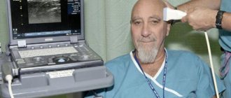
A brain biopsy is performed with a very thin and hollow needle. The purpose of the procedure is to select a portion of cells from a specific area. To access soft tissues, a minimal hole is made in the skull. The material is taken with a syringe and the resulting canal is sutured, which quickly overgrows.
Brain biopsy is the last method of research when MRI and computed tomography cannot make a definitive diagnosis. The results only add to an already disappointing verdict. For the patient, these data do not radically change the situation.
Complications
There is a small chance that important brain structures will be damaged during the biopsy. A small scar is formed in the area where the material is taken, which can later become a source of epileptic impulses and the cause of epileptic seizures. When performing a stereotactic biopsy, common surgical complications such as infection and bleeding, as well as negative consequences associated with the use of anesthesia, are possible. The preparation of the patient for surgery, contraindications and complications are described in more detail.
When is research needed?
Stereotactic brain biopsy helps to accurately determine the type of tumor. It is recommended for diseases: multiple sclerosis, Alzheimer's disease, hemorrhagic stroke. The method is indicated for meningitis and encephalitis.
Brain tumor biopsy is a relatively dangerous research method, so it is not suitable for many categories of patients. In practice, doctors try not to use invasive methods at all. People turn to him when the tumor in the head is quite large. And often the result of research gives either a chance for a cure, or a sentence of imminent death.
If a brain biopsy is performed, the consequences may cause the detected tumor to grow faster. With a benign neoplasm, re-growth of the pathology is observed in 50% of cases.
Varieties
Open brain biopsy is very rarely used. As an operation to remove a tumor is performed, the affected cells are examined at the same time. This type of research is quite complex and carries risks for the patient. The skull is open at the time of puncture sampling and there is a possibility of damage to the upper layers of the brain.
The stereotactic method is the least invasive. Modern equipment displays the entire process, eliminating unnecessary needle movements. The doctor controls every stage of the procedure.
Visual examination is only possible with an open biopsy. But MRI and computed tomography are added to stereoscopic, which makes it safer.
Conducting an open method
Let us describe how an open brain biopsy is taken. Before the procedure begins, the patient is given anesthesia. To access the brain, a small section of the skull is removed.
The open method is not performed separately; it is always done during surgery to remove tumors. Part of the skull must be restored, and this is a long process. The patient will be on sick leave for a long time after this procedure.
The open method poses a health risk, although it is used more often. The recovery period can last several months.
Brain biopsy: what it is, how it is performed, consequences
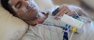
A brain biopsy is a diagnostic procedure that involves removing a small tissue sample (biopsy) for further study. The data obtained allows us to establish the necessary steps for subsequent treatment. Doctors decide on the methodology - whether surgery, chemotherapy, radiation therapy, etc. is necessary.
Taking material helps to determine the degree of malignancy of the tumor. In addition, this process is often prescribed to confirm Alzheimer's disease or Creutzfeldt-Jakob disease. Using a biopsy, healthcare professionals also diagnose multiple sclerosis and inflammatory processes in the brain.
In standard cases, this type of diagnostic study is carried out at the last stage and in conditions of insufficient information collected by other methods.
A biopsy becomes unnecessary: when the last stage of cancer development has already started, there is an acute period of inflammatory processes, as well as in case of blood clotting problems.
Experienced neurosurgeons use the following types of brain biopsies:
- Open.
- Stereotactic.
- Puncture.
Indications
The essence of a biopsy is to extract a certain type of tissue from the brain, the study of which will allow for a more accurate diagnosis of the disease. Since this method is invasive, it can only be performed strictly according to indications.
The procedure is carried out when:
- Neoplasms of unknown origin. In order to determine the severity of the ongoing disease, prescribe adequate treatment, and give the patient prognoses for subsequent life, the doctor needs to know whether the tumor located in the brain is benign or malignant. A biopsy will help answer this question.
- Cerebral toxoplasmosis. This pathological condition occurs as a result of the penetration of Toxoplasma into the cells of the human body. The only way to detect the parasite inside the brain is through a biopsy.
- Abscesses and bruises that occur between brain tissues.
- Multiple sclerosis.
- Hemorrhagic stroke.
- Alzheimer's diseases.
- Diseases of the brain of an inflammatory and infectious nature.
This diagnostic method is used for histological purposes to determine the changes that brain tissue has undergone during certain diseases.
Stages of the procedure
To understand how a biopsy is done in the brain area, you need to get an idea of the procedure. The preparatory stage includes a comprehensive examination, including laboratory tests, consultations with a cardiologist, phlebologist, and anesthesiologist. Several hours before the intervention, the patient does not take food or water.
It is important to promptly inform your doctor about any allergies to medications and regular use of medications that prevent blood clots. If the patient is taking thrombolytics before an invasive diagnostic test, therapy is temporarily suspended. Stages of the open procedure:
- Making an incision on the scalp.
- Formation of a hole in the skull.
- Removal of a fragment of skull bone tissue to form a trepanation window.
- Peeling of the meninges.
- Resection (partial or complete removal) of the tumor.
- Closing the trepanation window with bone tissue or a special plate.
After completion of neurosurgical procedures, a sample of neoplasm tissue is sent to the laboratory to study the cellular structure. The stages of performing a stereotactic biopsy in the brain region depend on the type of equipment used. The sequence of manipulations when using a coordinating stereotactic frame:
- The exact localization of the pathological focus is determined using neuroimaging (mainly MRI).
- Local anesthetics or general anesthesia are used.
- A stereotaxic frame is installed in the head area.
- An MRI localizer with contrast marks is attached (a special device for limiting the area of intervention).
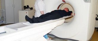
- A CT scan is performed.
- The results of MRI and CT studies are combined to achieve maximum accuracy when creating injection points.
- An incision is made into the skin of the head.
- A hole is drilled in the skull bone.
- A needle is inserted and moves under the control of an MRI or CT diagnostic complex.
- Material is being collected.
A study using a frame allows you to precisely approach the site of a tumor or other pathological process with an accuracy of 1 mm. The duration of the intervention is about 6 hours. One day after completion of the procedure, the patient is discharged from the hospital. Sequence of manipulations when using the navigation system:
- A study is carried out in MRI or CT format to obtain information such as the structural features of the patient’s brain, the location and size of the pathological focus.
- Neuroimaging results are transferred to the navigation system.
- A model of the brain is created in 3D projection.
- The image is studied to determine the optimal insertion point and needle trajectory.
- Anesthetics are administered to the patient.
- An incision is made on the skin of the head.
- A diagnostic hole is formed.
- Under the control of a navigation system, a puncture needle equipped with a camera and LED illumination is inserted into the cavity of the skull.
- Material is being collected.
A brain biopsy using a navigation system is carried out with minimal negative impact on healthy brain tissue, because calculations such as the shortest and safest path to the tumor site are previously performed. The doctor has the opportunity to choose a trajectory that goes around the areas responsible for vital functions, thereby significantly reducing the risk of complications.
The procedure time for a biopsy in the brain area using a navigation system is significantly reduced compared to intervention using a stereotactic frame. Reducing the duration of the procedure is associated with preliminary planning of actions.
During a puncture biopsy, an incision is made on the skin in the projection where the tumor is localized. Through a hole drilled in the skull bone, a puncture needle is inserted into the pathological focus to obtain a sample of the material. The stereotactic and puncture-type procedure is associated with minimal damage to the medulla, meninges, and bone tissue.
Types of biopsy
It was previously noted that certain indications are required for a doctor to prescribe a brain biopsy. The choice of the right type of biopsy will depend on the clinical picture, the pathology occurring in the patient, the severity of its course, as well as other characteristics of the body not related to the underlying disease.
There are three types of brain biopsies:
Source: https://neuro-orto.ru/diagnostika/biopsiya-mozga.html
Carrying out a minimally invasive method
Stereotactic intervention is performed using a frame and neuronavigation. Both methods are accurate compared to open. The classical method includes the first method. At the moment, the data obtained are the most accurate with the least invasion into the patient’s organ.
Before the procedure, an MRI is performed to determine the exact position of the tumor. Special contrast agents are used. When doctors have decided on the puncture site, they install the frame on the patient’s skull. It is secured with screws. A ring is installed on it, on which the needle will be placed.
The MRI localizer is turned on and a computed tomography is performed. The entire process is displayed on the monitor screen. Next, the surgeon drills the needle insertion site, having previously cut the skin. Biomaterial is collected and the incised area of skin is sutured.
The patient will have to spend a recovery period in bed. Doctors will periodically examine him to rule out complications from the operation.
Stereotactic biopsy
Stereotactic biopsy involves a minimally invasive procedure. The process of collecting brain tissue consists of the following steps:
- A person is put on a stereotactic frame, which allows him to hold his head in the desired position.
- Surgical intervention is accompanied by CT or MRI control. Using a modern device, the doctor can enter the results directly into a special program.
- A cut of up to 1 cm in size is made in a pre-prepared locus on the skull, after which the bone is drilled.
- A puncture needle, which is equipped with a microcamera and an LED, is inserted into the resulting hole. The recording camera transmits the image online. With its help, the doctor can examine in detail the formation and the required location for the puncture.
- A biopsy sample is taken and the hole is sutured.
Neuronavigation
This biopsy method also includes preliminary MRI and CT scanning before surgery. Based on the resulting three-dimensional image, the site of needle insertion is determined. The surgeon can also calculate the direction of passage of the instrument when collecting biomaterial. The patient is given anesthesia.
The patient's arms, legs, and head are securely fixed on the couch; the slightest unsuccessful movement can lead to the needle moving and entering the brain more than intended. The surgeon makes a hole with a needle, control is carried out by neuronavigation. At the end of the procedure, stitches are applied and a recovery period is required.
The method differs in that the patient does not feel anything at all. The computer helps the surgeon choose the least traumatic needle path for the skull and brain. The tumor is often located deep; the previous method makes it difficult not to affect the surrounding healthy tissue.
Neuronavigation studies not only the brain; it is used to obtain biomaterial from tumors in the spinal cord. However, doctors warn that both methods can have consequences for the patient's health. They are resorted to in extreme cases, when there is already an overgrown tumor.
Recovery period
After carrying out this diagnostic method, the patient must be under the supervision of medical personnel for a certain time. The duration of this time period is determined by the doctor, who takes into account the complexity of the procedure, the general condition of the sick person after it, and the presence of concomitant diseases in the patient. If we take into account the fact that a biopsy is a diagnostic method that is often prescribed for a pathology that is serious and dangerous to the patient’s health, restorative measures will be more aimed at treating the manifestations of the underlying disease, rather than the consequences of the procedure.
Tulpa V.V., doctor, medical observer
8, total, today
( 187 votes, average: 4.63 out of 5)
Pregnancy examinations
Blood test for tumor markers: types of tumor markers and interpretation of results
Related Posts
Negative consequences from research
A biopsy always has consequences. The level of reaction of body tissues is different for each person undergoing surgery, and it will be impossible to predict what kind of complication. The most common minor ailments are: bleeding, headaches due to swelling at the site of biomaterial collection.
There are more dangerous consequences: damage to brain cells, the patient may fall into a coma. Poor circulation at the surgical site will affect the functioning of the entire body. Seizures and disturbances in motor skills may occur. A weakened body becomes defenseless against infections, and chronic diseases become more active.
Modern equipment has significantly reduced the likelihood of problems occurring after a biopsy. But consequences still occur. Patients are reassured by the reliability of the instruments used. Doctors just cannot take into account the patient’s body’s reaction to the introduction of a foreign object into the brain tissue.
The inexperience of medical workers is the main factor that can cause complications. You can exclude it by contacting a trusted diagnostic center.
Brain biopsy: indications, contraindications, disadvantages of the procedure
A brain biopsy is a diagnostic procedure; it is performed when brain lesions are suspected, if other methods have failed. This study was previously considered quite dangerous and traumatic, but with the help of imaging equipment it was possible to reduce the risk of brain damage.
Indications for the purpose of manipulation
The procedure is performed to diagnose brain diseases. During a biopsy, a small amount of organ tissue is removed to examine and determine the problem. This is an invasive technique, so there must be strict indications for its implementation.
The procedure may be prescribed in the following cases:
- In a cancer patient with tumors of unknown origin. The study allows you to determine the severity of the disease, select the most appropriate therapy and make a prognosis. A biopsy will show what type of tumor it is.
- If a person suffers from cerebral toxoplasmosis. With this pathology, parasites - toxoplasma - enter the human body. Only a biopsy can determine their presence in the brain.
- In the presence of bruises and abscesses that have appeared in the tissues of the organ.
- If the patient has Alzheimer's disease, multiple sclerosis, or has had a hemorrhagic stroke.
- For inflammatory and infectious pathologies.
During the study, a brain biopsy is taken for histological analysis, during which pathological changes in the brain tissue are determined.
Contraindications
There are contraindications for performing a brain biopsy. The procedure cannot be performed:
- if the blood coagulation system is disrupted;
- if the pathological process develops in a part of the brain, penetration into which can lead to serious complications;
- in the presence of systemic diseases.
Methods for performing a biopsy of a tumor in the brain
There are different methods for performing a biopsy. The appropriate option is selected depending on what pathological process has affected the patient, the severity of its course and the individual characteristics of the body.
Research can be of the following types:
- Stereotactic brain biopsy. The procedure is classified as a minimally invasive technique, since during its implementation a small amount of material is taken for research. During the procedure, magnetic resonance imaging is used to monitor the process.
- Needle biopsy of the brain. This biopsy method involves removing material with a needle. To do this, the doctor drills a hole in the skull using a milling cutter. Through it they take part of the tissue.
- Open biopsy. This is an intervention that can cause serious harm to human health. It is performed under general anesthesia. The required particle is collected through a hole in the skull. To do this, the doctor removes a certain part of the skull.
After removing a section of tumor or tissue, the patient is given a rehabilitation period, during which complications can be avoided.
Method using a frame
The biopsy is performed using a frame. Before the procedure, an MRI is performed and the point through which the material will be taken is determined.
Before the operation, the main ring is attached to the head with special screws. Since the screws will be driven directly into the skull, these areas will be numbed. After this, a localizer and radiopaque markers are put on the ring.
The patient is placed on the operating table and a frame is attached to the ring, which contains the established coordinates of the pathological focus. The frame is adjusted so that its center is exactly at the target.
After this, the surgeon cuts the skin and makes a burr hole through which a biopsy needle is inserted. The trajectory of its introduction is calculated in advance. Using a needle, they take the material.
Methodology using a neuronavigation system
Before the procedure, magnetic resonance imaging is performed and the results are recorded on disk. They are transferred to the navigation system and converted into volumetric ones. This allows you to see pathological foci in different projections. Using the data, the surgeon plans a biopsy. It determines in advance the point through which the needle will be inserted.
After determining the entry point, the patient is given general or local anesthesia and secured on the operating table. Using a special pointer, a point is determined on the head, which is shown on the MRI results. After this, a hole is made and the necessary material is taken using neuronavigation.
Puncture method
A puncture biopsy is performed if the case is not severe. First, the patient is given anesthesia, after which a hole is drilled in the skull. A thin needle with a small video camera at the end and a lighting device is inserted through it. This allows you to control the biopsy process from start to finish.
Stereotactic method
This procedure does not require a long rehabilitation period; the patient can return to their normal lifestyle the very next day. First, a stereotactic frame is fixed on the head, and the trajectory of insertion of a special needle is calculated using a special program.
The stereotaxic needle has other functions. With its help, the tissues are moved apart, rather than pierced. Therefore, there is no risk of damage to important areas or vessels. A piece of tissue is taken with a needle and sent for examination.
Progress of the brain biopsy procedure
The entire biopsy procedure consists of the following steps:
- The skin is pierced using a special tube.
- After this, a hole is made using a cutter with a diameter of no more than two centimeters.
- A needle is inserted into the tumor, which pushes the soft tissue of the brain apart.
- They take part of the material, place it in formaldehyde and send it for research.
- Sutures are placed and an MRI is performed to ensure there is no damage.
During the operation, the patient may be under local or general anesthesia, depending on the type of biopsy.
Stages of open biopsy
The process of performing an open biopsy consists of the following sequence of actions:
- general anesthesia is given;
- perform craniotomy;
- a special syringe is used to suck out part of the material;
- cover the hole with bone or a metal plate.
This is quite dangerous, so most often they try to resort to minimally invasive techniques.
Disadvantages of Brain Biopsy
A biopsy carries certain health risks. The puncture method has a small risk of complications. Whereas with an open biopsy, the patient not only needs to recover for a long time after the procedure, but also may experience serious impairment of brain function due to damage during the study.
Due to incorrect and inaccurate implementation of the procedure, the following problems may arise:
- disturbances in the circulatory process in the brain;
- involuntary muscle contractions;
- secondary infection;
- severe blood loss;
- the tissue around the test site may swell;
- the patient may fall into a coma.
Modern techniques can significantly reduce the risk of developing such complications.
After the biopsy
After the procedure, the patient should be monitored by doctors. How long this is necessary is determined depending on the type of research.
Source: https://GolovaUm.ru/simptomy/biopsiya-mozga.html