4.38 out of 5 (8 Votes)
Often newborns have developmental difficulties, so that they do not develop into serious problems, an ultrasound scan of the baby’s brain is performed. To conduct a successful examination, an open fontanel between the bones of the skull is required.
Ultrasound examination is considered one of the most popular methods for diagnosing various pathologies. The examination is not difficult, does not require significant financial costs, is safe for patients, and allows one to obtain accurate information.
Ultrasound of the brain is performed in children, and not in adults, because with age, fontanelles grow between the bones of the skull, ultrasound radiation does not penetrate the brain, and sensors do not pick up signals.
This examination method is called echography because, when passing through soft tissue, signals in the frequency range from 0.5 to 15 MHz are reflected in different ways from substances with a different structure. Based on an analysis of existing reactions after comparison with normal results, doctors make a diagnosis.
The brain is examined in this way only in infants. The maximum age at which an ultrasound of the cerebral vessels is performed on a child is 1.5 years.
. The sensor is located between the frontal and parietal bones of the baby, but fontanelles are still present in the area of the temples and the back of the head, and later they heal.
What is the procedure?
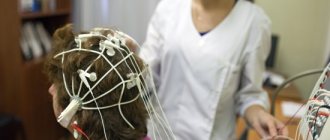
Neurophysiography means performing an ultrasound examination of the brain in newborns and children under 1 year of age. High-frequency vibrations are reflected from the brain and display its outline on a digital display.
The procedure is safe, does not cause pain, and allows you to obtain a lot of information. These circumstances make it possible to carry it out regularly. No need to administer anesthesia to the patient
, it is enough to keep the child still or wait until he falls asleep. Crying or screaming has no effect on the results.
This method of examination is considered most suitable for infants. It is carried out until the fontanel between the bones of the skull is overgrown.
Ultrasound of the brain in newborns makes it possible to familiarize yourself with all the individual structures of the organ. During a short procedure, doctors will be able to analyze brain processes and the general dynamics of their flow.
How much does the examination cost?
The cost of NSG in different institutions is approximately the same, but differs slightly. The wound is about 1 thousand rubles. If they also do Dopplerometry, then the cost increases to 1.5 thousand rubles. Baby examination is available to every family. It will help identify pathologies in the brain and avoid serious problems with the baby’s health.
If you have a strong and seemingly healthy baby, give him up to 2 months. NSG. Make sure everything is fine and raise the baby calmly. Timely prevention is important.
Indications
For a child in the first year of life, there are many indications for an ultrasound examination. This could be pregnancy and childbirth, suspicions of various diseases, congenital defects, etc. Determining pathology in the primary stages of development gives a better chance of recovery.
The procedure lasts a maximum of 15 minutes. To examine the head, a specialist attaches sensors to the anterior fontanelle and temple area. In rare examples, the occipital areas are examined. An ultrasound is performed to obtain reliable information about the state of the newborn’s brain.
.
The displayed data is compared with the normal state and a diagnosis is made. A total of 12 common diagnostic indicators are used. The neurologist takes into account the examination results when determining the therapeutic technique.
The doctor determines the condition of the wide blood vessels of the brain, identifies deformation of the walls, damage, plexus, neoplasms, and analyzes the structure of the tissues. The main parameters to study are the volume of the ventricles. Excessive indicators indicate violations.
Neurosonography indicators are determined by the week in which the baby is born. We list the normal indicators of ultrasound examination:
- The grooves and convolutions in the brain should be located symmetrically, the ventricles and cisterns should be clearly visible.
- The echogenicity index of the subcortical nuclei is normal.
- The anterior horn of the extreme ventricle is approximately 1-2 mm deep. Its size should not exceed 4 mm.
- There is no fluid in the gap between the hemispheres of the brain. The normal width is 2 mm.
- The choroid plexuses in the area of the ventricles are homogeneous.
- The size of the 3rd ventricle is 2-4 mm.
- The stem structures are located in their place.
- The length of the large tank is 3-6 mm.
A neurologist interprets the ultrasound results. He can prescribe the correct therapeutic technique for the baby. Monitor and determine trends in the development of brain tissue, identify possible diseases. The neurologist needs to evaluate the neurophysiography data and compare them with the identified symptoms.
Is neurosonography harmful for a baby?
For a long time, there was an opinion among the population that ultrasound examination procedures were harmful. Many expectant mothers refused to do an ultrasound during pregnancy, citing the fact that they did not want to “irradiate the child.” In fact, diagnosis using ultrasound is the most harmless research method.
Its action is based on the emission of high frequency sound waves. They are reflected from the surface, and the analyzed data in the form of a picture is displayed on the monitor. In living nature, bats and dolphins orient themselves in space in a similar way—by emitting ultrasound and analyzing the time of its reflection from obstacles. But you’re not afraid of exposing your child to radiation when visiting the dolphinarium, are you? Then why be afraid of a harmless, but such an important diagnostic procedure as neurosonography?
Read in this article:
At all times, the dream of healthy offspring has been the most cherished among parents, and if we are talking about newborns, then worries about their future health naturally arise even at the stage of pregnancy planning. If a child was born without any abnormalities, parents still want to make sure that their child will not face any hidden dangers in the future, but it is almost impossible to carry out routine studies, for example, brain tomography, on infants (for this the child would have to be put under anesthesia).
The development of modern medicine makes it possible for anxious parents to get a broad picture of the health of their newborns from the first days of life. This is possible thanks to a special study of the brain, which is called neurosonography of newborns, the interpretation of the results of which will tell a lot about the health of the baby. It has been widely used in medical practice since the 1990s.
Decoding
Often the transcript shows such diseases. Enlarged ventricles of the brain
occurs if their depth is higher than normal. This is a characteristic sign of hydrocephalus. Externally, in a newborn, this disorder is visible to the naked eye.
The head increases in size, the forehead protrudes forward, moles become larger. diagnosed after ulcers or infectious processes, problems with the development of the child. The child often has migraines, fatigue, undeveloped physical development, and has mental problems.
An increase in the subarachnoid space of more than 3 mm is accompanied by an increase in body temperature and a deterioration in appetite. This may indicate meningitis. The disease can develop as a result of hydrocephalus.
Blood vessel plexus cyst
are cells of the ventricular mucosa that secrete cerebrospinal fluid. A cyst is formed in the form of a cavity filled with something. They often do not manifest visual symptoms in the child. Such formations do not require treatment, since they resolve over time.
Blood vessels don't do their job well. If these areas are highly dense, brain cells atrophy in fragments, and crust formation is distorted.
If the examination results show the presence of diseases, the neurologist will prescribe prophylaxis or select an appropriate therapeutic technique. Medicines will help accelerate the closure of fontanelles.
If you have high ICP in a newborn, you should not use Aquadetrim. If there are too many abnormalities in brain development, the child will be hospitalized and complex therapy will be prescribed. Children who develop brain diseases should not be vaccinated.
An ultrasound of a child’s head allows you to examine:
- Ventricles of the brain
. In the process of visualizing their volume and contours, the specialist identifies hemorrhages, cysts, dropsy, and processes that contribute to the development of rickets. If diseases are eliminated in time, pathologies of intellectual and physical development will be avoided. - Condition of wide vessels
. Aneurysms are detected in the form of enlargement of arterial walls. As a result, blood flow to the brain becomes difficult. - Hemorrhages
caused by prematurity. This problem needs to be monitored as closely as possible, as the result may be the death of the baby. - Infections
that provoke. If the disease is detected in time, the child can fully recover. - Oncology
is detected in rare cases.
Method of implementation
Performing an ultrasound of a child's head does not require prior preparation. It is necessary for the baby to be in a calm state, it is better to let him sleep. In such a situation, he will not have any discomfort; you can use a toy to distract his attention.
An ultrasound of a child’s brain involves performing the following steps:
- The baby is placed lying down and held in the correct position. The procedure is performed within 10 minutes. The patient needs to be calmer during this period.
- Newborns are not given anesthesia. You need to hold the head so that it does not move.
- The baby's head is smeared with a gel that differs in composition from the product used for ultrasound in adults. This substance is hypoallergenic and strengthens the sensor signal by eliminating air between the device and the skin.
- A sensor device is applied to the treated area. The specialist moves them around the head, reads data from organs and tissues.
When and to whom is neurosonography indicated?
It is recommended that all newborns undergo neurosonography once to exclude congenital disorders in the structure and function of the brain. The study needs to be carried out for up to 12 months - until the fontanel is overgrown, since ultrasound can only penetrate through soft tissue. Neurosonography is usually prescribed at the age of 1-1.5 months. If it is necessary to assess the dynamics of the brain, the study can be repeated at intervals of 1-2 months.
Ultrasound of the brain is vital for the following categories of infants:
- Premature
- Low birth weight babies
- With an unusual head shape
- With birth trauma
- Those who have suffered hypoxia
- After a difficult birth
- With suspicion of intrauterine infection
- With symptoms of neurological diseases
- With developmental disorders of other organs and systems
Safety of the procedure
When performing an ultrasound of the brain in newborns, the safety of the procedure must be taken into account. A child’s fragile body can easily get injured
. Similar studies have been carried out frequently for children recently.
Previously, procedures to study the state of the brain were carried out less frequently. Equipment for using anesthetics has proven to perform well in practice. It is easy to understand that this type of diagnosis is complex and can provoke negative consequences.
New methods have made the work of doctors and the lives of patients much easier. The procedure makes it possible to identify various brain diseases in children.
The procedure is harmless and always brings good results. Allows you to identify pathologies at the primary stage of development and improve the baby’s health using conservative methods without traumatic surgical interventions.
When there are indications, ultrasound of the brain in children must be performed.
. Parents often do not consent to such procedures; as a result, it is not possible to determine in advance the development of dangerous pathologies. Such negligence often leads to negative results.
To make a correct diagnosis and identify the problem in time, a simple external examination of the child is not enough. In some cases, it is necessary to use ultrasound equipment for examination.
– ultrasound examination of the brain. This is a common diagnostic procedure prescribed to newborns to exclude possible head pathologies due to birth injuries, infections or congenital diseases.
Ultrasound is a painless procedure and does not harm the fragile child’s body, therefore, having received a referral for it, you should not panic, much less ignore it.
Ultrasound of the brain
In this section you will find a lot of interesting and useful information on the area of interest and receive practical advice. Our medical specialists prepared the materials, and we collected them into one library. Pediatrician, neurologist, doctor of the highest category. Natalya Avdeeva, pediatrician of the highest category.
The use of Site materials is governed by the laws of the Russian Federation and International legal norms. A prerequisite for use by the user, including in the form of viewing the content of the Site, is the user’s full agreement with the User Agreement and the Privacy Policy posted on the Site. By this action, the user confirms that he is familiar with all the terms of the Agreement and is fully aware of their meaning, as well as the possible consequences of violating these terms.
All information presented on the Site cannot be considered a public offer. The use of forms on the site by the user does not establish a relationship between the owner of the Site and the user and does not indicate acceptance by the owner of the Site of an order for the provision of services.
The information that the user provides through the form is used to answer the question asked and to contact the user. User reviews may be published on the website. Use of the services offered on the Site may require the creation of a user account (Personal User Account).
For subsequent login to the Site, unique authorized data is generated - login and password. This information is confidential and is not subject to disclosure, except in cases established by the legislation of the Russian Federation and this Agreement.
When creating a password, the user is recommended to choose a password of sufficient complexity to prevent it from being picked up by third parties. The user is personally responsible for maintaining the confidentiality of account information, including the password, as well as for any and all activities that are conducted on behalf of the account user. When creating an account, the user undertakes to provide only accurate and complete information about himself, as well as to keep this information up to date.
If false information is provided, the Site Administration has the right to block or delete the user’s Personal Account and refuse to further provide services through the Site’s services. The Site Administration has the right at any time to require the user to provide documents confirming the information specified by the User during registration.
Failure to provide such documents may be regarded by the Site Administration as reporting false data when registering the User and entail the consequences specified in clause. The User must immediately notify the Site Administration of unauthorized use of his account or password or any other violation of the security system.
The user confirms that when filling out forms on the Site, he acts on his own behalf and in his own interests, confirms his consent to transfer his personal data to the owner of the Site, by indicating them in response to requests from the Site interface, confirms the accuracy of the personal data provided. The risks associated with the consequences of the user providing false information are borne by the user in full.
The site administration has the right to change the terms of this Agreement unilaterally at any time, which come into force from the moment of their publication. The Site Administration is not responsible for damage that a user may receive when following links to other Internet resources posted on the Site.
The Site Administration is not responsible for damage caused to the user as a result of actions taken independently by him, guided by the information posted on the Site. The site administration has the right to disclose information about the user if the current legislation of the Russian Federation requires such disclosure. The site administration has the right to publish a question and answer with anonymized personal data.
Any use, appropriation, copying, distribution of information posted on the Site is not permitted and entails the application of penalties.
The user confirms that he is familiar with all clauses of this Agreement, the Personal Data Processing Policy and unconditionally accepts them. Personal data is not publicly available. The basis for the processing of personal data is: Art. During processing of personal data, the following actions are performed: collection; record; systematization; accumulation; storage; clarification update, change; extraction; usage; transmission distribution, provision, access; depersonalization; blocking; deletion; destruction.
The processing of personal data may be terminated at the request of the subject of personal data. Thank you very much for making an appointment with us; our administrator will contact you shortly to confirm the time of your appointment with a specialist. Thank you very much for registering with us, in the near future, our administrator will contact you to clarify the time of appointment with a specialist. Request a call Online recording. Clinic on Pr.
Pobeda, Main Library. Women Health. This is necessary so that the child learns to turn, sit down, stand up, and walk in time. Another alarming symptom for mom should be hand tremors.
Here, too, a full examination is necessary. Early initiation of treatment prevents the child from becoming disabled. For the first three months, the baby burps after almost every feeding. Table of alarming symptoms Pathologies Symptoms Actions Neurological abnormalities Twitching of the chin. Tongue protruding. Additional examinations may be required.
Pathology of the retina 1. The child turns blue when feeding or crying. Author: Khabibullina Zulfiya Kabirovna Pediatrician, neurologist, doctor of the highest category. The user undertakes: When quoting materials from the Site, a link to the Site is required. The agreement comes into force from the moment the user accepts its terms. Contact number:. I agree with the personal data processing policy and consent to the processing of personal data User Agreement.
I agree to the processing of personal data and agree to the processing of personal data Terms of use English. Your name:. Review text:. I agree with the personal data processing policy and consent to the processing of personal data Agreement. Thank you very much for your feedback.
Thank you very much for writing to us! Departure of a specialist:. Twitching of the chin.
Why do tests be done on newborns?
Neurosonography is designed to promptly identify or exclude suspicions of brain diseases in infants. Therefore, the list of indications for its implementation is very wide:
- lack of oxygen during the prenatal period or during childbirth;
- uncharacteristic shape of the head in a newborn;
- protracted or, conversely, too rapid labor;
- absence of the newborn's first cry;
- premature birth;
- trauma during childbirth;
- the child’s weight is less than normal (2.8 kilograms);
- low Apgar score (less than 7);
- the woman in labor had a caesarean section;
- obstetric manipulations were performed during childbirth (for example, fetal rotation);
- the child often spits up;
- seizures for unknown reasons;
- intrauterine infection;
- predisposition to genetic diseases;
- developmental delay;
- developmental defects during pregnancy;
- The examination is also carried out at the request of the parents without any medical indications.
Preparing for NSG in infants
for an infant – a simple procedure that does not require prior preparation.
It can be carried out while the baby is sleeping or awake; in some cases, the study is done even in intensive care, without removing the child from the box.
There is no need to follow a diet or a special daily routine before the ultrasound, but so that the baby does not worry and allows himself to be examined, it is better to feed him in advance.
The only strict prohibition is the use of creams or ointments on the child’s head; they impair the performance of the sensors, which, in turn, affects the quality of the study.
Reference! Neurosonography is performed without anesthesia or any additional drugs; the total procedure time will not take more than twenty minutes.
Conducting research in a hospital setting
Neurosonography examinations of young patients can easily be carried out in a hospital inpatient setting. This requires compliance with the necessary sequence of actions.
Preparing the baby for the procedure
Special preliminary preparation of the infant for this procedure is not required. It is enough that the child is not hungry or thirsty. In addition, if the child falls asleep, he does not have to be specially woken up. Moreover, this will greatly facilitate neurosonography for both medical personnel and parents. The results of the operation will become available immediately after the session, a maximum of a couple of minutes after it is performed.
Process
The very nature of the neurosonography process does not differ significantly from the procedure for performing ultrasound. The child is placed on a flat couch, after which the places where the sensors are attached to his head are lubricated with special compounds. Then comes the procedure itself.
The doctor gains access to the baby's brain through the fontanel located on the parietal part of the head, as well as through the foramen area at the back of the head. To decipher the results of the study, a sufficiently qualified doctor is required.
How is an ultrasound of the head performed on a child under one year old?
The peculiarity is that it is carried out only through holes in the skull, since bones do not allow ultrasound to pass through. Therefore, ultrasound is often performed on children under one year of age, preferably before the large fontanel closes.
The equipment sensors are installed in the area of the fontanel, less often in the temporal region or the occipital foramen, pre-treated with conductive gel.
The doctor observes a dynamic image on the screen, noting the condition of the structures, grooves and convolutions, ventricles of the brain, the presence of cysts or formations, and the condition of the membranes.
The examination ends with a snapshot and display of digital indicators, according to which the results are deciphered.
Normal indicators
The absence of violations is indicated by:
- symmetry of the location of brain structures;
- ventricles of normal shape and size with clear and even boundaries;
- immutability of the membranes of the brain;
- absence of foreign formations.
Digital indicators for newborns:
- the depth of the anterior horn of the lateral ventricle is from 1 to 2 millimeters;
- the depth of the body of the lateral ventricle does not exceed 4 millimeters;
- the size of the third ventricle is no more than 6 millimeters;
- the width of the gap between the hemispheres is no more than 2 millimeters;
- the size of a large tank is from 3 to 6 millimeters;
- The width of the subarachnoid space is up to 3 millimeters.
Important!
Only a specialist should interpret the ultrasound results. To avoid unnecessary panic, do not self-diagnose.
Ultrasound results are only part of the information; they are assessed in conjunction with external signs of pathologies and indications of other studies by a neurologist. In addition, minor deviations from the norm in infants both at 1 month and at 6 months can be age-related and will go away over time. Only a professional can judge this.
Neurosonography in the diagnosis of cerebral lesions in children with congenital viral infection
Ultrasound scanner HS60
Professional diagnostic tools.
Assessment of tissue elasticity, advanced 3D/4D/5D scanning capabilities, BI-RADS classifier, options for expert cardiological studies.
Introduction
In recent years, brain damage in the early stages of ontogenesis has attracted increasing attention from a wide range of specialists. It is the leading cause of perinatal mortality and accounts for 60-70% of neurological pathologies in childhood [3]. According to the literature, anomalies of brain development account for about 25% of all congenital malformations in newborns [2]. Numerous publications discuss the participation of viral infections in the formation of congenital malformations of the brain, meningitis, meningoencephalitis and other structural lesions of the central nervous system [1,2]. An etiological connection between congenital herpetic infection and intracranial hemorrhage has been established [5]. Echoencephalography allows you to assess the degree of brain damage due to congenital viral infection. However, the issues of visualization of structural changes in the brain during intrauterine viral infections are not sufficiently covered in the literature.
The purpose of this study is to identify the characteristics of structural changes in the brain in newborns with various intrauterine viral infections.
Materials and methods
50 children with congenital viral infection hospitalized in the departments of pathology, resuscitation and intensive care of the city clinical hospital N4 of Izhevsk were under dynamic observation. The etiological diagnosis was established on the basis of clinical, immunological (enzyme immunoassay) studies, indirect and direct immunofluorescence reactions. Virological examination using the immunofluorescence method determined the mixed nature of congenital viral infection in 100% of cases (viral-viral associations consisted of two or more antigens). Antigens of enteroviruses (Coxsackie A and B, entero 68-71, polio) were detected in 100% of cases, cytomegaly - in 86%, rubella - in 52.1%, herpes simplex - in 40%.
Ultrasound examination of newborns was carried out using modern ultrasound devices. A 5 MHz convex sensor was used for neurosonography. Scanning was carried out in standard planes: coronal, sagittal, parasagittal and axial. Analysis of the echogram included an assessment of the state of the brain parenchyma, the ventricular system, cisterns and subarachnoid space, the pattern of convolutions and sulci, pulsation of cerebral vessels and the presence of focal pathological formations. An echographic examination was carried out on observed children in the first week of life and over time.
A total of 100 echographic studies of the brain were performed, in which pathological changes were diagnosed in 36 (72%) children. Intracranial hemorrhages were found in 27 (84.37%) children: periventricular hemorrhages - in 19, intraventricular hemorrhages - in 8.
In 2 cases, periventricular hemorrhage was combined with intraventricular hemorrhage. Periventricular leukomalacia was detected in 3 (6.3%) patients. Congenital malformations of the central nervous system were diagnosed by ultrasound in 7 (21.9%) newborns: congenital hydrocephalus - in 5, partial agenesis of the corpus callosum - in 1, holoprosencephaly (semilobar form) - in 1.
On the echogram of a child with partial agenesis of the corpus callosum, narrow, far apart, anterior horns of the lateral ventricles with vaguely contoured walls and a concave outer edge were visualized. The third ventricle was dilated to 5 mm. The cavity of the septum pellucidum and the intercingulate groove were not visualized. A fan-shaped, radial arrangement of grooves was also observed (Fig. 1).
Rice. 1.
Partial agenesis of the corpus callosum: 1 - III ventricle, 2 - fan-shaped arrangement of convolutions.
9 children were diagnosed with stage I PVC; the subependymal hematoma was visualized as an oval-shaped hyperechoic structure in the projection of the caudate nucleus and caudothalamic notch, ranging in size from 2 to 7 mm in diameter. Periventricular hemorrhages of the second degree were detected in 5 children and were represented by hyperechoic areas (thrombi) more than 1 cm in diameter. We found grade III periventricular hemorrhages (Fig. 2) in 4 children; they were bilateral, with the presence of blood clots inside the lateral ventricles.
Rice. 2.
Stage III periventricular hemorrhage. Posthemorrhagic internal hydrocephalus due to expansion of all parts of the ventricular system.
Periventricular hemorrhages of IV degree were detected on neurosonography in only one of the observed children. The echogram visualized a hyperechoic formation with clear contours located above the body of the lateral ventricle (parenchymal hemorrhage); in the dynamics, a heterogeneous echostructure of this formation was noted with the subsequent formation of a porencephalic pseudocyst. With repeated ultrasound examinations, in 14 (38.9%) children, the subependymal hematoma decreased in size, its heterogeneity was noted, in 2 children the subependymal hematoma resolved. In 3 children, an anechoic structure of a round shape with clear, even contours (pseudocyst) formed at the site of the subependymal hematoma.
In 7 newborns, one or several (2-4) isolated cystic structures with a diameter of 2 to 10 mm, localized in the body and at the apex of the choroid plexuses of the lateral ventricles, were identified. These pathological changes were determined in one lateral ventricle in 5 children, in both lateral ventricles - in 2. Initial ventriculometry data corresponded to normal values in 5 (13.9%) patients. In 2 cases, an expansion of the interhemispheric fissure and subarachnoid spaces along the convexital surface of the brain was found, as well as a moderate symmetrical increase in the width of the frontal horns and the height of the bodies of the lateral ventricles (up to 7 mm). At the end of the observation period, in 4 (11.1%) patients, choroid plexus cysts had the same size, and ventriculometry data corresponded to the age norm. During dynamic observation, progressive expansion of the lateral ventricles (from 7 to 15 mm) was noted in 2 children. Further expansion of the interhemispheric fissure and subarachnoid spaces along the convexital surface of the brain from 5 to 8 mm was detected in 4 cases. In 1 patient, no choroid plexus cysts were detected during repeated echoencephalography.
In all children with periventricular hemorrhages we observed, subependymal cystic structures were also found, localized at the level of the caudothalamic notch, with a diameter of 3 to 8 mm. In the majority of newborns (88.2%), subependymal cysts were a single cavity with homogeneous or heterogeneous contents and in 4 cases they consisted of multiple fluid inclusions surrounded by an echo-positive rim (“honeycomb”). In 10 children, subependymal cysts were located in one hemisphere, in 7 children - in both hemispheres. In addition to the above-mentioned subependymal cysts, 3 patients were found to have septal structures in the anterior horns of the lateral ventricles and uneven distribution of cerebrospinal fluid. In all of these cases, the initial ventriculometry data corresponded to normal values. However, with dynamic echoencephalography, 7 (19.4%) children showed a moderate (up to 9 mm), symmetrical increase in the width of the frontal horns and the height of the bodies of the lateral ventricles; asymmetric moderate dilatation of the lateral ventricles - in 2 patients. With further observation, subependymal cysts decreased in size in the majority of children (70.6%) and were resorbed in 5 cases. Intraventricular hemorrhages were diagnosed in 8 (22.2%) cases: 6 premature and 2 full-term children. Unilateral IVH of grade I predominated (65%), bilateral were identified in 35% of cases. Posthemorrhagic dilatation of the lateral ventricles was found in all children. Ventriculomegaly of severe posthemorrhagic origin was detected in only 1 child and was accompanied by an increase in the depth of the bodies of more than 20 mm and expansion of all parts of the lateral ventricles (Fig. 3).
Rice. 3.
Severe ventriculomegaly, ventriculitis: 1 - hyperechoic suspension in the cavities of the dilated lateral ventricles, 2 - fibrin strands in the dilated lateral ventricles, 3 - thickening of the wall of the lateral ventricles (ependymatitis).
Viral meningitis and meningoencephalitis were diagnosed in 5 (10%) children, which were further complicated by the development of post-infectious ventriculomegaly and expansion of the subarachnoid space. During dynamic observation, neurosonography and computed tomography revealed internal hydrocephalus with brain atrophy in 1 child. Ventriculitis was diagnosed in 1 child with meningoencephalitis; the echogram showed thickening of the walls of the lateral ventricles, the presence of a hyperechoic suspension in the dilated lateral ventricles, and dilation of the choroid plexuses. When repeat scanning after sanitation of the cerebrospinal fluid, a decrease in ventriculomegaly and the disappearance of hyperechoic inclusions were observed.
Periventricular leukomalacia was found in 3 newborns and was accompanied by a zone of increased echogenicity surrounding both ventricles around the bodies and occipital horns. During dynamic observation, 1 child had cystic degeneration of the brain in an area of increased echogenicity with the formation of multiple periventricular pseudocysts ranging in size from 2 to 5 mm in diameter (Fig. 4 a, b). All children with periventricular leukomalacia subsequently had moderate symmetrical ventriculomegaly and expansion of the subarachnoid space.
Rice. 4.
Periventricular leukomalacia.
A)
Stage of formation of pseudocysts (arrows) in the coronal plane.
b)
Stage of formation of pseudocysts (arrows) in the sagittal plane.
In 25 (69.4%) newborns, echosigns of brain immaturity were revealed: weak expression of the gyri, visualization of the Verge's cavity and the cavity of the transparent septum in the form of mid-located anechoic structures, increased echogenicity of the Sylvian fissure and its visualization in the form of a “triangle”.
It should be noted that we have not established a clear connection between the number, location, dynamics of subependymal cysts and the data of a comprehensive virological and immunological examination, as well as clinical manifestations both in the early neonatal period and at the end of the observation period. The diagnosed echographic changes in the observed children were not specific and were polymorphic. Structural changes in the brain, such as intracranial hemorrhages and periventricular leukomalacia, occurred in all of the above viral monoinfections. Congenital malformations, meningitis and meningoencephalitis were diagnosed in children with mixed viral infection (enterovirus in association with rubella, herpetic and cytomegalovirus infection). The polymorphism and nonspecificity of structural changes in the brain can be explained by the mixed nature of the viral infection.
Literature
- Vatolin K.V. Ultrasound diagnosis of brain diseases in children. - M.: Vidar. - 1995. - 129 p.
- Geppe N.A., Nesterenko O.S., Nagibina N.S. and others. Malformations of the central nervous system in newborns with intrauterine infection // Pediatrics. - 1999. - N5. — P. 42-44.
- Domanin E.I., Volosnikov D.K., Maslennikova N.V. and others. Frequency of brain defects in newborns // Ross. Bulletin of Perinatology and Pediatrics. - 2000. - t.45, N2. — P. 28-31.
- Zubareva E.A., Neizhko L.Yu. Clinical neurosonography of newborns and young children // Clinical manual. Ed. V.V. Mitkova, M.V. Medvedev. - M.: Vidar. - 1997. - T.3. - P.9-72.
- Ozerova O.E., Kudashov N.I., Orlovskaya I.V. and other Ultrasound features of structural changes in the brain of newborns with intrauterine herpes-cytomegalovirus infection // SonoAce International. - 2000. - issue. 6. - P.44-49.
- Reuck J., Charrha A., Richardson E. Pathogenesis and evolution of periventricular leukomalacia in infancy // Arch. Neurol. — 1972, Vol. 27. - N9. — P. 229-236.
- Zorzi C., Angonese I. Subependimal pseudocysts in the neonate // Eur. J. Pediatr. - 1989. - Vol.148. — P.462-464.
Ultrasound scanner HS60
Professional diagnostic tools.
Assessment of tissue elasticity, advanced 3D/4D/5D scanning capabilities, BI-RADS classifier, options for expert cardiological studies.
What does the examination show and what pathologies does it reveal?
As a result of the study, pathologies can be identified. A number of them do not require additional intervention, but some need to be treated, so for a detailed interpretation of the ultrasound you should definitely contact a neurologist
.
Based on the results of neurosonography, diseases such as:
- Cysts are bubbles of fluid of different shapes and sizes. The cyst itself does not harm the child’s health and can resolve over time, but when it is detected, it is important to observe the dynamics of the development of the anomaly to prevent complications.
- Tumors are a serious problem. It must be said that they are not often observed in infants, but depending on their size, nature and location, they can pose a great danger.
- Hydrocele is a dangerous disease in which fluid accumulates in the ventricles of the brain. If not treated promptly, hydrocephalus seriously affects the child’s mental development and can lead to speech impairment. For this disease, ultrasound is prescribed repeatedly and consultation with specialists is required.
- Ischemic lesions are observed in premature infants. The dangerous consequences of ischemia are the death of brain cells, and in complex cases, of entire areas.
- Hematomas and hemorrhages are common in infants born before 36 weeks or who have suffered birth injuries. Such phenomena should be under constant medical supervision, because may be symptoms of other diseases.
- An increase in intracranial pressure may be a consequence of a displacement of any hemisphere of the brain or an excess of cerebrospinal fluid (cerebrospinal fluid).
- Aneurysms are expansion of the walls of various areas.
- Meningitis is a very dangerous infectious disease of the brain; for successful treatment it is important to diagnose it as early as possible.
What does neurosonography help identify?
When conducting an ultrasound examination, a specialist can see a number of pathologies:
- Thickening and changes in the shape of the membranes of the brain are the first signs of meningitis. At first, this infectious disease does not manifest itself in any way, so it is important to recognize the signs and begin treatment as early as possible.
- Brain hypoxia often develops in premature infants with undeveloped lungs. Having seen the lack of oxygen in the tissues, the doctor will help avoid the death of nerve cells.
- cysts and tumors compress areas of the brain and lead to epileptic seizures. Having identified their presence in time, you can help the child with therapy selected by a neurologist.
- aneurysms can be detected in the vessels - thickenings that disrupt proper blood flow and threaten the occurrence of hemorrhages.
- altered contours and sizes of the ventricles of the brain may indicate the formation of cysts, tumors inside them, as well as the presence of and.
By identifying dangerous diseases at an early stage and immediately starting their treatment, you will avoid delays in your baby’s development. As mentioned above, neurosonography allows specialists to assess the ratio of the lobes of the baby’s brain, learn about anomalies of its development, the presence of cysts, hemorrhages and serious diseases. If detected early, many problems can be corrected.
If your baby has any problems, there is no need to be too upset. With the results of the examination, you should go to a neurologist who, based on the data from the diagnostic protocol, will prescribe appropriate therapy for you.
The sooner you start taking medications, the faster your baby will recover and the disease will pass without consequences. It is important to completely trust the neurologist and strictly follow his instructions, because the brain is responsible for the development of the baby, any deviation in its functionality can have serious and even tragic consequences. Even if the instructions for the medicine describe many terrible side effects, this is not a reason not to start taking them, the described reactions will not necessarily appear in your baby.
Any treatment is a partnership between the doctor and the patient; trust the doctor and consult with him.
Harm of neurosonography for children's health
Ultrasound examination of the brain has been used in medicine since the 70s of the last century.
And, although some doctors insist on carrying out the procedure only as prescribed by a doctor, citing the negative thermal effects of ultrasonic waves, over almost fifty years of practice, no real dangers to the child’s body have been identified.
The procedure is absolutely painless, has no contraindications and gives quick results for analysis. Today, this is the most accessible way to diagnose abnormalities.
When carried out in a timely manner, neurosonography allows you to see abnormalities, avoid complications, and save the child’s health and life.
For the most accurate results, doctors advise performing neurosonography on infants at least three times: in the first two days, one month and three months.
. This is due to the rapid growth of the child and the impossibility of earlier (for example, intrauterine) diagnosis of certain diseases.
How safe is neurosonography?
Ultrasound of the brain is completely safe for children even at such a young age and is prescribed for infants a few days old. Ultrasound waves are not perceived by human hearing, so research with their help does not have a negative impact on the child’s brain. Unlike MRI, in which the influence of radiation can negatively affect the fragile body of the infant.
Equipment for neurosonography undergoes strict safety control during production. The new generation devices used for research at MEDSI fully comply with the requirements of the World Health Organization.
Ultrasound of the brain has no side effects or contraindications.