Why and to whom is an encephalogram prescribed?
In order to undergo an EEG for a child, parents should consult a neurologist. This is done if the following symptoms are observed:
- Frequent fainting.
- Seizures in which the baby loses touch with reality and does not respond to other people or is in a state of stupor.
- Suffered traumatic brain injury.
- The child talks about strange sensations that cause concern among parents.
The presence of any of these symptoms is an indication to consult a doctor. Periodic examination of the brain is carried out when the baby exhibits one or more symptoms from the list:
- convulsive syndrome;
- epilepsy (EEG as a diagnosis at the treatment stage);
- sleep disorder;
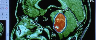
- signs of a possible brain tumor;
- traumatic brain injury (to determine the severity of damage, as well as the effect of prescribed treatment);
- recovery period after neurosurgery;
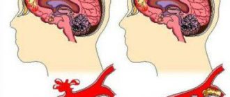
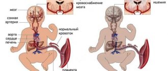
- circulatory disorders in the brain at an early age;
- inflammation of the brain - meningitis, encephalitis;
- pathologies in a newborn in the perinatal period;
- Cerebral palsy, autism;
- delayed mental and physical development of the baby for various reasons.
Electroencephalography in children is not only one of the most effective ways to determine the presence of various brain pathologies, this procedure is also absolutely harmless and can be prescribed at any age.
Dysrhythmia of background activity
It means irregular frequencies of electrical brain processes. This occurs due to the following reasons:
- Epilepsy of various etiologies, essential hypertension. There is asymmetry in both hemispheres with irregular frequency and amplitude.
- Hypertension - the rhythm may decrease.
- Oligophrenia – ascending activity of alpha waves.
- Tumor or cyst. There is an asymmetry between the left and right hemispheres of up to 30%.
- Circulatory disorders. The frequency and activity decreases depending on the severity of the pathology.
To assess dysrhythmia, indications for an EEG are diseases such as vegetative-vascular dystonia, age-related or congenital dementia, and traumatic brain injury. The procedure is also carried out in case of high blood pressure, nausea, and vomiting in humans.
Preparation for the procedure
It is a little more difficult to do an EEG for children than for adults, because not all children can sit quietly during the procedure. That is why it is important for parents to ensure that the minor does not create problems for doctors.
If the baby constantly shows aggression that cannot be controlled, then the doctor may advise you to start giving the child sedatives a few days before the procedure. Thanks to them, it will be possible to correct aggression and ensure that the baby sits calmly during the procedure.
Before the procedure, you should not give your child food or drinks that can affect the central nervous system. These include strong tea, coffee, chocolate and energy drinks. If the baby is forced to take medications, for example, anti-seizure medications, the specialist must be notified about this before the EEG. Some medications can distort data, which can lead to an incorrect diagnosis.
If there are earrings, hair clips, piercings or other metal objects on your head, they must be removed. Moreover, it is not recommended to wear even bodily jewelry, so as not to interfere with the procedure. The head should be washed before the EEG of the brain, and hair care products should not be used. There is no need to apply mousse, varnish, or foam, and it is also prohibited to braid hair or make dreadlocks. Before doing an EEG on a child, you need to feed him two hours before the procedure. If a person is hungry, then his blood sugar concentration will decrease, and this also affects the results. Separately, parents can be recommended to take their favorite toy with them so that they can distract the baby during the EEG. If the patient is in infancy, then it is best to do an EEG for the child during sleep.
When the subject is of conscious age, you need to first tell him about the procedure. It should be clarified that it is painless, and you should also warn how much time you will have to spend in a calm state, without making virtually any movements.
An EEG is not done for children if they suffer from acute infectious diseases, for example, ARVI. If the baby gets sick, then the procedure should be postponed for the required period. In cases where an examination has already been carried out previously, you need to take the previous results with you.
Types of ultrasound
There are three main types of ultrasound:
- Doppler ultrasound (USDG) is a study of the vessels of the neck, brain, head, etc. The main goal is to determine the patency of the vessel.
- Duplex ultrasound is so called because it includes two main functions - assessing the speed of blood flow in blood vessels and studying their anatomy. Briefly, this method provides visualization of blood clots, plaques, thickening of the vessel walls and their tortuosity.
- With triplex scanning, you can see the vessel against the background of tissue on the screen. In this case, mapping occurs, in other words, the vessel acquires different colors.
EEG IN CHILDREN
Carrying out an electroencephalogram in children has some peculiarities. It is difficult to administer to a small child under 1 year old. In this regard, the procedure is carried out while the baby is sleeping.
It is a little more difficult to conduct research for a child under 3 years old - he already understands a lot and needs to be persuaded and explained what they want from him. If the child willingly agrees, then the procedure is carried out while he is awake, otherwise you will also have to wait until he falls asleep.
Carrying out an EEG in children over 5 years of age, as a rule, does not cause difficulties; just talk to him.
Important. Start the explanatory process not in the doctor’s office, but at home, in a familiar environment, so that the child is ready and not afraid of an unfamiliar environment.
Contraindications
There are no special contraindications for the procedure, since it is performed even on newborns. If a child exhibits symptoms of a mental disorder or behaves aggressively, sedatives are administered to calm him down before the procedure begins.
It is not a contraindication, but it is worth paying due attention to the presence of damage to the epidermis in the places where the electrodes should be attached. They should not be installed on open wounds, severe rashes or sutures that have not yet healed after surgery.
What can REG of the head show?
Based on several characteristics read by the device - the magnitude and amplitude of the waves, the presence of an additional wave and its ratio in comparison with the descending main wave - it is possible to assess changes in brain activity, distinguish organic processes from pathological ones, and determine the area of brain damage (localization of the lesion).
After performing a REG of the brain vessels, when deciphering the results, the doctor will pay attention to:
- regularity of waves;
- characteristics of the top, what it represents, how it is rounded;
- what do downward and upward waves look like;
- determines the localization of the incisura, dicrotic tooth and the presence of additional waves.
What does the REG of the head show based on the description of the wave:
- Cerebral atherosclerosis is determined by the following indicators: smoothness of the wave, flattening of the apex and the absence of additional waves in the descending section.
- Atherosclerotic changes in cerebral vessels are characterized by a waveform in the form of an arch or dome.
- A decrease in arterial tone is determined by increased amplitude, high steepness, sharp peak of the wave, as well as when a large dicrotic wave with displaced localization is detected.
- An increase in arterial tone can be recognized by the low amplitude of the graphic image of the wave; the size of the anacrotosis is lengthened as its steepness decreases; additional waves are detected on the anacortex.
With vasospasm on the REG of the head, all vertices have a round shape.
In hypertension, the graphic lines are chaotic, different peaks are identified. Low and high amplitude of waves, additional waves appear in different parts of the graph, without a specific rhythm.
Interpretation of REG of brain vessels contains a definition of the type of behavior of vessels in a situation with or without load:
- The dystonic type is characterized by a predominance of reduced tone of the vascular walls and weak pulse filling. With dystonia, stagnation of venous blood in the vascular bed is possible.
- The angiodystonic type is similar to the first, but differs from the dystonic one in that the disturbance of blood flow is associated with mechanical changes in the vascular wall, which leads to a decrease in its tone. Poor blood circulation locally, at the site of the lesion.
- The hypertensive type of REG is characterized by increased tone of the vascular network, which can also cause difficulty in blood movement.
Important. The designated type in the REG interpretation of the vessels of the head is only a symptom that can be determined for many different diagnoses, and not a separate disease.
After performing rheocephalography of the cerebral vessels and receiving the transcript in your hands, you should not make diagnoses for yourself and begin treatment. These results are important for the doctor; in combination with other studies, they make it possible to establish an accurate diagnosis, which is a determining factor in the choice of treatment tactics.
DETAILS: Cerebral epiphysis cyst: signs, consequences, treatment
Examination stages
An EEG is performed on a child in several stages, during which practically nothing is required from parents. The entire procedure is supervised by a medical specialist, who will tell the child what needs to be done. To better understand the survey, it is useful to consider what steps it contains.
Procedure:
- The child will be taken to a specialized soundproof room where equipment and a couch are located.
- The minor will be asked to sit or lie down and then have a helmet with electrodes attached to their head. It comes in different types, most often resembling a fabric cap or rubber mesh.
- Before connecting the sensors, the scalp will be lubricated with a special gel; in rare cases, the epithelium will also be wiped with alcohol. Clips will be attached to the baby's ears that will not conduct current.
- Further actions depend on the patient's age. The babies are placed on the table or left in the mother's arms. You need to make him fall asleep, otherwise you won’t be able to get accurate results. The examination itself takes about 20 minutes.
- For older children, the procedure is performed a little differently. They need to sit on the couch and take a reclining position. The patient will need to relax and not turn his head, and there is no need to bend over. Such actions can significantly distort the result.
- Next, the specialist will record a background curve that records the state of the brain during calm and inactivity.
- After this, the child will need to open and close his eyes according to the doctor's command. Thanks to these actions, it will be possible to assess the processes of inhibition, as well as excitation.
- If the baby is already three years old, then the doctor will ask him to take a deep breath and exhale, or vice versa, breathe quickly. This test will help identify the presence of a tumor, epilepsy and other hidden disorders.
- At the last stage, the child undergoes photostimulation during an EEG. To do this, the baby will need to close his eyes, after which the doctor will present a light bulb to his head. It will light up and go out at a certain interval, and this test will be useful in identifying problems with the mental and mental development of the child.
The procedure is standard and usually does not cause any difficulties. In most cases it takes about 20 minutes, in extreme cases 30 minutes.
As an exception, the doctor can conduct special tests, for example, to check the reaction to sound, to record the functioning of the brain during deep sleep. The specialist may also ask the child to clench and unclench his fist, or take a light psychological test.
Much depends on the child’s condition, as well as what abnormalities he has or is suspected of having. The obtained indicators will help to understand whether everything is in order with the minor’s central nervous system, as well as whether therapy is helping. In some cases, the analysis is carried out not just once, but several times, if the situation requires it. All decisions regarding the examination should be made by the attending physician, and parents will only have to listen to his recommendations.
PREPARING A CHILD FOR EEG
For the examination, specific equipment will be placed on the child's head. Therefore, it is important to explain to an older child why this is required and that it is not scary at all.
- When performing the procedure on an infant, coordinate the choice of time for the study with his sleep schedule - the EEG will be performed during this period. Be sure to feed your baby before the session.
- For older children, imagine the procedure as an exciting adventure. This will allow you to safely attach the device to his head. Practice doing something similar at home so that your baby doesn’t get scared.
- To ensure peace of mind, take your favorite toys or books and use them to distract your baby if necessary.
- In some cases, the child will be required to do what the doctor says - open and close his eyes, undergo various tests, and breathe in a special way. Parents should warn their child against such situations.
- If the child takes any prescribed medications, be sure to use them according to the standard regimen before the study. However, the doctor who performs the EEG must know this in advance. The only exception is anti-seizure medications; they are discontinued three days before the EEG.
- The study is postponed if the baby has a cold. At the wrong moment, he begins to sneeze and cough, which will negatively affect the result.
- By the time the procedure begins, all foreign objects (earrings, hair clips) must be removed from the child and the hair must be loosened.
- By the time the procedure begins, the scalp and hair should be washed and dried, without a hint of any cosmetic substances.
- Before the procedure, the child should be calm, well-fed and well-rested.
- The manipulation is carried out in a soundproofed room, where there is a couch for the patient and EEG equipment.
- Immediately before the examination, an EEG device is placed on the child’s head. This is either a special cap with electrodes connected to it, or a rubber mesh on which the doctor will attach the sensors.
- Where these sensors contact the surface of the head, the skin is lubricated with gel.
- Special clips that are not electrically conductive are placed on the child’s ears.
- If the study is carried out on a baby, it remains in the mother’s arms or is placed on a changing table. The child should be asleep by now.
- The baby’s calm behavior during the procedure is an important point. It will play a special role in the interpretation of the results.
- The duration of the session is about 20 minutes.
- Older children do not lie down during the procedure, but are in a semi-sitting position. In this case, it is important to keep your head still - you cannot move it or tilt it forward.
Decoding the results
Usually the EEG results are ready the next day. The results are a paper with drawn curves or the same file on a computer. Based on the frequency, amplitude, rhythm of these curves and their jumps in certain areas, the doctor issues a conclusion, which is attached to the EEG results. The transcript must necessarily contain a description of the activity and dynamics of waves on the encephalogram, the interpretation of these results, as well as their correspondence to previously identified clinical symptoms.
The doctor must inform the parents of the results, clearly describe the results, and in case of any pathology, prescribe treatment or refer them to another doctor.
If there are any doubts about the EEG procedure, it can be done one more time or even several times, since it does not cause any harm to the child’s body. When going for the next procedure, parents should have the results of the previous one with them for comparison. This is necessary because at such an early age the nervous system is in constant development and results may vary significantly at different ages.
VARIETIES OF RHYTHM ACCORDING TO EEG RESULTS:
- Alpha rhythm - observed at rest over both cerebral hemispheres. If there is a disturbance in amplitude, frequency and amplification in any one hemisphere, then this indicates the presence of a neoplasm and indicates injury or hemorrhage. Increased amplitude, excessive activity in the crown and back of the head, increased reaction during deep inhalations indicate a delay in the child’s psycho-emotional state.
- The beta rhythm is normally present only in the frontal lobes. If a similar rhythm is detected in any other areas, it indicates a delay in mental development.
- Theta and delta rhythms normally differ only when the child is sleeping. In all other cases, these rhythms indicate dementia, psychosis or neurosis in the child.
- A flash (spike) is a confirmation of the presence of epilepsy (the diagnosis can only be reliably confirmed using an EEG).
ASSESSMENT OF THE PATIENT'S CONDITION
When the doctor begins to describe the electroencephalogram, he takes into account all the points and gives a conclusion. Negative indicators based on EEG results include:
- barrier symmetries in any hemisphere over 30%;
- manifestation of a wave with sinus rhythm;
- the presence of an arc-shaped rhythm;
- general rhythm increased by 50%;
- a beta wave with a sick amplitude indicates a traumatic brain injury;
- an amplitude of less than 50 volts means the child has a concussion.
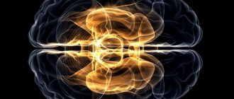
Electroencephalogram rhythms
An electroencephalogram of the brain shows regular rhythms of a certain type. Their synchrony is ensured by the work of the thalamus, which is responsible for the functionality of all structures of the central nervous system.
The EEG contains alpha, beta, delta, tetra rhythms. They have different characteristics and show certain degrees of brain activity.
Alpha - rhythm
The frequency of this rhythm varies in the range of 8-14 Hz (in children from 9-10 years old and adults). It appears in almost every healthy person. The absence of alpha rhythm indicates a violation of the symmetry of the hemispheres.
The highest amplitude is characteristic in a calm state, when a person is in a dark room with his eyes closed. When thinking or visual activity is partially blocked.
A frequency in the range of 8-14 Hz indicates the absence of pathologies. The following indicators indicate violations:
- alpha activity is recorded in the frontal lobe;
- asymmetry of the interhemispheres exceeds 35%;
- the sinusoidality of the waves is disrupted;
- there is a frequency scatter;
- polymorphic low-amplitude graph less than 25 μV or high (more than 95 μV).
Alpha rhythm disturbances indicate a possible asymmetry of the hemispheres due to pathological formations (heart attack, stroke). A high frequency indicates various types of brain damage or traumatic brain injury.
In a child, deviations of alpha waves from the norm are signs of mental retardation. In dementia, alpha activity may be absent.
Normally, polymorphic activity is within the range of 25–95 μV.
Beta activity
The beta rhythm is observed in the borderline range of 13-30 Hz and changes when the patient is active. With normal values, it is expressed in the frontal lobe and has an amplitude of 3-5 µV.
High fluctuations give grounds to diagnose a concussion, the appearance of short spindles - encephalitis and a developing inflammatory process.
In children, the pathological beta rhythm manifests itself at an index of 15-16 Hz and an amplitude of 40-50 μV. This signals a high probability of developmental delay. Beta activity may dominate due to the use of various medications.
Theta rhythm and delta rhythm
Delta waves appear in deep sleep and in coma. They are recorded in areas of the cerebral cortex bordering the tumor. Rarely observed in children 4-6 years old.
Theta rhythms range from 4-8 Hz, are produced by the hippocampus and are detected during sleep. With a constant increase in amplitude (over 45 μV), they speak of a dysfunction of the brain.
If theta activity increases in all departments, we can argue about severe pathologies of the central nervous system. Large fluctuations indicate the presence of a tumor. High levels of theta and delta waves in the occipital region indicate childhood lethargy and developmental delays, and also indicate poor circulation.
BEA - Bioelectric activity of the brain
EEG results can be synchronized into a complex algorithm - BEA. Normally, the bioelectrical activity of the brain should be synchronous, rhythmic, without foci of paroxysms. As a result, the specialist indicates which violations have been identified and based on this, an EEG conclusion is made.
Various changes in bioelectrical activity have an EEG interpretation:
- relatively rhythmic BEA – may indicate the presence of migraines and headaches;
- diffuse activity is a variant of the norm, provided there are no other abnormalities. In combination with pathological generalizations and paroxysms, it indicates epilepsy or a tendency to seizures;
- decreased BEA may signal depression.
Other indicators in the conclusions
How to learn to independently interpret expert opinions? Decoding of EEG indicators is presented in the table:
| Index | Description |
| Dysfunction of midbrain structures | Moderate disturbance of neuronal activity, characteristic of healthy people. Signals dysfunction after stress, etc. Requires symptomatic treatment. |
| Interhemispheric asymmetry | A functional disorder that does not always indicate pathology. It is necessary to organize additional examination by a neurologist. |
| Diffuse alpha rhythm disorganization | The disorganized type activates the diencephalic-stem structures of the brain. A variant of the norm, provided that the patient has no complaints. |
| Center of pathological activity | Increased activity in the area under study, signaling the onset of epilepsy or predisposition to seizures. |
| Irritation of brain structures | Associated with circulatory disorders of various etiologies (trauma, increased intracranial pressure, atherosclerosis, etc.). |
| Paroxysms | They talk about decreased inhibition and increased excitation, often accompanied by migraines and headaches. Possible tendency to epilepsy. |
| Reducing the threshold for seizure activity | An indirect sign of a predisposition to seizures. This is also indicated by paroxysmal brain activity, increased synchronization, pathological activity of midline structures, and changes in electrical potentials. |
| Epileptiform activity | Epileptic activity and increased susceptibility to seizures. |
| Increased tone of synchronizing structures and moderate dysrhythmia | They do not apply to severe disorders and pathologies. Requires symptomatic treatment. |
| Signs of neurophysiological immaturity | In children they talk about delayed psychomotor development, physiology, and deprivation. |
| Residual organic lesions with increased disorganization during tests, paroxysms in all parts of the brain | These bad signs are accompanied by severe headaches, attention deficit hyperactivity disorder in a child, and increased intracranial pressure. |
| Brain activity disorder | Occurs after injuries, manifested by loss of consciousness and dizziness. |
| Organic changes in structures in children | A consequence of infections, for example, cytomegalovirus or toxoplasmosis, or oxygen starvation during childbirth. They require complex diagnostics and therapy. |
| Regulatory changes | Fixed for hypertension. |
| Presence of active discharges in any departments | In response to physical activity, visual impairment, hearing loss, and loss of consciousness develop. Loads must be limited. In tumors, slow-wave theta and delta activity appears. |
| Desynchronous type, hypersynchronous rhythm, flat EEG curve | The flat version is characteristic of cerebrovascular diseases. The degree of disturbance depends on how much the rhythm hypersynchronizes or desynchronizes. |
| Slowing down the alpha rhythm | May accompany Parkinson's disease, Alzheimer's disease, post-infarction dementia, groups of diseases in which the brain can demyelinate. |
Online consultations with specialists in the field of medicine help people understand how certain clinically significant indicators can be deciphered.
CONTRAINDICATIONS
EEG is a method that differs favorably from some others precisely in the absence of absolute contraindications. In children with mental disorders, an anesthesiologist performs medicinal rehabilitation. Moreover, the drug is used for this purpose only after a laboratory examination.
Only a neurophysiologist should carry out the procedure and interpret the results. Even neurosurgeons and neurologists require detailed interpretation of the EEG. An incorrectly read EEG leads to an inaccurate diagnosis and the prescription of incorrect treatment. This will not only be ineffective, but even unsafe for the life of a small patient.
Electroencephalography - indications
An electroencephalogram of the brain is prescribed by a neurologist. Specialists in this profile are directly involved in the treatment of diseases of the nervous system. Speaking about what the EEG of the brain shows in young children, among the disorders to be examined, doctors name:
- dizziness;
- headache;
- fainting;
- insomnia;
- disruption of the endocrine system;
- neuroses (stuttering, nervous tics);
- nocturnal enuresis;
- delayed psychomotor and speech development;
- brain tumors;
- traumatic brain injuries;
- perinatal damage to the nervous system;
- cerebral palsy;
- vascular diseases of the head, causing cerebrovascular accidents.