Hello, dear readers. Today I want to talk about examining cerebral vessels using transcranial Doppler ultrasound.
Vascular pathologies are one of the most common causes of morbidity in the population, and this trend is increasing over the years.
Their development is partly provoked by a sedentary lifestyle and lack of physical activity, partly by an unbalanced diet and bad habits, and partly by stress and a violation of the general homeostasis of the body in combination with a hereditary predisposition.
General information about the Doppler method
The ultrasound ultrasound technique is based on two principles. It is based on “traditional” ultrasound, which is familiar to every person nowadays. The improvement of the method concerns the use of the Doppler effect, named after the physicist who discovered it. The essence of this phenomenon is that waves of various natures, including sound and ultrasonic ones, change their frequency when reflected from static and moving objects. And if a conventional ultrasound examination in B-mode shows the state of motionless tissues, then Dopplerography reflects the nature of the environment in motion. The new method allows you to obtain not only black and white images. With color mapping, dynamic objects can be highlighted in color.
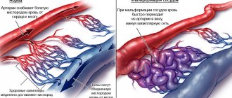
To put it simply, ultrasound shows the walls of blood vessels and adjacent tissues in the form of a picture, and ultrasound scanning is similar to a live broadcast of the movement of blood inside the vessels. Duplex Doppler sonography shows blood flow in black and white. There is also a triplex ultrasound, which allows you to visualize the movement of blood in a color image. In this case, the picture differentiates venous and arterial blood. The blood flow of the veins in the image is colored blue, the blood flow of the arteries is colored red.
For a more complete picture, Doppler ultrasound is usually combined with a B-mode study. The procedure begins with a traditional ultrasound, then the blood flow characteristics are clarified using Doppler scanning.
What is transcranial Doppler ultrasound of the brain?
This procedure helps determine not only the nature, but also the severity of the disorder. In addition to the blood supply to the brain, the study helps to assess the condition of the craniovertebral junction and the cervical spine.
Transcranial Dopplerography (sonography of cerebral vessels) is a method based on ultrasound scanning of cerebral vessels, determining their morphological and functional characteristics. Hemodynamic parameters (blood flow speed, volume, character) are assessed using the Doppler effect.
Why is the study prescribed? What does it show?
Doppler ultrasound can examine arteries, veins, and the capillary system. This procedure is prescribed for certain indications. The method helps to detect:
- pathological change in the shape of the vascular walls;
- the size of the lumen of blood vessels, their narrowing or expansion;
- cholesterol deposits, cholesterol plaques;
- thickness and tone of the vascular walls, visible sagging of the vessel wall during an aneurysm;
- blood consistency, blood clots, blood clots inside the vessels;
- the presence of an inflammatory process in the veins during congestion;
- abnormal nature of blood movement in the bloodstream (blood stagnation, flow turbulence, etc.);
- the speed of blood movement inside the studied vessels;
- deviations in the shape of capillary branches, the appearance of additional small vessels, “networks” in the studied body segment;
- malnutrition of the fetus during pregnancy;
- internal hemorrhages;
- compression, pinching of a vessel (for tumors, edema, uterine fibroids, difficult pregnancy, etc.).
All these indicators make it possible to identify many diseases affecting the circulatory system and not only it. During examination, pathologies are often diagnosed in tissues adjacent to the vessel, nearby organs, and dysfunctions of entire systems are detected.
The condition of blood vessels affects the central nervous system, spine, liver, kidneys, bones, joints. Vascular disorders lead to disruption of tissue nutrition, improper metabolism, deterioration of the general condition of the body, and weakening of its immune defense. Everything in the body is interconnected. Advanced blood vessel disease can lead to many undesirable consequences. And some diseases associated with the condition of blood vessels can even be fatal.
What parts of the body are examined?
There are different types of ultrasound examination depending on which areas of the body need to be examined.
Ultrasound scanning of the vessels of the neck and head is prescribed for migraines, fainting, tinnitus, sudden deterioration of vision, hearing, and coordination of movements. The reason for prescribing the procedure may be signs of hypertension, diabetes mellitus, osteochondrosis, or coronary artery disease. The study helps to identify a number of ailments: from vegetative-vascular dystonia, which not even everyone considers a disease, to life-threatening strokes. Information obtained using Doppler ultrasound also allows one to assess the risk of developing certain vascular diseases in healthy people.
In addition to transcranial Dopplerography, which studies the blood supply to the great vessels of the brain (neck and head), ultrasound dopplerography of the vessels of the upper and lower extremities is used. The most common reason for this procedure is varicose veins in the legs. In case of thrombosis, the vessels of the legs, as well as the arteries and veins of the abdominal cavity are examined. The condition of the jugular, renal, and other arteries of the abdominal region is carefully studied if diseases of the kidneys or genitourinary system are suspected. Pregnant women undergo Doppler ultrasound if they suspect kidney disease, placental pathologies, after falls, bruises, abdominal injuries, or vascular diseases.
Research Modes
The study uses several modes, depending on the specific area:
- scanning of the main arteries, which allows you to study vascular blood flow;
- duplex arterial and venous scanning, which gives the specialist a color two-dimensional sketch, which records the condition of the vessels in the neck, skull, and both lobes of the brain;
- triplex scanning, which complements the first two studies.
To display a comprehensive state of the brain vessels, main veins and arteries that provide blood flow, these modes are used simultaneously. Thanks to TCD of the vessels of the head and neck, a clinical picture is reliably obtained on a computer monitor using the radiation of an ultrasonic wave that penetrates the vessels and is reflected from the blood red cells, depending on the speed and direction in which they move.
The reflected waves are caught by special sensors and converted into an electrical signal, which forms a dynamic sketch. If there are blood clots, narrowing of the lumens and spasms, the flow of blood changes. This immediately gets a screen fix. To study the veins and arteries located in the brain, a sensor is installed on the bones of the skull, which have a minimum thickness.
Indications and contraindications for Doppler ultrasound
Doppler ultrasound is used in various fields of medicine: cardiology, neurosurgery, gynecology, phlebology, neurology, oncology and others. The procedure is prescribed for certain indications
- Numbness, swelling of the legs, weakness in the legs, cramps, lameness, pain and heat (coldness) in the legs are good reasons to examine the veins and arteries of the lower extremities.
- Severe headaches, head injuries, loss of consciousness, deterioration of memory, vision, hearing, hallucinations - all this raises suspicions of pathologies of the vessels that supply the central nervous system. For such symptoms, ultrasound scanning of the arteries and veins of the head and brachiocephalic vessels of the neck is performed.
- Ultrasound scanning of the renal vessels is necessary if the patient has pain in the kidneys, symptoms of renal failure, identified differences in the size of the kidneys, injuries to the bones and soft tissues in the lumbar region, development of hypertension with suspected complicated renal disease.
- Prematurity, disorders of the nervous system, and previous hypoxia are indications for prescribing Doppler sonography for newborns.
- Osteochondrosis of the upper spine, obesity, coronary heart disease, hypertension, angina pectoris, previous stroke or heart attack, vascular diseases in blood relatives is a reason to refer the patient for an ultrasound scan of the neck vessels.
- For pregnant women undergoing routine ultrasound, Doppler ultrasound may be prescribed unscheduled for kidney disease, diabetes, low hemoglobin levels, high blood pressure, benign uterine tumors, infections and late toxicosis. The threat of miscarriage, spontaneous termination of pregnancy, Rh conflict between the blood of mother and fetus, entanglement of the child with the umbilical cord, suspicion of placental abruption, incorrect presentation at a late stage - direct indications for an urgent ultrasound examination.
- The procedure is also indicated for all future patients under 18 and over 35 years old - an age when, according to statistics, the risk of pathologies is above average.
- Doppler scanning of blood vessels can be performed after surgical interventions to monitor the progress of the patient's treatment.
There are practically no contraindications to ultrasound-based diagnostic procedures. They are prescribed even to infants and very elderly people, not to mention all other age categories of patients. The study can be carried out even during sleep. The main conditions are immobility of the parts of the body being examined, cleanliness of the skin, and the absence of any acute inflammation. Doppler ultrasound is not prescribed only for exacerbation of inflammatory processes and skin diseases. In such cases, Doppler scanning is postponed until the current disease is cured.
Who is TCD indicated for?
A similar procedure is prescribed in such cases as:
- headaches of various etiologies;
- migraines and migraine-like attacks;
- dizziness that is associated with turning the head and changing body position;
- noise in the head and ears;
- feeling of lack of oxygen;
- suffocation;
- episodes of sudden dizziness and loss of consciousness;
- VSD;
- after TBI;
- neck pathologies;
- severe osteochondrosis;
- suspicion of abnormal development of the cerebral vascular system;
- clinical signs of disruption of the circulatory system in the brain.
Also, using TCD, you can diagnose coronary heart disease, myocardial infarction, cerebrovascular disease, transient ischemic attack, etc. Dopplerography is prescribed for people who have recently suffered a stroke, after which the physician needs to determine the degree of damage to the patient’s brain and blood vessels by the disease. A similar analysis is carried out for patients prone to motion sickness, with weather dependence and other external stresses. TCD should be mandatory for all people over 50 years of age. Especially for those who have relatives who have suffered a heart attack, stroke, arterial hypertension and atherosclerosis.
How does the procedure work?
A few hours before the ultrasound scan, it is necessary to avoid smoking, drinking strong tea and coffee, alcoholic beverages, and hot spices. It is necessary to adjust the intake of medications that affect the characteristics of the vascular sphere: vascular tone, their narrowing or dilation. It is better to cancel or interrupt taking antispasmodics (no-spa, etc. medications). The doctor usually warns about all this in advance when prescribing the procedure. If the patient is taking any medications, the doctor and ultrasound specialist must be informed about this.
The study is carried out using an ultrasound machine, which switches from B-mode to Doppler mode. The patient is required to take a horizontal or sitting position and, upon a signal from the health worker, remain motionless. The area of the body being examined is freed from clothing. A special gel is applied to the surface of the skin in this area to slide the ultrasonic sensor. Then, by rotating and moving this device, the area under study is visualized on the device monitor. The information is recorded for subsequent study by medical specialists.
The patient may be asked to change position (for example, tilt the head to examine the lateral vessels of the neck, raise an arm or leg). You may need to hold your breath and tense certain muscles.
The patient does not experience any painful sensations. The procedure itself lasts from 20 to 40 minutes, sometimes up to 1 hour. It depends on the complexity of the clinical case to be diagnosed. Sometimes 2-3 vessels are scanned, but no complications are detected. In other cases, it is necessary to find and examine several veins and arteries in different segments of the body, study neighboring areas, and the condition of their tissues. In addition, the ultrasound specialist needs to carefully record the results of the study.
Carrying out an examination - video
TCD of cerebral vessels does not require a special preparatory period. Before the examination, the patient is recommended to stop smoking and drinking alcoholic beverages.
Drinking coffee, tea and other drinks with a tonic effect is strictly prohibited. Treatment with vascular medications before a transcranial Doppler examination is prohibited, as they lead to a change in vascular tone, which adversely affects the reliability of the results.
Before the examination, the patient is placed on his back on a special couch. The examination site is the head, over which the doctor sits. A gel or ultrasound emission is first applied to it. Using transcranial Doppler, he touches certain points on the head, which makes it possible to locate the blood flow. During the manipulation, it is recommended, at the doctor’s request, to hold your breath, breathe frequently, turn your head, etc.
During the period of manipulation, the patient is strictly forbidden to talk and turn his head without the doctor's request . During the examination using transcranial Doppler, the patient does not experience any discomfort. Some patients complain of discomfort when briefly pressing on the neck, which is performed by the doctor several times during the examination. This manipulation is performed to assess vascular patency.
In accordance with the position of the acoustic window, the possibility of inspecting certain vessels is determined. The window is orbital, temporal and subcocipital. The temporal window is used to examine the middle, anterior and posterior cerebral arteries, and the carotid artery. To determine the condition of the vessels, the temporal region is examined.
The orbital window involves scanning the blood vessels through the eyeballs. With its use, the siphon of the internal carotid artery and the orbital arteries is assessed. The subcocytal window is located at the junction of the occiput and the spine. Allows examination of the basilar artery and intracranial segments of vertebral blood vessels.
There are several types of examination of cerebral vessels, which allows you to choose an acceptable option in accordance with the affected area.
Advantages and disadvantages of ultrasound scanning
Doppler ultrasound has many more advantages than disadvantages. The advantages of this diagnostic method include:
- non-invasive, painless;
- there is no need for special training;
- no need to administer contrast agents;
- there are no harmful radiations, such as from x-rays;
- there are no restrictions on the patient’s age;
- minimum contraindications for examination;
- speed of the procedure, efficiency of assessing the condition of blood vessels;
- information content, high accuracy of the information received.
The disadvantages of the method include:
- inability to capture the causes of dysfunction in the picture;
- insufficient information content in the study of venous pathologies, in which there remains a need to undergo computed tomography () or magnetic resonance imaging (MRI).
Average cost of transcranial Doppler ultrasound
The cost of the service varies. In government institutions, transcranial Doppler ultrasound is performed free of charge. In private clinics, the average price ranges from 2800–3300 rubles.
| City | Price in rubles |
| Moscow | 1800–4200 |
| Saint Petersburg | 2000–3750 |
| Kazan | 1900–3200 |
| Novosibirsk | 2000–3550 |
Transcranial Doppler sonography serves as an informative method for both routine and emergency diagnostics. The lack of special training, ease of implementation and accuracy make it indispensable for identifying blood supply disorders.
If you have ever undergone transcranial Doppler ultrasound, share your opinion in the comments.
How the results are assessed, data decoding
As a result of ultrasound examination, information is recorded in the form of a special protocol - a Dopplerogram. This is an image consisting of a spectrogram and an envelope curve. The specialist deciphers the results, evaluating each of these indicators separately, as well as in combination with each other.
Based on the Doppler ultrasound examination protocol, the doctor draws conclusions about the presence of diseases in the patient, the degree of their danger to life, and the risk of developing vascular diseases in a healthy person. The doctor’s conclusion is the reason to prescribe therapy for the detected disease. For relatively healthy people at risk, recommendations regarding prevention are usually given. This is a healthy lifestyle, giving up bad habits, changing your diet to a healthier one, and sufficient physical activity.