Previous Next In medicine, for a detailed study of blood vessels, a very informative diagnostic method has been used for more than 20 years - MR angiography of blood vessels. Alternative names: MRI of vessels, MRA, MRI angiography, MRI in angiography mode. There are the following types of angiography:
- MRI angiography of the veins of the head and neck - MR venography;
- MRI angiography of cerebral arteries - MR arteriography;
- MRI angiography with contrast.
Sometimes they are carried out together to obtain a more complete picture. Thanks to MRI of arteries and veins, it is possible to notice deviations in the vascular bed of any part of the body in a timely manner and prescribe effective treatment to the patient, thereby reducing the possibility of further development of the disease. It is a painless and safe type of examination without punctures, skin damage or radiation exposure.
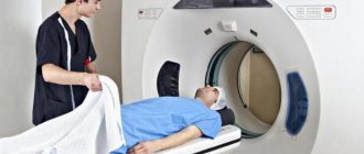
- MRI
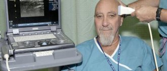
- Ultrasound
MRI tomograph:
Siemens Magnetom C
Type:
Open (expert class)
What's included in the price:
Diagnostics, interpretation of images, written report from a radiologist, recording of tomograms on CD + free consultation with a neurologist or orthopedist after an MRI of the spine or joint
What is vascular MRI angiography?
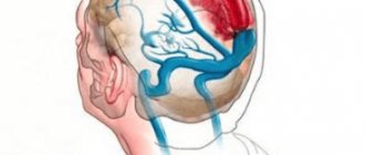
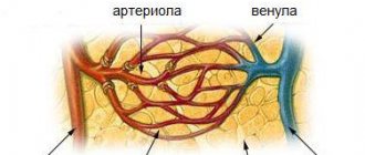
The human circulatory system includes the heart, blood vessels and lymphatic network. Vessels are usually divided into arteries, veins and capillaries. Arteries carry blood away from the heart, and veins are vessels that carry blood towards the heart. Capillaries are the most numerous and thinnest blood vessels in the body. They are located between arteries and veins, or connect veins to veins and arteries to each other. Gas exchange occurs in them. The vascular network permeates the entire body of the body from the brain to the lower extremities. Violation and failure of its work threatens a person with serious health problems, loss of capacity or threat to life. Vascular angiography allows doctors to monitor the condition of the vascular system.
The principle of obtaining an image of the vascular network on a magnetic resonance imaging scanner is based on the phenomenon of nuclear magnetic resonance. When the patient's body is placed in the machine and radiofrequency pulses are produced, the hydrogen atoms in the cells begin to vibrate. This resonance is captured by a computer tomograph, digitizes the data and, based on it, constructs three-dimensional images of the vascular bed. Since human blood contains a large amount of water, a strong resonance occurs, and the arteries and veins are very clearly visualized in the images.
Diagnostics
When preparing for an angiogram, both laboratory and instrumental methods are prescribed:
- Blood and urine analysis;
- Checking the functional state of the kidneys;
- ECG diagnostics;
- Fluorography;
- Examination by a therapist and anesthesiologist;
- Study of blood clotting ability, its Rh factor and group;
MRI angiography of vessels: which vessels can be examined?
Vascular MR angiography allows you to examine such areas of the human vascular system as:
- cerebral vessels
- neck vessels
- spinal vessels
- thoracic aorta, small arteries and veins
- abdominal aorta, small arteries and veins
- coronary vessels of the heart
- iliac arteries and veins
- vessels of the upper and lower extremities.
Initial appointment with a NEUROLOGIST
ONLY 1100 rubles!
(more about prices below)
Indications for MR angiography
Doctors prescribe magnetic resonance angiography of blood vessels on a tomograph if the patient exhibits the following symptoms:
- memory impairment;
- noise in ears;
- blurred vision;
- chronic migraine;
- sleep disturbance;
- dizziness,
- disorder of consciousness;
- intracranial pressure;
- weakening of attention;
- headache;
- brain contusion;
- before carrying out operations.
Contraindications for diagnostics
Angiography using a tomograph is performed according to indications at any age.
Unfortunately, some diseases may be contraindications for MRA diagnostic procedures. This type of diagnosis cannot be performed :
- in the presence of infectious or inflammatory diseases;
- in acute periods of mental illness;
- with existing heart failure;
- in case of allergy to iodine and iodine-containing drugs (when conducting a study using a contrast agent);
- with decompensated liver or kidney failure;
- if the patient’s body contains implants made of metal alloys, spoke-shaped structures for osteosynthesis, metal dentures;
- with an implanted pacemaker or insulin pump;
- in case of blood clotting disorders;
- in patients weighing more than 150 kg;
- during pregnancy and lactation.
It should be noted that structures made of titanium, copper, and ceramics located on the body (or inside it) are not contraindications to the procedure. All other metals (or alloys of them) under the influence of a magnetic field can move and cause damage to tissues and organs. If the patient has a fear of closed spaces (claustrophobia), the examination is performed under general anesthesia.
MRI or CT angiography, which is better?
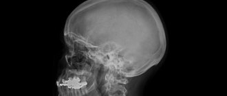
MRI and CT of vessels are the two main non-invasive methods for diagnosing angiopathologies at an expert level. The information content of magnetic resonance and computed tomography is very high, but each of them has its own disadvantages. The main disadvantage of vascular CT is the presence of radiation exposure to the body. This diagnostic method cannot be considered completely safe. The average radiation dose per scan can reach 5-7 mSv.
It is difficult to examine the vessels of the lower extremities using MRA. For this area, ultrasound of the vessels of the lower extremities or CT is better suited. Due to the low sensitivity of the MRI machine to weak blood flow, doctors may falsely diagnose pseudostenosis or occlusion during MR angiography. CT angiography of the vessels of the head will show an aneurysm better than MRI of the vessels of the brain.
With computed tomography, doctors have the opportunity to scan the vessels of almost the entire body at one time, which is impossible during MRA, where the examination is limited to certain areas of the body.
MR angiography, compared to computed tomography, has a longer duration of examination, which does not always allow examining patients in serious condition or young children who will require general anesthesia to remain immobile.
The advantage of MRI using the angiography protocol compared to CT is the absence of harmful effects on the body due to the absence of ionizing radiation and toxic contrast agent containing iodine.
The final decision about whether MRI or CT angiography is better should be made by the attending physician. His knowledge of the medical history, patient's health status and diagnostic goals will allow him to make the right choice between magnetic resonance or computed tomography angiography of blood vessels. If the patient himself decides that he needs to undergo vascular diagnostics for preventive purposes, then it is better to start his diagnostic path with a safe MRI of the vessels of the brain and neck or ultrasound of the vessels of other parts of the body.
Previous Next
MRA of cerebral vessels
Magnetic resonance angiography (MRA) of cerebral vessels
Introduction
MRA stands for magnetic resonance angiography. MRA of cerebral vessels is performed to evaluate disturbances in the arterial blood flow system to the brain.
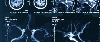
Three-dimensional (3D) time-of-flight (TOF) MRA angiography is the most common method used to evaluate the arterial blood supply to the brain.
MRA 3D TOF provides higher signal-to-noise and short image processing time. MRA of the brain is easy to perform and does not require contrast enhancement.
angiography ( TOF)
This is a technique for obtaining contrast between fixed tissues and blood flow by influencing the magnitude of magnetization.
The magnitude of magnetization from moving spins is very large compared to that from stationary spins. This leads to the formation of a high signal from moving blood spins and a reduced signal from stationary tissue spins. Time-of-flight (TOF) angiography uses the longitudinal vector of magnetization to manipulate images. The 3D sequence allows for thinner slices with a smaller voxel size (0.6-1.0 mm).
Indications for MRA of cerebral vessels:
- Aneurysm, stroke, vasospasm, vasculitis;
- Occlusion and stenosis of the internal carotid artery;
- Occlusion and stenosis of the cerebral artery;
- Occlusion and stenosis of the basilar artery;
- Arteriovenous malformation (AVM);
- Vascular atherosclerosis
Contraindications:
- Any electrical, magnetic, or mechanical activated implant (eg, pacemaker, insulin pump biostimulator, neurostimulator, cochlear implant, and hearing aids);
- Intracranial (intracranial) aneurysmal clips (except titanium);
- Pregnancy (if the risk outweighs the benefit);
- The presence of ferromagnetic surgical clamps or staples;
- Presence of a metallic foreign body in the eye;
- The presence of metal shrapnel or bullets in the body.
Preparing the patient for an MRI examination of the brain
- Before the scanning procedure, it is necessary to obtain the patient's written consent for the study;
- Ask the patient to remove all metal objects, including keys, coins, rings, plastic cards with magnetic stripes, jewelry, hearing aids, and hairpins;
- If necessary, provide an accompanying person for patients suffering from claustrophobia (for example, a relative or employee);
- Offer the patient earplugs or headphones with music for additional comfort;
- It is necessary to explain to the patient the essence of the procedure and the procedure for its implementation;
- Warn the patient to remain calm during the procedure;
- Note the patient's weight.
Position during MRA of cerebral vessels
- Lying on your back with your head forward (towards the magnet);
- Position the head in the head coil and immobilize it with pillows;
- For additional comfort, place bolsters under the patient's feet;
- The center of the laser beam is focused over the bridge of the nose.
Recommended protocols, parameters and planning
Localizer
The first step in sequence planning is to take images in 3 planes. Exposure times are less than 25 seconds, producing low-resolution T1-weighted images.
Series T2 turbo spin echo, axial cut
Planning of axial sections on the sagittal plane; The angular position of the block should be parallel to the knee and splenium of the corpus callosum. These sections should completely cover the brain from the crown to the level of the foramen magnum. Check the location of the block on 2 other planes. An appropriate angle must be obtained in the coronal plane with the head in a tilted position (perpendicular to the line connecting the third ventricle and the brain stem).
Options
| TR | T.E. | SLICE | FLIP | PHASE | MATRIX | FOV | GAP | NXA(AVRAGE) |
| 3000-4000 | 100-120 | 5mm | 130-150 | R˃L | 320X320 | 210-230 | 10% | 2 |
Time-of-flight angiography 3D ( time-of-flight, TOF)
Planning of axial 3D blocks on the sagittal plane; The angular position of the block should be parallel to the knee and splenium of the corpus callosum. These sections should completely cover the Circle of Willis from the level of the corpus callosum to the level of the foramen magnum. Check the location of the block on 2 other planes. An appropriate angle must be obtained in the coronal plane with the head in a tilted position (perpendicular to the line connecting the third ventricle and the brain stem). Applying saturation bands over the axial block will reduce artefacts from venous vessels in the images.
The Magnetization Transfer Contrast (MTC) technique is widely used in cerebral TOF MRA as a means of controlling image contrast. In TOF MR angiography, contrast between normal static tissue and blood flow is achieved through a series of T1 saturations of hydrophilic tissue, due to repeated excitation pulses. Additional suppression of immobile hydrophilic tissues using magnetization transfer pulses provides visualization of small vessels.
Options
3D - time-of-flight (TOF)
| TR | T.E. | FLIP | NXA | SLICE | MATRIX | FOV | PHASE | GAP | MTC |
| 30-40 | 5-9 | 25 | 1 | 0.7 mm | 256X256 | 250 | R˃L | -33% | ON |
Maximum Intensity Projection (MIP)
MIP is the most commonly used method for processing vascular MR imaging. MIP allows 2D image projections to be reconstructed from 3D data using a ray tracing algorithm that produces an image of white pixels as the maximum intensity signals of the region of interest.
MIP is the most commonly used post processing technique in MRI vascular studies. MIP reconstructs a 2D projection image from 3D data by a ray tracing algorithm, which produces an image of white pixels representing the highest intensity signal in that location within the examined volume.
CLICK ON THE ITEMS BELOW TO CHECK THE SCAN
What will MR angiography show?
MRI of arteries, sinuses and veins reflects a wide range of vascular abnormalities. Tomography in angio mode is capable of well visualizing:
- arteriovenous malformations;
- stenosis of the vascular bed
- thrombosis;
- vascular occlusion;
- bifurcations and kinks of blood vessels;
- vascular parietal tumors;
- acute circulatory disorder.
MR angiography allows you to find the cause of swelling of the legs and distinguish varicose ulcers from thrombotic ulcers. A non-invasive method for diagnosing blood vessels in medical centers in St. Petersburg is used quite often for planning surgical interventions.
How is MR angiography performed?
The magnetic resonance angiography procedure is atraumatic for the patient. The subject simply needs to remove all metal from himself and leave all electronic devices outside the MRI room, lie down on the tomograph table and not move during the scan. The MRI machine and medical specialists will do the rest themselves. During a tomographic session, two-way communication is always maintained with the patient. For contact with the doctor, a microphone is installed in the tomograph. In addition, the patient is given a signal bulb before the examination begins. If he begins to feel unwell or has a panic attack, he can press a signal bulb and a specialist will come to the rescue from the treatment room.
Based on the diagnostic results, the client in most medical centers receives a written x-ray report on the day of treatment, summarizing the presence of all disorders and pathologies, tomographic images and recommendations for further actions.
Special techniques for magnetic resonance imaging studies
MR diffusion
Diffusion is based on Brownian motion - the chaotic movement of small particles suspended in a liquid or gas, occurring under the influence of the thermal movement of molecules. In body tissues, Brownian motion is partially suppressed by tissue components - large molecules, intracellular structures, cell membranes and walls. In most tissues, diffusion is multidirectional (isotropic), but in well-structured tissues, such as the white matter of the brain, water diffusion is relatively directional. To obtain a diffusion-weighted image, an MR tomograph with a magnetic field induction of 1.5 or 3 Tesla is required. This is the most effective technique in determining the organization of the brain.
Perfusion magnetic resonance imaging
The principle of bolus injection of a tracer is the same for all perfusion diagnostic methods. A contrast agent (gadolinium compounds in the case of MRI) is injected intravenously and the area of interest is simultaneously scanned. As a bolus of gadolinium contrast agent passes through the vessel, it changes the magnetic field around it, reducing the signal intensity.
The degree of signal reduction is proportional to the level of blood flow. Since this process has a certain extent over time, it is possible to construct a curve of signal intensity versus time. The data is subjected to mathematical processing to determine regional circulating blood volume (rCBV), regional blood flow velocity (rCBF), mean transit time (MTT), and time to peak contrast concentration (TTP).
Dynamic research should be carried out in a short time. Thus, the bolus passes through the brain approximately 18 s after injection.
This method makes it possible to study blood flow in detail and build a map of the absolute volume of blood in the brain.
Spectral MRI
This technique is based on determining the distribution of a number of biologically active compounds. This makes it possible to clarify the diagnosis of hemorrhage; an increased lactate content and a decrease in acetylaspartate are detected (the predominance of the process of anaerobic glycolysis).
Functional MRI
This technique is based on the fact that cerebral blood flow and the activity of brain neurons are interconnected. If an area of the brain is currently active, then the blood supply to that area is increased.
What determines the price of MPA?
| Service | Price according to Price | Discount Price at Night | Price by Discounts During the Day |
| from 23.00 to 8.00 | from 8.00 to 23.00 | ||
| MRI of cerebral vessels (MR angiography) | 3300 rub. | 2400 rub. | 2900 rub. |
| MRI of neck vessels (MR angiography of the neck) | 3500 rub. | 2400 rub. | 2900 rub. |
| MRI of the brain and cerebral vessels | 6800 rub. | 4800 rub. | 5800 rub. |
| Comprehensive MRI examination of brain and neck vessels | 6600 rub. | 4800 rub. | 5800 rub. |
| MR arteriography | 3300 rub. | 2400 rub. | 2900 rub. |
What determines the cost of tomography?
Tomograph power
Applying Contrast
Personnel qualifications
Promotions and discounts
How to prepare for the procedure
Do not eat food 8-10 hours before the procedure, and do not drink water 4 hours before the examination. Immediately before the angiogram, the patient must remove all metal objects, as they can distort the picture. If the patient is anxious, an injection with a sedative is given.
After the procedure, the patient is given bed rest during the day. The patient's condition is monitored. Body temperature is measured and the puncture site is examined if contrast is administered. A day later, the bandage is removed, if there are no hemorrhages at the puncture site and the person’s general satisfactory condition, he is discharged.
Author of the article: Neurologist Konstantin Olegovich Makheev.