Currently, there is an increase in the incidence of cancer of various organs throughout the world. The human central nervous system is no exception. Tumors of the sella turcica, which include craniopharyngioma, are quite common. This neoplasm is detected annually in an average of one to two people out of a million people.
Why this pathological condition develops, how it manifests itself, what the treatment methods are, you need to know in order to take timely measures and avoid complications.
Causes of pathology
Few people can boast that they have never experienced a headache. This is one of the most common pain syndromes that affects the population. The diagnosis of craniopharyngioma is most often detected in children between 5 and 14 years of age.
There are several causes of the disease:
- Hereditary factor.
- Transference of serious illnesses during pregnancy. These include kidney pathologies, tuberculosis, and diabetes.
- Early toxicosis during pregnancy can affect the development of a benign tumor.
- Intrauterine infections that led to the development of fetal intoxication.
- Radioactive radiation to which a woman was exposed during pregnancy.
The complexity of the disease is that the cause that provoked its appearance is almost impossible to establish.
Symptoms
Symptoms of craniopharyngioma do not appear at the initial stage of tumor formation. After rapid progression, the following signs appear:
- various visual disorders due to compression of the optic nerve;
- severe and prolonged headaches;
- severe nausea;
- endocrine disorders;
- dysfunction of the adrenal glands;
- problems with sex life in men;
- absence of monthly menstruation in women;
- obesity;
- lethargy, slowness;
- memory problems;
- epileptic seizures;
- urinary incontinence.
The patient may experience growth problems (dwarf syndrome), excessive accumulation of fat deposits. The pathological process occurs individually, from several months to ten years.
Symptoms
The main sign of the presence of brain craniopharyngioma is compression of the brain structure. This leads to increased intracranial pressure, which causes a number of symptoms, ranging from headaches to the development of mental retardation. Symptoms of a benign tumor differ in adults and children. The main signs are as follows:
- Retarded growth and development is the most striking manifestation of the presence of a tumor in childhood. The child has delayed development both in growth and in physical and mental indicators. Most often, it is these signs that help determine the presence of craniopharyngioma.
Due to tumor growth, the development of the anterior pituitary gland slows down. This leads to a decrease in hormone production. As a result, there is a delay in physical and mental development, and frequent bone fractures in the child. In situations with adolescents, puberty may occur very early or, conversely, too late.
- Headaches – this symptom is observed in all patients, regardless of age. The pain occurs with increasing intensity, while the interval between attacks decreases. Most often, pain is associated with increased intracranial pressure.
- Obesity – 20% of patients with brain tumors have obesity and diabetes insipidus.
- Visual impairment - this symptom refers to the early manifestation of pathology. It occurs in almost all patients in more than 90% of cases. Both children and adults suffer from vision impairment. The danger lies in the fact that symptoms are detected already at the stage of optic nerve atrophy. At this stage, the irreversible process of loss of visual function is already underway.
- Mental disorders are typical for adult patients. In this case, changes occur unnoticed by the patient himself.
The most dangerous phenomenon is the growth of cranipharyngioma into the area of the cerebral ventricles. This can lead to serious sleep disturbance.
Almost all patients whose illness lasts for a long period experience a decrease in intellectual abilities. This is due to the fact that the formation damages the brain and leads to disruption of higher cortical functions.
...and how does it manifest itself?
This tumor may not manifest itself for quite a long time; the first signs most often appear closer to 10 years or later. The process usually proceeds slowly with the appearance of neurological symptoms during the formation of cysts and gradually acquires the character of a sluggish chronic disease.
Neurological symptoms depend on the size, growth pattern of the tumor and the effect on certain areas of the brain. Main clinical manifestations:
- attacks of headache , which are not relieved by analgesics and are accompanied by vomiting (cause - impaired outflow of fluid from the left ventricle compressed by the tumor and increased intracranial pressure);
- visual disturbances (compression of the chiasm causes various types of narrowing and loss of visual fields, rarely - one-sided blindness);
- pituitary insufficiency (underdevelopment and dysfunction of the genital organs, retardation in physical development, short stature);
- endocrine disorders (obesity, diabetes insipidus with diabetes up to 10 liters per day, galactorrhea - secretion of milk from the mammary glands);
- sleep disorders;
- increased blood pressure;
- sometimes psychopathic manifestations - schizophrenia-like states, depression, epileptic seizures;
- lesions of the cranial nerves - trigeminal neuralgia, disturbances of smell or other manifestations.
Diagnostic methods
The diagnosis of craniopharyngioma is made only after a thorough comprehensive examination of the patient. The patient should receive consultation from a number of specialists, including a cardiologist, neurologist, endocrinologist, and ophthalmologist.
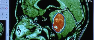
The X-ray method is used to determine the size of the tumor and wall erosion. The patient is required to undergo tests to determine hormone levels and determine the functioning of the thyroid gland. Additional research methods are MRI and CT. With their help, the specialist obtains an image of brain tissue in layers. This allows you to determine the exact location of the tumor.
Types of brain craniopharyngioma
This type of tumor is always benign, but some types can cause very serious complications. As a result of special diagnostics, the neurosurgeon will determine exactly what type of craniopharyngioma you have:
- soft with minor cystic cavities;
- cystic - one or two formations filled with fluid;
- rocky, thoroughly impregnated with calcium salts;
- mixed, combining the characteristics of the above types.
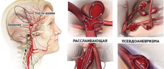
There is also a classification based on the location of the tumor. The infundibular type of tumor is usually located in the third ventricle and most often affects the oculomotor nerve. Suprasellar craniopharyngioma can cause destruction of the skull bones, damage to the internal trunk and optic nerve. Stem craniopharyngioma is located along the stalk of the pituitary gland and causes changes in the structure of the vessels at the base of the brain.
Treatment
Therapy for brain craniopharyngioma involves radical removal of the tumor in the pituitary gland in order to eliminate compression and restore function. The operation is not always possible to carry out in full. The difficulty is associated with the severity of access to the localization of cysts.
Surgical intervention can be of several types:
- Palliative surgery is an effective option, but often becomes a temporary solution.
- Shunt surgery - used for the development of dropsy, reduces intracranial pressure. Most often, bypass surgery is considered as the initial stage of treatment, followed by removal of the tumor.
- Aspiration of cysts – this must be performed as an emergency. Aspiration is the installation of an Ommaya system into the cyst, which will provide control over it. Most often, the system is used for further irradiation of the cyst.
- Radical intervention - today in medicine this type is considered the most effective. The tumor capsule is completely removed. If it is not possible to completely remove the capsule during the operation, radiation therapy is prescribed after surgery. Most often, radical removal is combined with radiation techniques. This helps relieve the patient from intracranial pressure and prevent tumor growth.
If the location of the tumor allows, surgery is performed through the nasal passages. This type of operation reduces the risk of possible complications, and also reduces the postoperative recovery period.
If the patient is diagnosed with a large cystic formation, then open implantation of an intracystic catheter is performed.
What does it look like?
Craniopharyngioma can be of two types in its structure: a neoplasm made of dense tissue and a cystic one, consisting of cavities with yellow or amber liquid. The diameter of this tumor is on average 2 - 3 cm; larger sizes are rare. Over time, the tumor may undergo structural changes.
In dense layers, necrosis, liquefaction and melting of the tissue begins. As a result, cysts appear with fluid containing large amounts of protein, cholesterol crystals, fatty acids and salts. Histological studies show that the tumor contains accumulations of cells of varying degrees of differentiation - epithelium of embryonic and epidermal origin in the form of papillomas or adamantines.
The tumor originates from the remnants of the pituitary tract, so it is usually located directly in the pituitary gland (usually above it) and can grow in various directions:
- forward to the site of the optic chiasm, located in the anterior wall of the third ventricle of the brain;
- upward to the region of the third and lateral ventricles;
- up and along the sides of the sella turcica (it contains the pituitary gland);
- into the cavity of the sella turcica;
- behind the sella turcica.
This division is of practical importance for choosing an operative approach during surgical treatment of a patient.
Possible complications
The issue of radical surgical intervention is still actively discussed among neurosurgeons. This concerns the possibility of selecting a more optimal method in cases with children and adolescents.
Many neurosurgeons are critical of this method due to the risk of damage to the hypothalamus, as well as a high percentage of relapses. Complications may include infection, bleeding and injury to soft tissue, nerves and blood vessels.
If the craniopharyngioma is located in a very hard-to-reach place or the person has concomitant diseases for which surgical intervention is prohibited, neurosurgeons prescribe only radiation therapy.
Previously, the course of treatment consisted of radiation therapy over several weeks. In this case, the head area was irradiated. The procedures took place five times a week. The disadvantage of this method is the inability to completely remove the tumor. All that happens is that its growth stops.
Today, most clinics use a more innovative method - radiosurgery . This includes Gamma Knife and Cyber Knife. The essence of the method is that the brain is exposed to beams of radiation at different angles using a special apparatus. Unlike surgery, the effect of radiosurgery is not observed immediately, but only after several weeks.
The big advantage of this method is the reduced risk of complications. In addition, the procedures are performed without anesthesia. Radiosurgery is characterized by the precise direction of the beam, which is adjusted taking into account MRI readings. After this treatment method, the patient does not require a recovery period.
1.What are craniopharyngiomas?
Craniopharyngiomas
- benign neoplasms, most often formed in the hypothalamic-pituitary region of the brain, or more precisely, above the sella turcica (suprasellar).
The tumor is formed by embryonic cells of the so-called Rathke's pharyngeal pouch. Craniopharyngiomas are characterized by the presence in their structure of both cystic and solid (solid) components. For this reason, as the tumor grows, cysts may form in it, filled with yellowish-brown liquid contents with a high percentage of cholesterol and protein.
In most cases, craniopharyngioma is found in children 5-14 years old, but can also develop in adult patients.
A must read! Help with treatment and hospitalization!
Postoperative period
During the postoperative recovery period, the patient is prescribed:
- Drugs that help relieve swelling and reduce inflammation in the soft tissues of the brain.
- Diuretic drugs.
- Since the presence of a benign tumor cannot exclude the occurrence of relapses, a course of radiation therapy is prescribed after surgery.
If necessary, after the recovery period, hormonal therapy may be prescribed.
Residual effects from a removed tumor may remain. The patient may experience decreased vision, increased daily urine volume, decreased potency, and menstrual irregularities.
Treatment of craniopharyngioma in children, surgery
The child's oncologist will determine the specific course of treatment based on several factors, including age, general health and medical history, the type, location and size of the tumor, and the severity of the disease.
Treatment for craniopharyngioma includes:
- Neurosurgery. Surgery is almost always the first step in treating craniopharyngioma. Doctors try to remove as much tumor as possible without damaging the brain. Even if the tumor is completely removed, the tumor may grow back. In some cases, the surgeon cannot remove the entire tumor with surgery. This is associated with the risk of damage to other tissues of the head. These include the optic nerve, hypothalamus and carotid artery. The surgeon will remove as much of the tumor as possible.
- Radiation therapy. High energy waves are used to damage or kill the tumor. Focused radiation treatment can be used to treat tumors that cannot be removed by surgery. Radiation is usually given daily over a 6-week period. Preference is given to proton radiation therapy. Radiation is given only to the area of the tumor, as it usually does not spread to the brain and spine.
In some cases, your child may need experimental chemotherapy if the tumor comes back after radiation.
note
Patients who have had their pituitary gland removed due to a diagnosis of craniopharyngioma will require hormone replacement therapy. This means that removal of the tumor invariably leads to partial or complete hormonal deficiency of the pituitary gland. These children will require lifelong hormone therapy, which is prescribed by an endocrinologist.
Clinical trials, or studies evaluating new treatment approaches, are a chance for many children with brain tumors to be cured completely.
Forecasts
The prognosis for future life after cerebral craniopharyngioma depends on the success of the operation and the size of the tumor. Lethal outcomes during surgical intervention are no more than 10%. Most often this occurs due to damage to the hypothalamus.
Up to 85% of operated patients survive the surgical treatment and recovery period. Statistics are taken over five years. Relapses are possible throughout the entire period. The most dangerous complications are those that can arise within a year after surgery.
Almost all patients should be registered with an endocrinologist and regularly undergo hormonal therapy.
Prevention of harmful effects on pregnant women in the first trimester is used as preventive measures for the occurrence of the disease. Any types of intoxication and exposure to radiation must be excluded. Early registration of a pregnant woman plays an important role. This will allow us to exclude the possibility of a tumor at the initial stage.
If the occurrence of cerebral craniopharyngioma cannot be avoided, it is important to make a diagnosis in the early stages of the disease. This will help to avoid complications and prescribe timely surgical treatment.
Much attention should be paid to the patient’s quality of life after surgery for cerebral craniopharyngioma. Survival can only be ensured if a person undergoes a comprehensive examination by a number of specialized doctors. Monitoring of the patient should be lifelong.
Diagnostics
The simplest and most accessible method is skull radiography. The presence of petrification in the chiasmal-sellar region, changes in the shape and size of the sella turcica serve as reliable criteria, which, together with clinical manifestations, make it possible to make a diagnosis.
Changes that are detected using MRI or CT are quite characteristic and in the vast majority of cases allow us to clarify the diagnosis. Each method has its own advantages; CT provides more complete information about the structure of the tumor, the density of the cysts and the presence of areas of calcification. MRI differentiates petrification worse, but tumor measurements and its topography are more clearly revealed.