Vascular formations, especially in such an important location as cerebral structures, are relatively common. According to statistics, this kind of disorder is present in almost 3% of the world's population.
There are even more cases when the pathological condition is just developing, taking shape, but the prerequisites already exist. As a rule, diagnosis is greatly delayed, since the disease hardly makes itself felt until the critical moment.
It turns out that the patient understands what is happening so late that the treatment itself becomes dangerous.
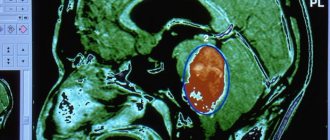
A cerebral aneurysm is a wall protrusion of large arteries of cerebral structures, which is accompanied by a decrease in the elasticity of the internal walls of the vessel, a change in the density and strength of tissue at the local level.
The condition is dangerous and has its own code I67.1 according to ICD-10. If nothing is done, death from aneurysm rupture and massive hemorrhage cannot be avoided. The only question is how soon this will happen.
Unfortunately, the pathological process either does not produce any symptoms, or the signs are so scant that it is impossible to discern such a dangerous condition right away.
The prospects for recovery directly depend on the stage of the disorder and the moment of initiation of therapy. There is absolutely no time to hesitate. It's a matter of life and death.
What is an intracranial aneurysm?
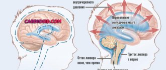
When an aneurysm occurs, its cavity fills with blood, gradually expanding, putting pressure on nearby brain tissue and nerves, which affects the person’s condition.
The main danger of the formation is that it can rupture under pressure. As a result, blood flowing from the vascular bed fills the brain tissue adjacent to this area. Hemorrhage occurs in the subarachnoid (subarachnoid space) or directly in the substance of the brain.
This occurs due to a violation of the structure of the vessel:
- Under the influence of various factors, changes in the internal elastic layers create the prerequisites for the formation of protrusion.
- With further destruction of the muscle layer, the elastic membrane undergoes changes; integrity is maintained only at the base.
- The thinnest section is the dome, consisting of an inner layer - the intima. This is where the damage occurs.
This pathology can occur in any part; it often forms at the site where the artery branches; this is facilitated by turbulence in the blood flow in this area.
Causes
As for specific provocateurs of the pathological process:
- Congenital anomalies, mutations. They occur relatively often, but the degree of change is so small that it is very difficult to suspect deviations. Not always, but it happens.
- Complicated anamnesis. If there was at least one elder in the family with a similar problem, the risks automatically increase by 50-70%. It is worth taking a closer look at the people of the side branch: brothers, sisters. The probability increases by a quarter or a little less. It makes sense to undergo regular examinations with a neurologist or therapist; this is an important factor in prevention.
- Encephalitis. A type of inflammation of brain structures.
- Meningitis. The same thing, but of a different origin. Both pathological processes lead not only to an aneurysm, but also to immediate destruction of nerve tissue, deficiency, and focal symptoms. Be it dementia or speech, hearing, or vision disorders.
- Brain injuries. TBI. From concussions to open injuries, penetrating wounds and more. The probability is calculated based on a specific pathological process.
- Smoking. Nicotine and harmful substances affect blood vessels, their strength, and the elasticity of the internal lining.
- Alcohol consumption.
- Drug use.
- Arterial hypertension. Stable increase in pressure in the bloodstream. At any stage, it increases the likelihood of a pathological process.
- Heart diseases. IHD, angina pectoris.
- Tumors. They can compress blood vessels, thereby reducing their regenerative capabilities and adaptive potential.
- Atherosclerosis of cerebral vessels.
- Vasculitis. Autoimmune inflammation of the internal lining of blood vessels. Potentially lethal disorders. They manifest themselves in dozens of ways. Extremely dangerous in themselves. They require treatment under the supervision of a rheumatologist and as quickly as possible.
The causes of aneurysm are vascular, autoimmune, traumatic, cardiogenic pathologies.
If there is at least one of these disorders, and the aneurysm has not yet formed, the patient is classified as a high-risk group and is closely monitored.
Causes of development of cerebral aneurysm
At the present stage, a holistic picture of the development of the disease is still at the development stage. Violation of the vascular wall can have a congenital or acquired etiology; it is believed that a combination of several factors leads to its development.
Congenital causes:
- The reason for the formation is often congenital changes in the muscle layer of the arteries or a lack of collagen in the tissues. The high sensitivity of blood vessels to mechanical stress contributes to their thinning.
- Changes are caused by hemodynamic disturbances associated with increased pressure or uneven blood flow due to narrowing of the cardiac aorta. Often found in children.
- An intracranial aneurysm develops during the formation of arteriovenous anastomoses. That is, arterial blood, bypassing the capillaries, is released under pressure into the calmer venous blood flow. The vessels cannot withstand the stress and become deformed.
Secondary causes that change the structure of blood vessels are also named:
- arterial hypertension;
- Carotid artery aneurysm is 90% provoked by atherosclerosis;
- past infectious diseases;
- head injuries;
- oncology;
- use of drugs, especially cocaine;
- smoking and alcohol;
- Sometimes the cause of the development of intracranial aneurysm is the use of hormonal drugs.
It has been proven that women develop protrusion much more often than the stronger half of humanity. Aneurysms are often companions of cancerous formations; they accompany primary and metastatic tumors in the head and neck, and also appear in children with congenital tumors.
Development mechanism
The formation of the pathological process is based on a group of common pathogenetic factors. Which ones:
Congenital vascular anomalies
They occur more often than you might think. Another thing is that such conditions and characteristics of the body do not always lead to something as dangerous as an aneurysm.
We are talking about a pathological process that is formed in the prenatal period.
Whether this is due to a mutation or something else does not have much clinical significance. Since this does not affect the consequences in any way, and there is no point in clarifying the issue yet: prevention of genetic abnormalities is not yet possible.
Previous brain injuries
Cerebral structures are not only nerve tissue, but also an abundance of blood vessels. When the fibers are damaged, the arteries themselves undergo changes.
This can have a very negative impact, although it is impossible to guarantee the development of an aneurysm. These are just theoretical risks.
It takes some time: a year or two to monitor the patient’s condition.
Tissue irradiation
Radiation causes cell destruction and changes at the molecular level. The result is decay, gradual or rapid. Depends on the dose.
The result is softening of the vascular wall, tissue destruction at the local level.
Attention:
Especially often this consequence occurs against the background of radiation therapy.
Patients who deal with radiation as part of their professional duties are also at risk. Submariners, nuclear power plant employees and others.
Past infectious pathologies
Mostly viral. For example, some forms of herpes, as has already been proven by theorists of medical science. There are also risks from bacteria, for example, after meningitis or encephalitis.
Atherosclerosis
Chronic pathology. It is typically characterized by the deposition of cholesterol on the walls of blood vessels, the inner lining of the arteries of the brain.
Gradually, fats turn into full-fledged plaques, clogging the blood flow.
Pressure at the local level is growing significantly. The vessels are not designed for such loads. A slow or rapid degenerative process begins.
If nothing is done, an aneurysm will form within a few years.
Toxic destruction of vascular tissue
For example, with systematic smoking, consumption of alcohol or drugs. It makes sense to give up the bad habit as quickly as possible.
How quickly the change will develop is difficult to say. How to predict whether it will happen at all. But there are such risks and they are more than real.
Genetic predispositions
If there were patients with aneurysms in the family, the likelihood of a pathological process becomes several times higher.
You need to look at relatives of the direct, ascending line. That is, father, mother, grandparents are involved in the probabilistic analysis.
These are the basic mechanisms. As a rule, they do not intersect. Although certain combinations are possible. For example, long-term smoking causes injury.
The risk of developing an aneurysm in this case will be prohibitively greater, since the pathogenetic factors are numerous.
Types of cerebral aneurysms
Several types of aneurysms form in the brain, they are classified:
- type;
- appearance;
- place of localization.
As for the form, there are 2 types:
- Saccular aneurysm is the most common form, develops in adults, and in appearance resembles a round sac filled with blood. It is also called berry because it looks like a fruit hanging from a stem.
- A spindle-shaped aneurysm develops due to expansion of the walls on both sides of the vessel.
By location and frequency of occurrence on a specific vessel, the statistics are as follows:
- the anterior cerebral artery is the most common type, accounting for 45% of all recorded facts;
- Carotid artery aneurysm occurs in 32% of cases;
- pathology of the middle cerebral vessel is less common - 20%;
- several formations simultaneously appear on different vessels, recorded in 13% of episodes;
- the vertebrobasilar artery undergoes pathological changes by 5%.
The size of the aneurysm determines the symptoms, risks of rupture and hemorrhage. The size classification is as follows:
- Miliary, that is, small, are considered enlargements whose dimensions do not exceed 3 mm. The risk of rupture is no more than 3%.
- Common ones include aneurysms that do not exceed 11 mm in diameter. The risk of rupture increases sharply to 40%.
- Formations are called large when their sizes are in the range from 11 to 25 mm. With a value of 15 mm, the probability of a breakthrough is 90%.
- When the size of the aneurysmal protrusion is more than 25 mm, it is considered gigantic, but blood clots accumulate in the cavity and they rupture less frequently.
It must be remembered that the danger of hemorrhage cannot be excluded with any type of formation, regardless of symptoms.
Symptoms of an aneurysm
70% of patients are unaware of the presence of the disease; this happens when the size of the aneurysm is small. Until it enlarges or ruptures, symptoms may not appear.
If the brain aneurysm does not grow, no changes in the condition occur, but as the process progresses, the volume of the protrusion increases and it compresses adjacent tissues. Then the first signs of an aneurysm appear:
- general weakness occurs;
- painful sensations appear in the eye area;
- vision loses clarity, objects are distorted, there is double vision;
- pupils are constantly dilated;
- sometimes there is strabismus;
- one part of the face becomes numb, or paralysis occurs, the facial nerve suffers;
- ptosis develops (involuntary drooping of the eyelid);
- hearing problems;
- the person cannot tolerate bright light;
- sometimes symptoms are characterized by speech disorders;
- anxious mental states.
It is not necessary that all the symptoms will appear at the same time; their features are related to the location of the pathology, so even individual signs should alert a person and become a reason for an urgent visit to a doctor.
If a cerebral aneurysm ruptures, the sensations become more vivid:
- a sudden headache occurs, characterized by severity and intensity;
- nausea and vomiting appears;
- the neck muscles are tense;
- convulsions and loss of consciousness are not excluded;
- in severe matings, coma occurs.
Sometimes the attack is preceded by headaches for several days or weeks, often accompanied by nausea and vomiting.
Symptoms of an aneurysm depend on the part of the brain where it is located, for example, on the basilar artery, pain is distributed over half of the head.
Prevention
Prevention is the best cure for brain aneurysms. With this care, you reduce the likelihood of an aneurysm forming and rupturing.
No smoking
Smoking increases the chances of aneurysm problems and also brings various other health risks.
Not to drink
Alcohol, especially if consumed in large quantities or very frequently, puts people at risk of developing and rupturing aneurysms, which can cause serious problems.
Eat well
Food is essential to reduce the likelihood of health problems. Avoid foods with a lot of sodium or fat, as exaggeration of these substances weakens the circulatory system, which in turn increases the likelihood of aneurysms.
Take care of your blood pressure
If you have high blood pressure, be careful with it. Listen to your doctor and try to keep your blood pressure at the proper level. High blood pressure can cause an aneurysm to form and rupture.
Beware of stress
Stress can cause increased blood pressure, which can lead to a cerebral aneurysm rupturing.
Regular tests
From the age of 45, undergo regular examinations. Because brain aneurysms rarely show symptoms before they rupture, preliminary testing is the only way to find one.
Complications
The consequences are influenced by the location of the aneurysm, the degree of bleeding during rupture, age and general condition of the body, and the time interval between the attack and the provision of medical care. Often an aneurysm in the brain is discovered for the first time when it ruptures, so the prognosis is not encouraging:
- up to 15% die before doctors have time to provide assistance;
- when a person manages to save a life, the consequences are reflected in the functioning of the brain; 12% are left with persistent neurological abnormalities.
The consequence of hemorrhage is the formation of a hematoma, which causes serious complications:
- cerebral vasospasm develops;
- recurrent rupture is possible; it develops in 4% of patients on the first day;
- the occurrence of cerebral ischemia, fatal in 17% of cases;
- fluid accumulated in the brain causes dropsy or hydrocephalus in a quarter of people.
Timely assistance and subsequent rehabilitation are important here in order to avoid the serious consequences of an attack.
What threatens the rupture of a dilated vessel?
There may be situations when it is possible to diagnose dilation of the artery in the vessels of the head, in which removal is not yet required. During this period, you must follow the doctor’s recommendations, and not try to be treated with folk remedies or turn to charlatans for help. If you avoid medical supervision and miss the moment when the aneurysm increases in size, then the risk of rupture is very high. The main dangers will be:
- bleeding in the brain (stroke);
- cerebral vasoconstriction (angiospasm and tissue ischemia);
- accumulation of fluid in the head (hydrocephalus).
Any of these options can cause rapid death or coma with a slow decline in vital processes.
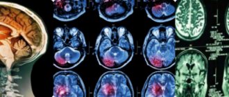
Diagnostic methods
Diagnosis of an aneurysm is carried out using several methods:
Angiography
A popular method for diagnosing an aneurysm is angiography, which makes it possible to qualitatively assess the condition of the vessels, determine the shape, degree and location of the pathology. Angiography is an X-ray examination using contrast agents. Before it is carried out, a special illuminating drug is injected into the femoral artery with a catheter, which helps to identify the aneurysm and its condition. Angiography is used to diagnose aneurysms in children.
Angiography is not carried out in the only way; there is:
- Digital angiography. The data is subsequently processed by computer, resulting in high-quality images. The dosage of the contrast agent is reduced and it is injected into a vein.
- Color angiography allows you to conduct research quickly, since one image reflects the entire process.
- 3D angiography reconstructs the projection of the image in a volumetric format.
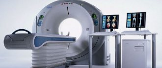
CT scan
The method is painless. During the examination, X-rays are taken, then the data is processed by a computer. The program helps to identify problems in the vascular system of the brain, identify areas of dilation, blockage and narrowing of blood vessels, see ruptures and hemorrhages.
Magnetic resonance imaging
The examination is abbreviated as MRI. This diagnosis allows you to obtain a three-dimensional image of the arteries of the brain using electromagnetic waves. The picture on the computer screen gives a clear idea of the condition of the veins and arteries.
Cerebrospinal fluid puncture
It is used when the doctor suspects that an aneurysm has ruptured. In this case, the analysis is taken by puncturing the spinal trunk with a special needle. The fluid is examined for the presence of blood, since after hemorrhage it enters the spinal cavity.
Is it possible to prevent the disease?
Preventive measures are ineffective in the presence of congenital pathology or hereditary predisposition. However, even in this case there is no need to provoke an early onset of the disease, for which you need to:
- no smoking;
- maintain an active lifestyle;
- maintain a constant and optimal sleep and work schedule;
- avoid surges in blood pressure;
- undergo regular examination by a doctor with CT or MRI.
In the case of vascular problems due to atherosclerosis or injury, much depends on the timeliness of diagnosis and compliance with the doctor’s advice. It should be clearly understood that no folk remedies or conservative measures will help get rid of a slowly growing aneurysm.
The danger of a vascular cerebral aneurysm is the extremely high risk of rupture with hemorrhage, which can cause sudden death of a person. It is not always possible to diagnose a slight enlargement of the artery; the period of increase in size can be long, so sometimes the patient ends up in the hospital too late. If it is possible to detect dilation of the vessel in a timely manner, then the optimal treatment method will be a vascular or neurosurgical operation, thanks to which it is highly likely that the life and health of the sick person can be saved.
Treatment
Each specific case requires an individual approach; the choice of treatment is influenced by:
- aneurysm size;
- location;
- the possibility of its rupture;
- symptoms;
- age;
- general condition of the body;
- presence of concomitant diseases.
The doctor must take into account the risks associated with the treatment. What options exist:
- Surgery remains the only productive and main method of eliminating the anomaly; it is also used to remove aneurysms in children.
- Drug therapy is used when surgery is not possible or there are contraindications.
- With a slight expansion, the specialist chooses a wait-and-see approach, observing and monitoring the condition of the formation.
Self-treatment of an aneurysm is unacceptable; it is deadly.
Surgery
Surgery is indicated if the cerebral aneurysm enlarges and there is a risk of rupture. There are several options for performing the operation:
- Aneurysm clipping. Open intracranial intervention involves removing the aneurysm from the bloodstream while preserving the carrier and surrounding vessels. The neck of the formation is blocked with a clip, then the blood is removed. After which the aneurysm dies and is replaced by connective tissue. Clipping an aneurysm does not allow surgery when the formation is located in the deep parts of the brain.
- Strengthening the walls of blood vessels. In this case, the damaged part is wrapped in surgical gauze or muscle tissue from the patient. Subsequently, connective tissue grows in this place and a kind of capsule is formed. Disadvantages include the likelihood of bleeding and rejection of artificial material.
- Endovascular embolization. A modern and least traumatic way to eliminate an aneurysm. The operation makes it possible to reach hard-to-reach places in the brain; sometimes it is done when an aneurysm ruptures or with subarachnoid bleeding. Embolization does not require craniotomy (opening the skull); manipulations are carried out under angiography control. During the operation, a catheter is inserted into a distant vessel on the thigh and advanced to the site of the protrusion. Next, a microspiral is placed in its cavity, which causes blockage of the aneurysm and gradual death. The duration of hospitalization for endovascular embolization is no more than 5 days.
- Clipping or endovascular embolization methods are indicated for operating on children.
- There is a possibility of complications, especially when the operation occurs during an exacerbation. Hypoxia of blood vessels, spasm, bleeding cannot be ruled out; during embolization, perforation of the aneurysm occurs.
Classification
The pathological process should be divided according to three bases: size, location and shape.
By size
Based on the criterion of the volume of altered tissue, the following types of pathological process can be distinguished:
- Miliary or microaneurysms. They represent the initial stage of the disorder or a full-fledged variety of it. They do not reach three millimeters in size. But they still pose a danger.
Therefore, it makes sense to check the patient every six months. If there is no dynamics as such, medications are prescribed, special living conditions and a diet are prescribed.
However, a microvariety may be a transitional variant. That is, as was said, the initial stage of the disorder. Then you cannot do without surgical correction.
- Small ones. Size up to 10 mm. They occur much more often. They pose immediate risks to the life and health of the patient. It is from this variety that the pathological process begins.
Treatment is strictly necessary. It is carried out under the supervision of a vascular neurosurgeon.
- Average. Up to 1.5 cm in diameter. These are serious entities. Enough physical stress is enough for the structure to rupture and lead to bleeding. Therapy is prescribed on an emergency basis. It is carried out strictly in stationary conditions.
- Large ones. 16-25 mm. Such forms are relatively rare. The size of the vascular formation leads to enormous risks of complications. The structure can rupture due to minor mechanical activity.
- Macroaneurysms. Over 2.5 cm in diameter. The danger lies not only in the possibility of rupture. Also, such a vascular formation compresses the surrounding tissues, leading to neurological deficits. Treatment is urgent, surgery is needed.
By localization
Regarding the second criterion, location:
- Anterior aneurysms. Located in the area of the parietal lobes. They are especially common. They do not pose great difficulties in terms of access, therefore the chances of recovery and a good course of the postoperative period are high.
- Medium varieties. Localized in the area of the parietal and temporal lobes. Access needs to be worked out as the issue is somewhat more complex.
- Localized in the area of the vertebrobasilar region. That is, closer to the occipital lobes.
- Located in the area of the internal carotid artery. It is difficult to reach such an aneurysm straight away. The correction method needs to be worked out. The task is not the easiest; it requires the participation of an experienced and qualified neurosurgeon.
The location must be clarified in order to carry out a competent and safe operation for the patient.
This classification is rather arbitrary, since an aneurysm can mature in almost any part of the brain.
By shape
The third basis for classification is the actual form of education. The following types of pathological structures are distinguished:
- Saccular. They occur most often. And account for more than 80% of the total number of situations. Externally they look like lateral wall protrusions of the artery, strictly in one direction. Such swelling became the basis for calling the aneurysm saccular.
- A fusiform change develops much less frequently. As the name suggests, the artery bulges in both directions. That is, there are no healthy, unaffected areas left.
A pathological process of this kind is much more dangerous. Because there is no restraining or compensating factor. Both sides of the vessel are involved in the breach and are at risk of rupture at any moment.
Doctors use all three classifications to describe the pathological process and plan treatment.
How much does the operation cost?
How much it will cost to operate an aneurysm is related to:
- with the venue;
- severity of the lesion;
- carrying out diagnostic measures;
- selection of consumables.
In Germany and Israel, embolization has been done for a long time and successfully, but the prices there are more expensive. Abroad, you will have to prepare from 25 to 40 thousand euros for surgical intervention alone, not counting consultations and diagnostics. Moreover, this does not depend on the method of removing the aneurysm. In Russia, all types of surgical interventions are also performed, but their cost is several times lower.
Therapeutic treatment
Treatment of cerebral aneurysm is carried out using conservative therapy. The method does not remove the anomaly, it makes it possible to eliminate the negative impact of certain factors, contain the increase, and reduce the negative impact on the patient’s condition:
- medications are prescribed to stabilize blood pressure;
- Calcium channel blockers are used as a prophylactic against cerebral vasoconstriction;
- painkillers and anticonvulsants are used;
- antiemetics;
- medications are prescribed to reduce the acidity of gastric juice.
The doctor will choose the optimal dosage after a detailed examination of the condition of the blood vessels.
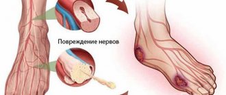
Stroke and its signs
Blood formation on the vessels of the brain leads to their thinning, as a result of which the wall of the aneurysm breaks through and blood enters the medulla. Hemorrhage under the arachnoid mater is called “subarachnoid”. It requires urgent hospitalization and surgery, since the mortality rate among patients with this diagnosis is high.
With a ruptured aneurysm and hemorrhage, severe symptoms occur. It is impossible to ignore them, these are:
- Severe headache. This pain occurs due to the fact that blood entering the brain tissue irritates the nerve fibers of the meninges.
- The compressive effect of the resulting hematoma on the brain tissue is expressed in various neurological abnormalities: photophobia, stiff neck, pain in the muscles of the neck, back, and legs.
- The patient begins to feel sick and vomits even with an empty stomach. This is also due to the effect of blood on the medulla.
- A person may faint because, as a result of a hematoma in the brain, intracranial pressure increases so much that normal blood circulation in the structures of the brain substance becomes impossible. Oxygen starvation occurs.
Rehabilitation period
Recovery after surgery is a difficult and lengthy period, since the consequences are severe. Hemorrhage affects the neurons of the brain - the mechanisms responsible for almost all body functions. The cells where the rupture occurred are damaged and atrophy. To prevent their complete death, it is necessary to take urgent measures for rehabilitation:
- recovery takes place in a specialized center or sanatorium;
- the patient is provided with care and nutrition, and has the opportunity to move around, usually in a wheelchair.
- a person learns to speak again, take care of himself, gradually restores physical capabilities, and adapts to society.
How long the recovery period will last depends on the extent of the consequences, usually its length is one and a half years. In children the process is faster. The patient is prescribed:
- medications;
- physiotherapy sessions;
- electrical stimulation of muscles;
- acupuncture;
- massage;
- physiotherapy.
Patients gradually regain cognitive skills, memory and speech.
Folk remedies
During the rehabilitation period, the consequences can be quickly eliminated and folk recipes will help you recover:
- Elderberry will remove excess fluid, cleanse blood vessels, soothe, relieve headaches, and also acts as a tonic. Grind 20 g of plant root and add a glass of water. After boiling, leave for several hours. Drink 30 g three times a day.
- Using the same recipe, a decoction of dill seeds is prepared.
- To strengthen the walls of blood vessels and prevent the re-development of an aneurysm, it is recommended to add hawthorn berries to herbal tea.
- Vitamin supplements help prevent loss of vascular elasticity. To prepare the composition, take a couple of lemons and oranges. Citrus fruits are passed through a meat grinder. 40 g of honey are stirred in the gruel. After a day, the mixture can be consumed 50 g daily.
- The root of the evasive peony can relieve tension and support blood vessels. 20 g of plant material is poured with boiling water (250 g), infused and used 5 times a day, 20 mg each.
- You can buy peony tincture at the pharmacy; drink 25 drops three times a day.
Traditional recipes help support the patient using natural means and reduce the negative effects of medications on the body.