How to create new neural connections in the brain: several effective ways
Synaptogenesis is easier to enhance. And this can be done when the brain is not yet fully formed (up to 25 years). But even in adulthood, new synapses can be formed. Below we will look at how.
Those in a cleaner environmental environment will be able to grow more new synapses than those growing in a standard or poor environment. Although most studies analyzing the phenomenon of synaptogenesis have been conducted in rats, there is reason to believe that many of the same methods may be effective in humans.
An increase of 25% means an increase in the total number of neural connections and propagation pathways. Therefore, for better development of synapses, it is necessary to improve the environment. If it is impossible to change your place of residence, then you need to take care of the cleanliness of the house. [R, R, R, R, R]
Learning new things is known to be good for the brain. However, not all “learning” is capable of inducing synaptogenesis. If you want to grow new synapses, you need to do motor learning or learn something new that involves movement (like juggling, table tennis, etc.).
Your brain must coordinate complex limb movements with specific activities. It doesn't have to be acrobatic training. Every time you engage in motor learning (like figuring out how to make a certain shot in table tennis), new synapses are formed. [R, R, R]
Phospholipids
Phospholipids are key components of synaptic membranes. Based on this, the researchers hypothesized that administration of phospholipid precursors could lead to synaptogenesis. Among them:
- Uridine. It is a "building block" for RNA that indirectly helps in the process of memory formation. Uridine can work together with choline to promote the formation of new synapses.
- Docosahexaenoic acid (DHA). This is an Omega-3 fatty acid. It improves cognitive function and overall mental health. Contained in fish oil and krill oil.
- Choline. It is an essential nutrient that serves as a precursor to acetylcholine. Acetylcholine is necessary for synaptogenesis, as well as a number of other neurophysiological functions. The best source of choline is eggs.
It is believed that antidepressants can induce both neurogenesis (the formation of new brain cells) and synaptogenesis. It is already well known that SSRIs can induce neurogenesis, but whether they also help in the formation of new synapses remains unclear.
Spadin (Spadin). Spadine is a novel antidepressant peptide that inhibits a potassium channel called “TREK-1.” In rodent studies, Spadine administration was found to increase multiple biomarkers of synaptogenesis, including PSD-95 and synapsin-1. Researchers later confirmed that after treatment with Spadine, the number of synapses in rodents increased significantly. [R]
Ketamine. It provides quick relief from symptoms of depression. Preliminary evidence suggests that ketamine's unique mechanism of action is capable of inducing synaptogenesis. It acts on a cell signaling pathway called "mTOR". When the mTOR pathway is activated, synaptogenesis occurs in the prefrontal cortex.
NMDA modulators. Other drugs are being developed that act on NMDA (glutamate), similar to ketamine, without the psychomimetic effects. In other words, pharmaceutical and biotech companies are trying to isolate the antidepressant mechanisms of ketamine while eliminating the possibility of dissociative side effects such as hallucinations or delusions.
Another way to grow new synapses is to change your behavior. The study documented that female birds experienced behavioral changes when treated with testosterone. The researchers noted that 51% of the synapses formed in a region of the forebrain nucleus called the “robustus archistriatalis” (RA). Increases in synapse size and number of vesicles per synapse are associated with behavioral changes.
Changes in behavior are known to alter neural activation and pathways. Learning a new motor skill is associated with synaptogenesis. It is possible that a change in behavior over a period of time, regardless of what is causing the change in behavior (eg drugs, environment, etc.), can lead to synaptogenesis. [R]
Increased hormone levels can induce synaptogenesis. In particular, increasing testosterone levels in men promotes the formation of new synapses, while increasing estradiol and progesterone levels in women may have the same effect. Some studies suggest that decreased production of various sex hormones may inhibit synaptogenesis.
Estradiol. Estradiol is known to increase synaptic plasticity in the hippocampal region of female rats. One study reported that decreased estradiol levels led to synapse loss. Increasing estradiol levels increased synapse formation in the hippocampus of females. But inhibition of estradiol production in males does not affect synaptogenesis.
Progesterone. In a study conducted on rats, the use of progesterone allowed synaptogenesis in a specific area of the hippocampus. This synaptogenesis helped rats with nerve recovery after stroke (global cerebral ischemia).
Testosterone. Based on a study analyzing the effects of testosterone administration in animal models of multiple sclerosis (MS), testosterone appears to enhance synaptogenesis. It also helps maintain neurons in the cerebral cortex. Increased synaptogenesis was associated with improved excitatory synaptic function. [R,R,R,R,R]
One study found that transcranial low-level laser therapy stimulated the formation of new synapses. The researchers initially tested low-level laser therapy based on the finding that traumatic brain lesions in animal models improved by applying near-infrared light to their heads. This near-infrared light reduces the size of brain lesions, minimizes inflammation, and induces neurogenesis.
Testing of low-level laser therapy was carried out in rodent models at intervals of one or three treatments per day. Neurological function was significantly improved for the laser-treated rodents compared to the untreated control group. This improvement occurred within 2 weeks and was characterized by a significant increase in synaptogenesis as a result of upregulation of synapsin-1.
The laser treatment simultaneously increased BDNF (brain-derived neurotrophic factor), another important component of mental health. Although this study was conducted in rodent models, the results were significant. It is conceivable that similar synaptogenic effects may occur with low-level laser therapy in humans. [R]
If you want to optimize your brain's ability to form new synapses, you must learn to increase your BDNF levels.
BDNF is responsible for helping the brain form both excitatory and inhibitory synapses. When BDNF production is low, synapses weaken. When BDNF is produced at high levels, existing synapses are strengthened through long-term potentiation. [R, R]
Various proteins
The production of endogenous proteins helps stimulate the formation of new synapses. Proteins that have been documented as drivers of synaptogenesis include: Netrin, Osteopontin, and Thrombospondin.
Netrin. This protein is thought to promote the formation of neural circuits in the mammalian forebrain, especially during periods of peak synapse formation. Researchers have shown that Netrin-1 is able to develop synaptogenesis in cortical neurons of rats and mice. Specifically, Netrin-1 increases the number and strength of excitatory synapses between cortical neurons.
Osteopontin. It is a protein molecule that improves synaptic reorganization and repair after brain injury. It is thought to promote synaptogenesis through its action on synaptin-1.
Thrombospondin. It is a protein that is secreted from glial cells and promotes synaptogenesis while maintaining synapse stability. It is also believed to help in the reconstruction of synapses after traumatic brain injury or exposure to various medications. [R, R, R]
When choosing between familiar and new behavior, most people will give preference to the first option. Why? From many men and women you can hear the following phrase: “In my mind I understand everything, but I can’t help myself. I tell myself that the current situation does not suit me at all, but I continue to behave as I did before!” Paradox? No! It's all about the already formed neural connections!
The stronger the neural connection, the more synapses are formed (a synapse is the point of contact between two nerve cells), and the more powerful and effective the electrical signals between the nerve cells included in this connection become. The more synapses are formed, the more active and efficient they begin to work.
Brain synaptic connections and believing neurons
The brain consists of approximately one hundred billion neurons of several hundred types, each of which has a cell body, a descending axon, and numerous dendrites and axon terminals radiating to other neurons and forming approximately a thousand trillion synaptic connections between these one hundred billion neurons. The numbers mentioned are staggering. One hundred billion neurons are 1011, or a one followed by 11 zeros: 100000000000. The connections of a thousand trillion are a quadrillion, or 1015, or a one followed by 15 zeros: 1000000000000000. There are about as many neurons in the human brain as there are stars in the galaxy The Milky Way is literally an astronomical number! The number of synaptic connections in the brain is equivalent to the number of seconds in 30 million years. Think about this for a minute. Start counting the seconds in the manner of "one one thousand, two one thousand, three one thousand..." When you get to 86400, you get the number of seconds in a day, when you reach 31536000, the number of seconds in a year when you finally reach one trillion seconds, then you you think it's been around 30 thousand years. Now repeat this 30,000-year count one thousand more times and you will count the number of synaptic connections in your brain.
The number of synaptic connections in the brain is equivalent to the number of seconds in 30 million years.
Of course, a large number of neurons provides significant computing power (like adding chips or memory cards to a computer), but the actions are carried out in the individual neurons themselves. Neurons have an elegant simplicity, yet they are beautifully complex machines for processing electrochemical information. Inside a neuron at rest there is more potassium than sodium, and the predominance of anions, negatively charged ions, creates a negative charge inside the cell. Depending on the type of neuron, when a tiny electrode is inserted into its body at rest, we will get a reading of -70 mV (millivolt - one thousandth of a volt). At rest, the cell membrane of a neuron is impermeable to sodium but permeable to potassium. When a neuron is stimulated by the actions of other neurons (or by electrical manipulation by curious neuroscientists armed with electrodes), the permeability of the cell membrane changes, sodium enters the cell and thus the electrical balance shifts from -70 mV to zero. This phenomenon is called excitatory postsynaptic potential
, or EPSP.
A synapse
is a tiny gap between neurons, hence the term
postsynaptic
means that the neuron on the receiving side of the signal crossing the synaptic gap fires to reach its firing potential.
In contrast, if the stimulation comes from an inhibitory neuron, the voltage shifts negatively, from -70 mV to -100 mV, making the neuron less likely to fire. This phenomenon is called inhibitory postsynaptic potential
, or IPSP. Although there are hundreds of different types of neurons, we can classify the majority as either excitatory or inhibitory according to the type of action.
If the EPSP rises to a sufficient value (as a result of multiple firings of one neuron after another or multiple connections with other neurons), then the permeability of the cell membrane of the neuron reaches a critical value
, sodium rushes into it, causes an instant surge of voltage up to +50 mV, it spreads throughout the cell body and gradually descends along the axon to the terminals.
With the same speed, the neuron voltage again decreases to –80 mV, and then returns to –70 mV at rest. This process of the cell membrane becoming permeable to sodium and the corresponding change in voltage from negative to positive, moving along the axon to dendrites and synaptic connections with other neurons, is called action potential
.
More often we use the expression “the cell is excited.” The increase in EPSP is called summation
.
Two types are known: (1) temporal summation
, in which two EPSPs of one neuron are enough for the receiving neuron to reach a critical point and excite;
and (2) spatial summation
, in which two EPSPs from two different neurons appear simultaneously and are sufficient for the receiving neuron to reach a critical point and fire.
This electrochemical change in voltage occurs rapidly, sodium permeability spreading sequentially along the axon from the cell body to the terminals, and this phenomenon, as you would expect, is called propagation
. The speed of propagation depends on two conditions: (1) the diameter of the axon (the larger, the faster) and (2) the myelination of the axon (the larger the myelin sheath covering and insulating the axon, the faster the impulse propagates along it).[102]
Note: if the critical point of excitation of the neuron is not reached, it is not excited; if the critical point is reached, the neuron fires. This system works on the “either-or”, “all or nothing” principle. Neurons do not fire “slightly” in response to weak stimuli or “strongly” in response to strong stimuli. They either get excited or they don't get excited. Therefore, neurons transmit information in one of three ways: (1) by firing frequency
(number of action potentials per second), (2)
location of firing
(which neurons are fired), and (3)
number of firing
(how many neurons are fired). Therefore, neurons are said to be binary in action, like the binary symbols of a computer, 1 and 0, corresponding to an on or off signal, passing or not passing through the neural pathway. If we consider these "on or off" neural states as one type of mental state, where one neuron gives us two such states (on or off), then when processing information about the world and the controlled organism, the brain has 2 × 10¹⁵ possible options to choose from . Since we cannot comprehend all this number with our minds, we can say that the brain is in all respects an infinitely large information processing machine.
How do individual neurons and their action potentials create complex thoughts and beliefs? The process begins with the so-called neural binding
.
"Red Circle" is an example of combining two input signals ("red" and "circle") into one perceptual object, the red circle. Neural signals from muscles and sensory organs merge, moving "upstream", through convergence zones
- areas of the brain that combine information contained in different neural signals (from the eyes, ears, senses of touch, etc.) so that we ultimately got an idea of the object as a whole, and not of countless fragments of the image. Looking at the upside-down photograph of President Obama in Chapter 4, we first perceive the face as one whole before we begin to notice that something is wrong with the eyes and mouth; As already explained, the reason is that two different neural networks operate at different speeds: first, the perception of the face as a whole occurs, then - the details of this face.
However, binding is a much broader phenomenon. There can be many objects perceived by different senses, and they all must be connected together in the higher regions of the brain to gain meaning. Large parts of the brain, such as the cerebral cortex, coordinate signals from smaller parts of the brain, such as the temporal lobes, which in turn integrate neural events from even smaller parts of the brain, such as the fusiform gyrus (for recognizing faces). This reduction occurs all the way down to the single-neuron level, where highly selective neurons (sometimes called “grandmother neurons”) fire only when subjects see someone they know. There are neurons that fire only when an object moves from left to right across the observer's field of view. There are other neurons that fire only when an object moves from right to left across the observer's field of view. And there are third neurons that have an action potential only when they receive EPSP signals from other neurons that fire in response to the diagonal movement of objects in the visual field. This is how the binding process occurs in neural networks. There are even neurons that fire only when we see someone we recognize. Caltech neuroscientists Christoph Koch and Gabriel Kreiman, together with UCLA neurosurgeon Itzhak Fried, discovered, for example, a single neuron that fires when a participant is shown a picture of Bill Clinton and no one else. Another works only if the participant is shown a photo of Jennifer Aniston, but only her, without Brad Pitt.[103]
Of course, we are not aware of the workings of our electrochemical systems. What we actually experience are subjective states of thoughts and feelings that arise from the integration of neural events, which philosophers call qualia
. But even qualia themselves are a type of neural coupling effect, combining signals from countless “lower order” neural networks. It really comes down to the electrochemical process of a neuron action potential, or neurons firing and communicating with each other to transmit information. How do they do this? Again, thanks to chemistry.
The connection between neurons occurs in the incredibly tiny synaptic gap between them. When the action potential of a neuron rushes along the axon and reaches its terminals, it causes the release of minute portions of chemical transmitter substances (CTS) into the synapse. The CTEs received by connecting neurons act as EPSPs, changing the voltage and permeability of the postsynaptic neuron, thereby causing it to fire and propagate its action potential down the axon to the terminals, where it releases its CTEs into the next synaptic gap, and so on along the line of the neural network. When we stub our toe, the pain signal travels from the pain receptors in the tissues of our toe all the way up to the brain, which detects the pain and transmits the signal to other parts of the brain, which send additional signals to the contracting muscles so that we pull our foot away from the unfortunate obstacle. It all happens so quickly that it seems almost instantaneous.
There are many types of CTV. The best known are catecholamines
, including
dopamine, norepinephrine (norepinephrine)
and
adrenaline (epinephrine)
.
CTBs act on the postsynaptic neuron like a key on a lock. If the key approaches and turns, the neuron fires; otherwise, the door remains locked and the postsynaptic neuron remains unexcited. After the excitation process occurs, most unused CTE is returned to the presynaptic neuron, reused elsewhere, or destroyed by monoamine oxidase (MAO) in the process of so-called first uptake
.
If too much CTB is present in the synaptic gap, then the remainder is absorbed into the postsynaptic neuron in a process of second uptake
.
Drugs affect synapses, CTE release, and subsequent uptake processes. For example, amphetamines accelerate the release of CTE into synapses, thereby speeding up the process of neural communication, which is why they are called speed
("speed"). Reserpine, which was once a common prescription for psychosis, destroys CTE vesicles in the presynaptic neuron, so MAOs destroy them before use, ultimately slowing down the functioning of neural networks, controlling manic states, hypertension and other symptoms of nervous system hyperactivity. Cocaine blocks the first absorption, so CTEs simply linger in the synapse and promote accelerated excitation of neurons, bringing neural networks to a state of excitement - remember Robin Williams with a microphone in front of an audience; in fact, Williams himself attributes much of the success of his comedies in the 1980s to his own cocaine addiction. As one of the most common CTEs, dopamine plays a critical role in smooth communication between neurons and muscles, and when it is insufficient, patients experience loss of motor regulation and uncontrollable trembling. These manifestations are called Parkinson's disease, one of the treatments for which is L-dopa, a dopamine agonist that stimulates its production.
How do we build the entire system from bottom to top, starting with transmitter chemicals like dopamine and linking the signals into a single belief system? Through behavior. Let me remind you that the primary function of the brain is to control the body and help it survive. One way to do this is through associative learning, or patterning. This is the link between neural action potential and human actions.
Dopamine, the drug of faith
Of all the chemical transmitters sloshing around our brains, dopamine appears to be the most directly linked to the neural correlates of belief. In essence, dopamine plays a critical role in associative learning and in the brain's reinforcement system, which Skinner discovered through his method of conditioning, whereby any reinforced behavior tends to be repeated. By definition, reinforcement is something that serves as a reward for the body, that is, it induces the brain to force the body to repeat that behavior in order to receive another positive reinforcement. Here's how it happens.
The divided brainstem, one of the most evolutionarily ancient brain regions found in all vertebrates, contains cavities or pouches with approximately 15,000 to 24,000 dopamine-producing neurons on each side, whose long axons connect to other brain regions. These neurons stimulate the release of dopamine whenever the reward received is greater than expected, resulting in the individual repeating the specific behavior. The release of dopamine is a form of providing information, a message to the body: “Do it again.” Dopamine creates a feeling of pleasure that accompanies solving a problem or achieving a goal; as a result, the body wants to repeat the same behavior, be it squeezing a barbell, pressing a key, or manipulating a lever of a mechanism. You get a response (reinforcement), and your brain gets a dose of dopamine. Behavior – Reinforcement – Behavior. Repeating sequence
.
However, the dopamine system has its pros and cons. On the plus side, dopamine is related to a bundle of neurons the size of a peanut located in the middle of the brain called the nucleus accumbens
(NAcc) – the nucleus accumbens, which is known to be associated with reward and pleasure.
In essence, dopamine appears to fuel this so-called reward center of the brain, which is involved in the “high” that both cocaine and orgasm produce. The "pleasure center" was discovered in 1954 by James Olds and Peter Milner of McGill University, who accidentally implanted an electrode into the NAcc of a rat and found that the rodent became highly aroused. The scientists then designed a machine that, when the rat pressed a bar, created a small amount of electrical stimulation in the same area of the brain. The rats pressed the bar until they collapsed in exhaustion, even forgetting about food and water.[104] The same effect has since been found in all mammals participating in the experiments, including in people who underwent brain surgery and received NAcc stimulation. They described their sensations with the word “ orgasm
.”[105]
This
is a typical example of positive reinforcement!
Unfortunately, the dopamine system also has disadvantages, namely the development of addiction. Addictive drugs act as a reward signal to dopamine neurons. Gambling, pornography, and drugs such as cocaine can trigger a dopamine response in the brain. Addictive ideas, especially unsuccessful ones
ideas such as those promoted by cults, such as those promoting mass suicide (think Jonestown and Heaven's Gate), or religions that encourage suicide bombing (think of the attacks of 9/11 and 7/7).
An Important Caution About Dopamine
: Neuroscientists make a clear distinction between “preference” (pleasure) and “desire” (motivation), and there is currently lively debate about whether dopamine actually promotes pleasure or motivates behavior. Positive reinforcement can lead to repetition of a behavior because it produces a pleasant feeling (preference, or pure pleasure from receiving a reward) or an unpleasant feeling if the behavior is not repeated (desire, or the motivation to avoid the anxiety of not receiving a reward). The first reward is associated with the pure pleasure of, say, orgasm, the second - with the anxiety that the addict feels when receiving the next dose inspires doubt. The studies I cited above support the pleasure assumption, but newer research is leaning toward motivation.[106] Neuroscientist Russell Poldrack of the University of California, Los Angeles, told me that the new findings imply “a role for dopamine rather in motivation.” than in pleasure itself, while the opioid system appears to play a central role in pleasure." For example, he points out that “you can block the dopamine system in rats, and they will still enjoy rewards, but will not want to try to get them.”[107] This is a subtle but important difference, but central to our understanding of the neural correlates of belief. The point is that dopamine reinforces actions, beliefs and patterns and is thus one of the primary “belief drugs”.
The connection between dopamine and faith was established in experiments conducted by Peter Bragger and his colleague Christina Moore at the University of Bristol, England. Investigating the neurochemistry of superstition, magical thinking, and belief in the paranormal, Bragger and Moore found that people with high dopamine levels are more likely to find meaning in coincidences and to perceive meaning and patterns where there is none. For example, one study compared twenty people who said they believed in ghosts, gods, spirits, and conspiracies with twenty participants who said they were skeptical about such beliefs. All participants were shown a series of slides of human faces, some of which were normal and some were “mixed up”, for example, some with eyes, ears or noses referring to other faces. In the next experiment, existing and randomly composed words were flashed on the screen. Overall, the scientists found that believers were much more likely than skeptics to mistake a confused face for a real one or a made-up word for an ordinary one.
Dopamine is the drug of faith. People with high dopamine levels are more likely to find meaning in coincidences and discern meanings
and patterns where there are none.
In the second part of the same experiment, Bragger and Moore gave all forty participants L-dopa, a drug given to Parkinson's patients to increase dopamine levels in the brain. After this, the slide show with faces and words was repeated. The rush of dopamine caused both believers and skeptics to perceive “confused” faces, as well as invented words, as ordinary. This suggests that patterning may be associated with high levels of dopamine in the brain. Interestingly, L-dopa had a stronger effect on skeptics than on believers. In other words, increased dopamine levels seemed to reduce the skepticism of skeptics more effectively than it increased the faith of believers.[108] Why? Two possible explanations come to mind: (1) perhaps the level of dopamine in believers is already higher than in skeptics, which means that the latter feel its influence more acutely; or (2) perhaps the tendency of believers to pattern is already so high that their dopamine effect is lower than that of skeptics. Additional research has shown that people who profess to believe in the paranormal, compared with skeptics, showed an increased tendency to see “regularities, or patterns, in noise”[109] and to attribute meaning to arbitrary connections they perceive to exist.[110]
Hear a signal in the noise
So what exactly does dopamine do to increase belief? One theory, promoted by Moore, Bragger, and their colleagues, is that dopamine increases the signal-to-noise ratio, or the amount of signal your brain detects in background noise. This is the error detection problem associated with patterning. The signal-to-noise ratio, in essence, is the problem of patterning - the search for significant patterns in both meaningful and meaningless noise. Signal-to-noise is the ratio of patterns that your brain detects in background noise, whether the patterns are real or imaginary. How does dopamine influence this process?
Dopamine enhances the ability of neurons to transmit signals from one to another. How? Acting as an agonist (as opposed to an antagonist), or a substance that enhances neuronal activity, dopamine binds to specific sites on receptor molecules in the synaptic cleft of neurons, like CTB that normally binds to them.[112] This increases the level of neuronal firing due to recognition. pattern, meaning that the number of synaptic connections between neurons is likely to increase in response to the perceived pattern, thereby imprinting the perceived patterns into long-term memory through the actual physical growth of new neural connections and the strengthening of old synaptic connections.
The dopamine rush causes increased pattern detection; Scientists have found that dopamine agonists not only promote learning, but in high doses can also provoke symptoms of psychosis, such as hallucinations, possibly related to the fine line between creativity (selective patterning) and madness (indiscriminate patterning). It all depends on the dose. If it is too large, Type 1 errors are likely to occur, false positives, in which we see connections where there really are none. If the dose is too small, type 2 errors occur, false negatives, in which we miss real-life connections. It's all about the signal-to-noise ratio.
Previous14Next
Classifications of synapses
The main elements of an electrical synapse (ephaps): a - connexon in a closed state; b — connecton in the open state; c — connexon embedded in the membrane; d—
monomer
connexin, e -
plasma membrane
; f — intercellular space; g - a gap of 2-4 nanometers in the electrical synapse; h -
hydrophilic
connexon channel
- chemical is a place of close contact between two nerve cells, for the transmission of a nerve impulse through which the source cell releases into the intercellular space a special substance, a neurotransmitter, the presence of which in the synaptic cleft excites or inhibits the receiver cell.
- electrical (ephaps) - a place of closer contact between a pair of cells, where their membranes are connected using special protein formations - connexons (each connexon consists of six protein subunits). The distance between cell membranes in the electrical synapse is 3.5 nm (the usual intercellular distance is 20 nm). Since the resistance of the extracellular fluid is low (in this case), impulses pass through the synapse without delay. Electrical synapses are usually excitatory.
- mixed synapses - the presynaptic action potential produces a current that depolarizes the postsynaptic membrane of a typical chemical synapse where the pre- and postsynaptic membranes are not tightly adjacent to each other. Thus, at these synapses, chemical transmission serves as a necessary reinforcing mechanism.
The most common are chemical synapses. Electrical synapses are less common in the mammalian nervous system than chemical ones.
Pathways
The nervous system has its spheres of influence throughout the body. With the help of conductive fibers, nervous regulation of systems, organs and tissues is carried out. The brain, thanks to a wide system of pathways, completely controls the anatomical and functional state of every structure of the body. Kidneys, liver, stomach, muscles and others - all this is inspected by the brain, carefully and painstakingly coordinating and regulating every millimeter of tissue. And in case of failure, it corrects and selects an appropriate model of behavior. Thus, thanks to the pathways, the human body is characterized by autonomy, self-regulation and adaptability to the external environment.
A pathway is a collection of nerve cells whose function is to exchange information between different parts of the body.
- Association nerve fibers. These cells connect various nerve centers located in the same hemisphere.
- Commissural fibers. This group is responsible for the exchange of information between similar centers of the brain.
- Projection nerve fibers. This category of fibers articulates the brain with the spinal cord.
- Exteroceptive pathways. They carry electrical impulses from the skin and other sensory organs to the spinal cord.
- Proprioceptive. This group of pathways carries signals from tendons, muscles, ligaments and joints.
- Interoceptive pathways. The fibers of this tract originate from internal organs, blood vessels and intestinal mesenteries.
The mechanism of functioning of the chemical synapse
A typical synapse is axo-dendritic chemical. Such a synapse consists of two parts: presynaptic, formed by the club-shaped extended end of the axon of the transmitting cell, and postsynaptic, represented by the contacting portion of the plasma membrane of the receiving cell (in this case, a portion of the dendrite).
Between both parts there is a synaptic cleft - a gap 10-50 nm wide between the postsynaptic and presynaptic membranes, the edges of which are strengthened by intercellular contacts.
The part of the clavate extension axolemma adjacent to the synaptic cleft is called the presynaptic membrane. The section of the cytolemma of the receiving cell that borders the synaptic cleft on the opposite side is called the postsynaptic membrane; in chemical synapses it is prominent and contains numerous receptors.
In synaptic expansion there are small vesicles, so-called synaptic vesicles, containing either a mediator (a substance that mediates the transmission of excitation) or an enzyme that destroys this mediator. On the postsynaptic, and often on the presynaptic membranes, there are receptors for one or another mediator.
When the presynaptic terminal is depolarized, voltage-sensitive calcium channels open, calcium ions enter the presynaptic terminal and trigger the fusion of synaptic vesicles with the membrane. As a result, the transmitter enters the synaptic cleft and attaches to receptor proteins of the postsynaptic membrane, which are divided into metabotropic and ionotropic.
The former are associated with the G protein and trigger a cascade of intracellular signal transduction reactions. The latter are associated with ion channels, which open when a neurotransmitter binds to them, which leads to a change in membrane potential. The mediator acts for a very short time, after which it is destroyed by a specific enzyme.
For example, in cholinergic synapses, the enzyme that destroys the transmitter in the synaptic cleft is acetylcholinesterase. At the same time, part of the transmitter can move with the help of carrier proteins across the postsynaptic membrane (direct uptake) and in the opposite direction through the presynaptic membrane (reverse uptake). In some cases, the mediator is also absorbed by neighboring neuroglial cells.
Two release mechanisms have been discovered: with complete fusion of the vesicle with the plasmalemma and the so-called “kiss-and-run”, when the vesicle connects to the membrane, and small molecules exit from it into the synaptic cleft, while large ones remain in the vesicle . The second mechanism is presumably faster than the first, with the help of it synaptic transmission occurs when the content of calcium ions in the synaptic plaque is high.
The consequence of this structure of the synapse is the unilateral conduction of the nerve impulse. There is a so-called synaptic delay - the time required for the transmission of a nerve impulse. Its duration is about 0.5 ms.
The so-called “Dale principle” (one neuron - one transmitter) has been recognized as erroneous. Or, as is sometimes believed, it is more precise: not one, but several mediators can be released from one end of a cell, and their set is constant for a given cell.
Interaction with neurotransmitters
Neurons of different locations communicate with each other using electrical impulses of a chemical nature. So, what is the basis of their education? There are so-called neurotransmitters (neurotransmitters) - complex chemical compounds. On the surface of the axon there is a nerve synapse - the contact surface. On one side there is a presynaptic cleft, and on the other there is a postsynaptic cleft. Between them there is a gap - this is the synapse. On the presynaptic part of the receptor there are sacs (vesicles) containing a certain amount of neurotransmitters (quanta).
When the impulse approaches the first part of the synapse, a complex biochemical cascade mechanism is initiated, as a result of which the sacs with mediators are opened, and quanta of mediator substances smoothly flow into the gap. At this stage, the impulse disappears and reappears only when the neurotransmitters reach the postsynaptic cleft. Then biochemical processes are activated again with the opening of the gates for mediators and those, acting on the smallest receptors, are converted into an electrical impulse that goes further into the depths of the nerve fibers.
Meanwhile, different groups of these same neurotransmitters are distinguished, namely:
- Inhibitory neurotransmitters are a group of substances that exert an inhibitory effect on excitation. These include: gamma-aminobutyric acid (GABA);
- glycine.
- acetylcholine;
History of discovery
- In 1897, Sherrington formulated the idea of synapses.
- Golgi and Ramón y Cajal received the Nobel Prize in 1906 for their research into the nervous system, including synaptic transmission.
- In 1921, the Austrian scientist O. Loewi established the chemical nature of the transmission of excitation through synapses and the role of acetylcholine in it. Received the Nobel Prize in 1936 together with H. Dale.
- In 1933, the Soviet scientist A.V. Kibyakov established the role of adrenaline in synaptic transmission.
- 1970 - B. Katz (Great Britain), U. v. Euler (Sweden) and J. Axelrod (USA) received the Nobel Prize for their discovery of the role of norepinephrine in synaptic transmission.
"Wiring"
Neural connections of the brain are the wiring of the nervous system. The functioning of the nervous system is based on the ability of a neuron to perceive, process and transmit information to other cells.
Information is transmitted through a nerve impulse. Human behavior and the functioning of his body completely depend on the transmission and reception of impulses by neurons through processes.
A neuron has two types of processes: axon and dendrite. A neuron always has one axon; it is through it that the neuron transmits impulses to other cells. It receives an impulse through dendrites, of which there may be several.
Many (sometimes tens of thousands) axons of other neurons are “connected” to the dendrites. The dendrite and axon contact through a synapse.
Functions of a neuron
Despite its relatively simple structure, the neuron has many functions, the main ones of which are the following:
- perception of irritation;
- stimulus processing;
- impulse transmission;
- formation of a response.
Functionally, neurons are divided into three groups:
In addition, another group is functionally distinguished in the nervous system - inhibitory nerves (responsible for inhibiting cell excitation). Such cells resist the propagation of electrical potential.
Receptors
Receptors are remembered every time they talk about drug or alcohol addiction. Why does a person need to be guided by the principle of moderation?
A receptor on the postsynaptic membrane is a protein tuned to transmitter molecules. When a person artificially (with drugs, for example) stimulates the release of transmitters into the synaptic cleft, the synapse tries to restore balance: it reduces the number of receptors or their sensitivity. Because of this, the natural levels of concentration of transmitters in the synapse cease to have an effect on neural structures.
For example, people who smoke nicotine change the sensitivity of receptors to acetylcholine, desensitization (decreased sensitivity) of the receptors occurs. The natural level of acetylcholine is insufficient for receptors with reduced sensitivity. Because Acetylcholine is involved in many processes, including those associated with concentration and a sense of comfort; a smoker cannot obtain the beneficial effects of the nervous system without nicotine.
However, receptor sensitivity is gradually restored. Although this may take a long time, the synapse returns to normal and the person no longer requires outside stimulants.
Neuron and synapses
The gap between the dendrite and the axon is a synapse. Because the axon is the “source” of the impulse, the dendrite is the “receiver”, and the synaptic cleft is the site of interaction: the neuron from which the axon comes is called presynaptic; the neuron from which the dendrite comes is postsynaptic.
Synapses can form between an axon and a neuron body, and between two axons or two dendrites. Many synaptic connections are formed by the dendritic spine and the axon. The spines are very plastic, have many shapes, and can quickly disappear and form. They are sensitive to chemical and physical influences (injuries, infectious diseases).
At synapses, information is most often transmitted through mediators (chemicals). Transmitter molecules are released on the presynaptic cell, cross the synaptic cleft and bind to membrane receptors of the postsynaptic cell. Mediators can transmit an excitatory or inhibitory (inhibitory) signal.
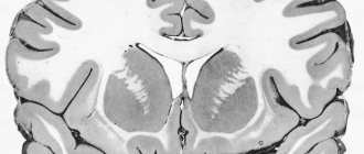
View gallery
Neural connections in the brain are the connection of neurons through synaptic connections. Synapses are the functional and structural units of the nervous system. The number of synaptic connections is a key indicator for brain function.
View gallery
Links
- Savelyev A.V. Methodology of synaptic self-organization and the problem of distal synapses of neurons // Journal of problems in the evolution of open systems. - Kazakhstan, Almaty, 2006. - T. 8, No. 2. - P. 96-104.
- Eccles D.K. Physiology of synapses. - M.: Mir, 1966. - 397 p.
| Neurons (Gray matter) | |
| Afferent nerve/Sensory neuron | |
| Efferent nerve/Motor neuron | |
| Synapse | |
| Touch receptor | |
| Neuroglia | |
| Myelin (White matter) | |
| Connective tissue | |
This page was last edited on May 5, 2020 at 6:41 pm.
Development of neural networks
Long-term changes in neural connections occur in various diseases (mental and neurological - schizophrenia, autism, epilepsy, Huntington's, Alzheimer's and Parkinson's diseases). Synaptic connections and internal properties of neurons change, which leads to disruption of the nervous system.
The activity of neurons is responsible for the development of synaptic connections. “Use it or lose it” is the principle underlying the neural networks of the brain. The more often neurons “act”, the more connections there are between them; the less frequently, the fewer connections. When a neuron loses all its connections, it dies.
Some authors also express other ideas that are responsible for regulating the development of neural networks. M. Butz associates the formation of new synapses with the brain’s tendency to maintain a “habitual” level of activity.
When the average level of neuronal activity drops (for example, due to injury), neurons build new contacts, and neuronal activity increases with the number of synapses. The opposite is also true: as soon as the level of activity becomes greater than the usual level, the number of synaptic connections decreases. Similar forms of homeostasis are often found in nature, for example, in the regulation of body temperature and blood sugar levels.
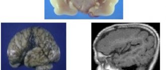
Henry Markram, who is involved in a neural brain simulation project, highlights the industry's prospects for studying the disruption, repair and development of neural connections. A team of researchers has already digitized 31 thousand rat neurons. The neural connections of a rat's brain are shown in the video below.
The cradle is not the beginning
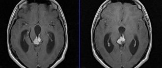
Scientists report that 70% of a person’s neurons die before birth. This occurs due to the rapid development of the fetus from a tiny embryo to a full-fledged baby in just nine months. The same thing happens throughout people's lives - they constantly lose neurons, and every year there are fewer and fewer of them. The question “how many neurons are in the human brain at birth” is very difficult to answer. It all depends on individual developmental characteristics and genetics. If we assume that, on average, by the age of three, a child's brain is 80% similar in volume to an adult, then the estimated number of neurons ranges from 65–80 billion. It should also be taken into account that the strength of synapses (areas that connect neurons to each other) is constantly growing from the moment of birth.
Neuroplasticity
The development of neural connections in the brain is associated with the creation of new synapses and modification of existing ones. The possibility of modifications is due to synaptic plasticity - a change in the “power” of the synapse in response to the activation of receptors on the postsynaptic cell.
A person can remember information and learn thanks to the plasticity of the brain. Due to neuroplasticity, disruption of neural connections in the brain due to traumatic brain injury and neurodegenerative diseases does not become fatal.
Neuroplasticity is driven by the need to change in response to new living conditions, but it can both solve a person's problems and create them. A change in synapse power, for example, when smoking, is also a reflection of the plasticity of the brain. Drugs and obsessive-compulsive disorder are so difficult to get rid of precisely because of non-adaptive changes in synapses in neural networks.
Neuroplasticity is greatly influenced by neurotrophic factors. N.V. Gulyaeva emphasizes that various disorders of neural connections occur against the background of a decrease in the levels of neurotrophins. Normalizing the level of neurotrophins leads to the restoration of neural connections in the brain.
Optimization of neurotrophin levels cannot yet be achieved by direct delivery to the brain. But a person can indirectly influence the levels of neurotrophins through physical and cognitive stress.
Is it possible to return what was lost?
As practice shows, it is better to take care of all neurons (and the body as a whole) from youth. Over the years, connections weaken, and it’s time not to give up, but to start straining the brain with information, training it, thinking, playing logic games and charades, solving crosswords.
It's definitely worth giving up bad habits. They prevent the body from developing to its full potential and dull the senses. As well as the feeling of love, care, joy and so on.
You should not try to bring back neurons by introducing stem cells. There will be an effect, but a risky one. It is unknown how many neurons will return to the human brain, but the sad consequences are known. Let these be just experiments for now. Until now, doctors cannot figure out why, when stem cells are administered to animals, 30–40% of cases end in the development of malignant neoplasms.
Cognitive load
Reviews of research show that exercise improves mood and cognition. Evidence suggests that these effects are due to changes in BDNF levels and improved cardiovascular health.
https://www.youtube.com/watch?v=IvpJUxH6i8M
High levels of BDNF were associated with better performance in spatial abilities, episodic and verbal memory. Low levels of BDNF, especially in older adults, have been correlated with hippocampal atrophy and memory impairment, which may be associated with cognitive problems that occur in Alzheimer's disease.
When studying possibilities for treating and preventing Alzheimer's, researchers often talk about the indispensability of physical exercise for people. Thus, studies show that regular walking affects the size of the hippocampus and improves memory.
Physical activity increases the rate of neurogenesis. The appearance of new neurons is an important condition for relearning (acquiring new experiences and erasing old ones).
Neural connections in the brain develop when a person is exposed to a stimulus-enriched environment. New experiences are the key to increasing neural connections.
A new experience is a conflict when the problem is not solved by the means that the brain already has. Therefore, he has to create new connections, new patterns of behavior, which is associated with an increase in the density of spines, the number of dendrites and synapses.
Learning new skills leads to the formation of new spines and destabilization of old spine-axon connections. A person develops new habits, and old ones disappear. Some studies have linked cognitive disorders (ADHD, autism, mental retardation) with spinal abnormalities.
The spines are very plastic. The number, shape, and size of spines are associated with motivation, learning, and memory.
The time required to change their shape and size is measured literally in hours. But this also means that new connections can disappear just as quickly. Therefore, it is best to give preference to short but frequent cognitive loads over long and rare ones.
Alcohol is the enemy of neurons... Yes or no?
Alcohol is a legal drug that helps cause “dopamine” addiction: if you drink, it will become easier, the body will be relaxed; If you feel bad, take a drink so that you feel good again. And so on ad infinitum. More precisely, before the first signs of alcoholism. By the way, drinking in the morning so that the day starts easier is already the first sign of them. Alcohol then actively changes a person's brain activity and functioning. Actually, the human appearance is also changing for the worse. As a result, it is difficult to calculate how many neurons in the human brain are killed by alcohol. It depends on how a person uses it and in what doses. There is even an opinion that cell death does not occur; ethanol simply destroys connections between neurons and various parts of the brain. And these connections are reversible. As soon as a person gives up the addiction, they will recover. Nevertheless, regular consumption (and even more so abuse) of alcohol-containing drinks can lead to diseases that destroy the brain. So there is still a certain amount of truth in the supposed myth that alcohol kills the brain.
Lifestyle
- omega-3 (fish, flax seeds, kiwi, nuts);
- curcumin (curry);
— flavonoids (cocoa, green tea, citrus fruits, dark chocolate);
— B vitamins;
— vitamin E (avocado, nuts, peanuts, spinach, wheat flour);
- choline (chicken meat, veal, egg yolks).
Most of the listed products indirectly affect neurotrophins. The positive effects of diet are enhanced by physical exercise. In addition, moderate calorie restriction in the diet stimulates the expression of neurotrophins.
Eliminating saturated fats and refined sugar is helpful for restoring and developing neural connections. Foods with added sugars reduce neurotrophin levels, which negatively affects neuroplasticity. And the high content of saturated fats in food even inhibits brain recovery after traumatic brain injury.
Among the negative factors affecting neural connections: smoking and stress. Smoking and long-term stress have recently been associated with neurodegenerative changes. Although short-term stress can be a catalyst for neuroplasticity.
The functioning of neural connections also depends on sleep. Perhaps even more than from all the other factors listed. Because sleep itself is “the price we pay for brain plasticity” (Sleep is the price we pay for brain plasticity. Ch. Cirelli - Ch. Cirelli).
Summary
How to improve neural connections in the brain? The following have a positive effect:
- physical exercise;
- challenges and difficulties;
- good sleep;
- balanced diet.
Negatively affects:
- fatty foods and sugar;
- smoking;
- long-term stress.
The brain is extremely plastic, but “sculpting” something out of it is very difficult. He doesn't like to waste energy on useless things. The fastest development of new connections occurs in a situation of conflict, when a person is not able to solve a problem using known methods.