What is MRI
Magnetic resonance imaging (MRI) is a test that scans the human body using strong magnetic fields and radio waves to produce high-quality images that help diagnose a wide range of human conditions.
People of any age are susceptible to joint diseases. There are many methods by which these diseases are diagnosed. However, modern doctors prefer diagnosing patients using an MRI . There is no alternative examination method that could compare in the accuracy of the results with magnetic resonance imaging.
Benefits of MRI
The main advantage of magnetic resonance imaging scanners is the high accuracy of the results. Some devices can examine every 2 centimeters of the body, which allows you to see a tumor, hernia or any other pathology in detail. By the way, only an MRI can show a tumor located under the bone tissue - X-rays, ultrasound and other devices cannot do this.
An important advantage of MRI is that a three-dimensional image is displayed on monitors. Any area of interest can be viewed from all sides. During the procedure, the patient does not receive radiation, so MRI is considered harmless.
How does an MRI machine work?
The operating principle of the device is based on nuclear magnetic resonance. The damaged area of the body is scanned using magnetic waves, and the computer, using the data obtained, recreates the image in three dimensions and different projections.
Open type tomograph Closed type tomograph
For further processing, the images are loaded into special programs. The advantage of this method is that it allows the diagnosis of pathologies at the earliest stages, when there are no external manifestations of the disease.
The effectiveness of MRI of bones and joints is due to the fact that waves are reflected from tissues of different densities at different frequencies, because the higher the density of the tissue with which the waves interact, the greater the resonance.
At first, only low-density tissues were examined using this method, but doctors soon discovered that the device also copes well with diagnosing diseases of hard tissues.
Thus, with the help of MRI, it is possible to check the condition of both bones and cartilage, and soft muscle tissue along with ligaments and menisci.
In our country, X-ray machines have been used for a long time to study joint diseases. However, this device is already considered obsolete because it gives less accurate results and, in addition, causes harm to the patient due to radiation exposure. Moreover, using an X-ray examination, it is impossible to detect arthritis or aseptic neurosis in a person in the early stages.
It follows from this that the most important advantage of magnetic resonance imaging over other research methods is that it does not harm the patient’s body. Thanks to this, even pregnant women and infants can undergo the procedure.
Contraindications and restrictions
Such examination is prohibited for people who have any metal implants or pacemakers. When exposed to an electromagnetic field, the stimulator can significantly speed up the heart rate. MRI can also be used with caution in people with heart failure and other heart pathologies.
MRI can be called a unique diagnostic method. After all, when it is carried out, the human body is not endangered and does not experience stress. In very rare cases, a person has to be given anesthesia; sometimes, for the greatest accuracy of data, a contrast agent is injected into the body, which is harmless.
Like any diagnostic method, magnetic resonance therapy has its own numerous points of application, but may not be as informative for others.
Based on existing experience in using MRI in diagnosing human internal diseases, a list of pathologies recommended for diagnosis using this study was compiled.
Brain diseases:
- Verification of ischemic and hemorrhagic strokes (to determine the possibility and availability of surgical elimination of intracerebral hematoma).
- Diagnosis of brain tumors, monitoring after their treatment, diagnosis of relapses.
- Diagnosis of degenerative brain diseases, monitoring changes during treatment.
- Diagnosis of inflammatory diseases of the brain of any origin (some of the changes characteristic of viral and prion diseases, including HIV, are diagnosed with a high degree of reliability); Anomalies of cranial-vertebral joints.
- Convulsive attacks, sudden onset epilepsy.
- Encephalopathy.
- Secondary arterial hypertension.
- Fresh (up to 3 days) traumatic brain injury, necessarily after a previous CT scan).
- Search for metastases of other tumors in the brain.
We suggest you read: Rheumatoid arthritis in children (juvenile): symptoms, treatment and causes of joint disease
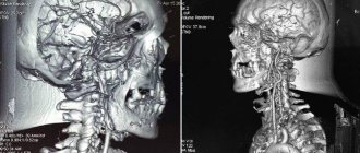
Diagnosis of vascular diseases of the brain and neck:
- Anomalies and various variants of normal development of vessels of this localization.
- Diagnosis of changes in the structure of the vessel wall (arteriovenous malformations, aneurysms).
- Symptoms of deterioration of blood supply to the brain (vertebrobasilar insufficiency).
- Functional assessment of the degree of insufficiency of blood supply to the brain through the vertebral arteries, intracerebral vessels when they are narrowed, with an assessment of the usefulness of the venous outflow.
- Observation of the dynamics of postoperative changes in blood vessels (contrast from compounds of the chemical element gadolinium is used).
- Vascular changes in clinical manifestations of hypertension, migraine.
Temporomandibular joints:
- Functional assessment of joints in malocclusion.
- Before and after orthodontic surgery.
Eyes and other organs located in the orbits:
- Tumors of the soft tissues of the orbit (oculomotor muscles, optic nerve, blood vessels, fatty tissue).
- Injuries and inflammatory changes in organs filling the orbit.
- Diseases of the lacrimal glands of various origins.
Intracranial air sinuses and organs of the oropharynx:
- Acute and chronic inflammatory diseases of the nasal sinuses.
- Assumptions about the presence of a tumor in this area.
- Planned plastic surgery in this area.
Soft tissues of the neck:
- Tumors of the thyroid gland and soft tissues to obtain information about the degree of invasion into adjacent structures.
- Unreasonable enlargement and/or pain of regional lymph nodes.
Spinal cord and spinal column:
- Assessment of degenerative changes in the spine (osteochondrosis, osteoporosis).
- Determination of the degree of pinching of the spinal cord and spinal nerves at their exit points in the intervertebral canals.
- Congenital and acquired anomalies of the development of the spinal cord and spinal bones.
- Diagnosis and verification of demyelinating, inflammatory and tumor diseases of the spinal cord.
- Assessment of the preservation of anatomical structures and functional capacity of the spinal cord after spinal injuries.
- Verification of primary spinal tumors with possible metastasis.
Other bones and joints:
- Diagnosis and verification of traumatic, inflammatory and tumor changes in bones and soft tissues that make up the joints.
- Determination of the degree of traumatic injury to muscles and tendons.
- Complex dislocations of the shoulder joint.
- Assumption of necrosis of the femoral head during its fracture.
- Study and assessment of the functional state of any of their bone-tendon joints if a space-occupying process is suspected to determine its cause.
- Osteoarticular manifestations of systemic diseases of internal organs;
- Sports joint injuries.
Examination of the organs of the retroperitoneal space and abdominal cavity:
- Diseases of the liver, intrahepatic and extrahepatic bile ducts, gallbladder.
- Verification between primary liver cancer and liver metastases with and without additional contrasts.
- Some diseases of the small and large intestine.
- Assessment of the functional state of the pancreas and diagnosis of its diseases of any origin.
- Diagnosis of changes in the spleen due to cysts, injuries, tumor and hematological pathologies.
- Differential diagnosis of kidney and ureter diseases.
- Assessment of changes in the lymph nodes of the abdominal cavity and retroperitoneal space.
Examination of the female pelvic organs:
- Anomalies and pathology of development of the vagina, uterus and appendages.
- Benign diseases of the cervix and body of the uterus, appendages.
- Diagnosis of endometriosis.
- Malignant and germ cell tumors of the body of the cervix and body of the uterus, appendages.
- Inflammatory and adhesive processes in the pelvic cavity.
- Changes in lymph nodes.
- Detection of varicose veins in the walls of the pelvis with and without thrombophlebitis.
- Rectal tumors.
- Determination of the degree of spread of tumor processes with the formation of abscesses and fistulas between the pelvic organs.
Examination of the male pelvic organs:
- Various diseases of the prostate gland.
- Anomalies of testicular development.
- Diagnosis of bladder diseases.
- Anomalies in the development of testicular vessels and seminiferous tubules (varicocele, etc.).
- Inflammatory, tumor diseases of the testicles.
- Diseases of the scrotum.
- Tumors of the rectum with determination of the degree of its germination to neighboring organs.
- Inguinal and femoral hernias.
Breast examination:
- Verification of inflammatory, tumor-like and neoplastic processes in the gland.
- Determination of the degree of tumor invasion into neighboring tissues and damage to lymph nodes.
- Monitoring postoperative treatment and post-traumatic changes.
Diseases of the thoracic cavity:
- Verification of effusion processes into the pleural cavity.
- Peripheral lung cancer.
- Diagnosis of changes in the vessels of the chest cavity (forming aneurysms).
- Mediastinal tumors.
Heart diseases:
- Carrying out coronary angiography.
- Assessment of the preservation of myocardial contractile function in chronic ischemia and acute myocardial infarction.
- Assessment of the valve apparatus in the presence of defects.
Diagnosis using tomography has a number of contraindications, which vary depending on the type of study.
There are no absolute contraindications to CT diagnostics. CT and MSCT are undesirable when:
- the patient is overweight (more than 120 kg, weight restrictions are determined by the tomograph model),
- in his stomach and intestines there is a barium solution left over from previous studies,
- he cannot hold his breath during the scan, as required by regulations.
A contraindication is a plaster cast or metal structure located in the area being examined. In addition, CT/MSCT is not performed in the presence of severe claustrophobia and inappropriate behavior of a person.
Due to the fact that the examination involves radiation exposure, the decision to perform the procedure on pregnant women, women during lactation and young children should be made individually, after considering each specific case. You cannot do CT and MSCT in the first trimester of pregnancy, since at this time the organs of the unborn child are being formed.
If it is necessary to use a contrast agent for CT examinations, the following contraindications should be taken into account: renal failure, allergic reaction to the components of the contrast agent, thyroid disease, severe diabetes mellitus.
Magnetic resonance imaging is prohibited if the patient has:
- Cardiac stimulator.
- Ferromagnetic middle ear implants.
- Large metal implants, ferromagnetic Ilizarov devices.
It is possible, but not advisable, to perform an MRI if:
- Insulin pump.
- Non-ferromagnetic inner ear implants.
- Prosthetic heart valves.
- Implanted neurostimulators.
- Clips installed on vessels (except for cerebral vessels).
- Tattoos that are inked with metal.
Also a contraindication for magnetic resonance imaging of organs and systems is:
- First trimester of pregnancy.
- Heart failure in the stage of decompensation.
- Pathological panic of the patient in a confined space (claustrophobia).
- Severe physical condition of the patient associated with his illness.
- The need to monitor a person's basic vital signs.
- Emotional instability, mental deviations of the patient.
Contraindications for positron tomography are:
- Pregnancy.
- Breast-feeding. When performing positron tomography during lactation, breastfeeding must be stopped for a day so that the radiopharmaceutical is completely removed from the body.
- Diabetes mellitus (if the study is carried out using fluorodeoxyglucose). The procedure is possible provided that the attending endocrinologist adjusts the glucose level.
- Renal failure - in this case, the retention of the radiopharmaceutical in the body leads to distortion of PET data.
- Infectious diseases.
- A serious health condition in which a person cannot remain in a motionless lying position for 1 hour.
- Chemotherapy (PET scan can be performed 12 days after the end of chemotherapy).
- Radiation therapy (PET scan is allowed 3 months after treatment).
- Recovery period after surgery (2 months).
Tags: magnetic resonance, feature, conduction, method, tomography
About the author: admin4ik
« Previous entry
Diagnosis of various joint diseases
For bone damage, tendon sprains and other tissue disorders, doctors often use Magnetic Resonance Imaging. The resulting images show in detail the cartilaginous structure along with the tendon system located next to it.
In addition, discs, joint capsules and bursae, menisci and intra-articular vessels can be easily seen. Below we describe which joints are examined using MRI, as well as the possible results of such examinations.
- Shoulder and elbow . Using magnetic resonance imaging, you can detect any injuries and pathologies: dislocations, fractures and cancer. Disturbances in the functioning of blood vessels are found in the shoulder joints. If you examine the elbow joint using MRI, then it is possible to detect malignant formations, malfunctions in the blood circulation, dangerous inflammations or mechanical injuries.
- Wrist joints and hands . These areas cannot be accurately examined without MRI. The photographs obtained during the scan are used by surgeons and oncologists.
- Knees . In this area, magnetic tomography can detect processes associated with tissue inflammation (arthritis, arthrosis, benign tumors, rheumatoid lesions). In addition, mechanical damage to the knee joint can be diagnosed. Images can be obtained in all planes.
- Maxillofacial . All mandibular joints are also subjected to magnetic tomography. It is used here if the patient has problems with chewing processes, facial and articulatory functions and other important options of the maxillofacial joint. Using the method of studying the type of disease described in the article, you can also find out about the factors that caused it.
- Ankle and hip. If a patient has pathological conditions associated with the ankle or hip joints, accompanied by changes in bone and near bone tissue, then MRI is used here too. After all, it is capable of detecting almost all types of pathologies at the initial stages.
Is it possible to understand the MRI results yourself?
In fact, the ability to decipher MRI abbreviations alone is not enough to make a diagnosis. Usually, the disease is detected by comparing the indicators of a healthy organ with the indicators of the examined one. You also need to take into account a number of other factors, such as individual characteristics and possible, albeit minimal, errors.
Decoding MRI of the brain
Brain MRI protocol
deciphered by a radiologist, but only the attending physician - a neuropathologist or neurosurgeon - can determine which diagnosis should be made. In the image attached to the extract, the specialist also sees certain changes and pathologies.
Interpretation of MRI of the abdominal cavity
Abdominal MRI
is prescribed most often in cases where, after other examinations, there are still some doubts. Also to identify pathologies of blood and lymph flow, oncological changes in tissues. An MRI of the abdominal cavity is interpreted by a doctor according to the general principle: identifying deviations from the norm, the focus of the disease and the degree of damage.
Interpretation of MRI of the spine
When examining the spine, the doctor already roughly suggests a diagnosis. During the collection of anamnesis and other tests, the location of the lesion is approximately determined. MRI of the spine
serves to make an accurate diagnosis and, during decoding, to clarify the details and severity of the pathology.
#!MRTseredina!#
Indications
Magnetic tomography is necessary in the following cases:
- when symptoms characteristic of the development of various joint pathologies appear: tumors and swelling, difficulty moving, pain;
- if necessary, an accurate formulation of the diagnosis;
- if you suspect arthritis or arthrosis;
- with osteochondropathy;
- in the presence of injuries (both new and old) and joint dislocations;
- when a malignant neoplasm is detected in bones, cartilage or soft tissues;
- for diseases with autoimmune symptoms in case of joint damage;
- when diagnosing osteomyelitis.
Deciphering MRI
Often, after undergoing tests or examination, patients peer at the report sheet, trying to predict the diagnosis. Some studies provide this possibility. However, the more complex and extensive the diagnostic method, the more difficult it is to understand something in the conclusion. The MRI discharge sheet is usually replete with a lot of professional abbreviations and requires mandatory qualified decoding.
What diseases can be seen on MRI?
Thanks to its informative content, MRI can detect various changes in organs and tissues. A set of indicators is needed to make a diagnosis. You should not try to decipher an MRI yourself, as this process requires a certain set of knowledge in different areas. The same symptoms and identified changes can be indicators of different diseases, and only a doctor is able to make one or another diagnosis. Unlike CT, MRI can detect diseases not only of bones and hard tissues, but also of the vascular and nervous systems. MRI easily detects inflammation, obstruction of blood flow or bleeding, tissue changes, cysts, swelling, tumors and metastases. MRI, when interpreted correctly, is the most universal method of obtaining information about the affected organ or tissue.
Contraindications
- You are a woman in the first months of pregnancy.
- Your body contains implants, cochlear devices, pacemakers that contain metal parts, prosthetics consisting of titanium, and you cannot have an MRI of bones and joints if you have tattoos.
- You have been diagnosed with heart failure at the stage of decompensation.
- You are claustrophobic.
- You have mental health problems.
How to do an MRI of joints
You do not need any preparation before undergoing the procedure, although some medical institutions require their patients to be examined on an empty stomach.
Next, you will learn how an MRI of joints is done. At the very beginning of the examination, you are placed inside a device that resembles a tube with an open top and bottom.
The magnetic field created by the device activates the nuclei of hydrogen atoms in your body. This leads to the emergence of a resonating effect in them and the radiation of energy of different frequencies by the nuclei, which is read by the apparatus for producing images.
However, this does not have any consequences for the cells, because after the end of the examination they return to their normal state. The computer can only process the received information and form a high-quality three-dimensional image of the examined area of the body.
Please note once again that X-rays are harmful to the patient’s health, and the magnetic field does not pose any danger and does not have a negative effect on the body.
If you are interested in the question of how often you can do MRI of joints , then know that magnetic tomography can be performed as many times as necessary during the course of treatment. After all, as soon as the procedure is completed, the nuclei in your cells will return to their normal state.
The higher the degree of damage to the examined area and the greater the number of pathologies present, the longer the tomography time will be. An MRI of one area lasts about a third of an hour. However, this time will increase to 40 minutes if contrast is used.
What you need to know about the survey results
You need to remember that in clinics magnetic resonance imaging is performed both with and without a doctor. The following is a description of two cases:
- If the doctor is absent during a patient's magnetic tomography, a technologist interacts with the patient and takes images of the highest quality possible. The doctor himself gets involved in the work at the stage of deciphering the received photographs. It is the doctor who draws up a conclusion based on the results of the examination - this takes him from forty to sixty minutes.
- If the doctor is present during the examination, then in this situation the doctor begins to decipher the photographs as they appear. The advantage of this method of examination is that the patient can receive images within a quarter of an hour after the tomography. This method is usually used if a person has a severe case of the disease.
How does the procedure with contrast work?
In some cases, the patient is recommended to undergo an MRI of the head with contrast. This helps to obtain a more accurate image of the vessels. The examination process differs from the usual only in the introduction of a special chemical substance into the patient’s blood. Contrast is administered some time before the start of diagnosis. The specialist must monitor the body’s reaction to the administered drug.
During administration, a person may experience a coolness or a metallic taste in the mouth. In some cases, the injection may be painful.
Next, the patient is also placed in a tomograph and scanned. The key to a successful result will be the immobile state of the patient. If a hyperactive child is being tested, anesthesia may be required.
The procedure is absolutely painless. Immediately after its completion, the person will be able to return to their usual lifestyle. An MRI with contrast will help the doctor better see the vascular pattern and any brain abnormalities.