Internal hydrocephalus
Classic signs of fetal internal hydrocephalus on ultrasound are an increase in the size of the lateral ventricles of the brain and the third ventricle.
Fetal hydrocephalus is most often recorded starting from the second planned ultrasound scan during pregnancy, although even at this time the pathology is not easy to determine, especially in the initial stages. The most “sensitive” and reliable criterion is considered to be an increase in the height of the body of the cerebral ventricles by 0.2-0.3 cm compared to the normal state.
The width of the bodies of the lateral ventricles of the fetal brain is normally 7.5 mm. With an increase of more than 1 cm, ventriculomegaly is recorded - isolated expansion of the ventricles. The progression of intrauterine hydrocephalus is accompanied not only by an increase in the width of the lateral ventricles, but also by the expansion of the frontal and occipital horns, the third and fourth ventricles of the brain.
External hydrocephalus
The ultrasound picture of external hydrocephalus is characterized by the following features:
- increase in the size of the large tank over 1.1 cm;
- an increase in the interhemispheric fissure over 0.5 cm at the level of the frontal lobes of the brain, with a simultaneously recorded decrease in its echogenicity;
- detection of subarachnoid spaces of the fetal brain, which are normally absent at this stage of pregnancy.
Distinctive signs of intrauterine hydrocephalus can be recorded on ultrasound, starting at approximately 20 weeks - the beginning of the 2nd trimester screening period
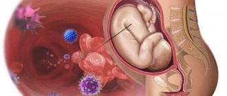
Hydrops fetalis
Hydrops fetalis is a polyetiological pathological condition in neonatology and pediatrics, which is characterized by the intrauterine development of generalized edema, accumulation of fluid in the chest and abdominal cavities, as well as in the pericardial sac. The overall prevalence is 1:1-14 thousand newborns, while the immune form occurs 1 time in 3,000 births. Despite the relatively high prevalence of AB0 and Rh conflicts between mother and fetus in the population, the development of an immune form of hydrops fetalis against this background rarely occurs. This is due to the possibilities of modern diagnostics, intrauterine treatment and prevention of hemolytic disease of newborns. In more than half of the cases (55-60%), the etiology remains unknown. The prognosis for this pathology is always serious. The mortality rate for the non-immune form is very high and is about 70-80%, while for the immune form it is about 20%.
Physiological hydrocele of the testicles in newborns and other forms of the disease
In newborns, the following variants of hydrocele are distinguished according to their anatomical structure:
- Hydrocele communicating with the abdominal cavity.
- Hydrocele, non-communicating or encapsulated.
- Funiculocele is an accumulation of fluid around the spermatic cord, with the distal area overgrown.
This is interesting
Hydrocele of the processus vaginalis can occur not only in boys. Girls also have a vaginal process of the peritoneum; at birth it also closes and is called the Nuka canal. In girls, it is also possible that this canal does not close for various reasons, with the formation of hydrocele phenomena in this area.
Hydrocele of the testicle appears in a baby if it is congenital, usually immediately, in the first days after birth. The appearance of the scrotum is characteristic due to its increase in size.
If the scrotum is enlarged on one side, then they speak of a unilateral hydrocele, if on both sides, then they speak of a bilateral hydrocele.
Pain as a symptom is not typical for testicular hydrocele, but it can appear if the hydrocele is tense, as well as with complications.
A mild hydrocele of mild severity, which resolves within a year after birth, can be interpreted as physiological hydrocele of the testicles in newborns.
Diagnosis of hydrocele in newborns
To make a diagnosis of hydrocele, it is necessary to differentiate it from an inguinal hernia. If a loop of intestine penetrates the scrotum, a characteristic gurgling sound can be detected upon palpation.
A hernia without signs of strangulation will slide and be easily reduced into the inguinal canal.
- A hydrocele can also decrease in volume with pressure, but this happens slowly, due to the flow of fluid into the abdominal cavity.
- A non-communicating hydrocele will not shrink in size when pressed.
- In the case of hydrocele of the spermatic cord, a large fluid formation can be detected in the area of its projection.
To diagnose testicular hydrocele, diaphanoscopy, or transillumination of the testicle using a light beam, is performed.
- For this purpose, special non-heating lamps are used.
- The transparent liquid has sufficient permeability to light, and in the case of accumulation of serous exudate, the scrotum will take on a characteristic appearance, translucent and painted in red-yellow shades due to the skin and blood vessels.
- The hernial sac, being more dense, will not transmit light as well as liquid. Also, transparency will be low with pyocele, or purulent inflammation, hematocele and testicular tumor.
To confirm the diagnosis and obtain accurate data on the condition of the processus vaginalis of the peritoneum, the degree of communication with the abdominal cavity and the amount of fluid accumulated in the membranes of the testicle, the patient undergoes an ultrasound examination.
Causes of hydrops fetalis
Hydrops fetalis is a heterogeneous disease. Often the exact etiology cannot be established, but the most likely causes of its development are known. The non-immune form can be caused by mutations (Down syndrome, Shereshevsky-Turner syndrome, triploidy and tetraploidy), genetic diseases (G-6-FDG deficiency, α-thalassemia, Noonan and Pen-Shockey syndrome, etc.), conduction disorders and congenital heart defects (ASD, VSD, supraventricular and ventricular tachycardia), pathologies of the great vessels (thrombosis of the superior and inferior vena cava, arteriovenous shunting), anomaly of the chest structure (diaphragm hernia and asphyctic dysplasia), TORCH infections (toxoplasmosis, CMV, syphilis, etc. ), dysplasia of the genitourinary system (congenital stenosis or aplasia of the ureter, hydronephrosis), pregnancy pathologies (placental transfusion syndrome, chorioangioma, eclampsia). The pathogenesis of non-immune hydrops fetalis is based on the child's heart failure, hypoproteinemia and anemia, which reduce the oncotic pressure of the blood.
In addition to all of the above reasons for the development of hydrops fetalis, one can identify risk factors that increase the risk of the non-immune form. These include pregnancy before 16 or after 40 years, multiple and post-term pregnancy, macrosomia. The male sex is considered more prone to hydrops fetalis. The main cause of immune hydrops of the fetus is incompatibility of mother and fetus by blood group (0 (I) in the mother and A (II) or B (III) in the child) or Rh factor (mother - Rh+, and child - Rh-), as a result, increased hemolysis of the child’s red blood cells occurs and hemolytic disease develops. At the moment, this option is extremely rare due to generally accepted diagnostic measures in obstetrics.
Classification and symptoms of hydrops fetalis
Hydrops fetalis has two etiological forms:
- Immune fetal hydrops. As a rule, it develops against the background of hemolytic disease of the newborn. Currently, with early diagnosis and treatment, this form of hydrops fetalis practically does not occur, despite the relatively high prevalence of Rh and ABO conflicts. The prognosis is relatively favorable.
- Non-immune form. Polyetiological group. This variant of fetal hydrops can be caused by congenital heart defects and disruption of its conduction system, genomic and chromosomal mutations, TORCH infections, various dysplasias, pregnancy pathologies, etc. In most cases, it has a fatal outcome.
Clinical manifestations of fetal hydrops are determined from the moment of birth. The general condition of the child is usually severe or very serious (more often). In such children, a disproportionately large head is revealed due to a significant increase in the cerebral part of the skull. Due to general muscle weakness, the child takes the frog position. The sutures of the skull are open, the fontanelles protrude, and the bones have a pliable consistency. A large volume of cheese-like lubricant and a lot of vellus hair are detected on the body, general swelling and pallor are detected. Subcutaneous fatty tissue is poorly expressed, which is why body temperature is very labile and highly dependent on the environment. With hydrops fetalis, boys may experience cryptorchidism, and girls may experience underdevelopment of the labia. Physiological reflexes (including sucking and searching, Moro) are suppressed. Heart rate is labile (120-160 beats/min), blood pressure is below normal (50-65 mm Hg). Tachypnea is observed, alternating with periods of apnea. Severe hepatosplenomegaly, hydrothorax, ascites, and hydropericardium are often observed. In some cases, transient hypothyroidism and pollakiuria may develop.
Hydrocephalus in the fetus - hydrocephalus of the brain
Hydrops fetalis is characterized by an increase in the volume of cerebrospinal fluid in the ventricular system of the brain, internal and external spaces containing cerebrospinal fluid.
Fetal hydrocephalus accounts for 40% of the total mass of central nervous system pathologies developing in the perinatal period (from 22 weeks of intrauterine life to 7 days after birth).
The prevalence of hydrocephalic syndrome in the neonatal period (from the moment of birth to the 28th day of extrauterine life) is about 4 cases per 1 thousand newborns.
Causes of pathology
Primary (true) hydrops fetalis in pregnant women is often caused by a hereditary predisposition provoked by genetic mutations.
In 30% of cases caused by chromosomal mutations, an increase in the diameter of the fetal head is observed during gestation; in 50% of cases, hydrocephalic syndrome develops 3 months after the birth of the child.
The secondary form is the result of a violation of the outflow of cerebrospinal fluid. The secondary form of hydrops in the fetus during pregnancy occurs as a consequence of many reasons:
- Inflammatory and infectious diseases suffered by the mother during pregnancy, including viral infections (ureaplasmosis, chlamydia, toxoplasmosis, CMV - cytomegalovirus infection).
- Intraventricular hemorrhages of traumatic or hypoxic-ischemic etiology (25-70% of cases).
- Congenital malformations of the brain (anomaly of the development of the cerebral aqueduct in 43% of cases, anomaly of the development of the foramina of Magendie, Luschki in 3-5% of cases).
- Inflammation of the leptomeninges (arachnoid and soft membranes).
- Platybasia (flattening of the base of the skull, depression of the lower part of the occipital bone into the cranial fossa from behind).
Cases of maternal infection with infectious agents are especially dangerous in the early stages of pregnancy, when brain structures begin to form. Hydrocephalus can develop as a consequence of Arnold-Chiari syndrome (an abnormality in the formation of the skull, in which there is a decrease in size or deformation of the part containing the cerebellum).
Diagnostics
The diagnosis is established based on the results of an instrumental examination, according to the opinions of specialists.
At the same time, a mother's blood test is performed, which shows the titer of antibodies to infectious agents (to identify viral and bacterial infections that occur latently).
The indication for treatment is severe ventriculomegaly (increase in the size of the ventricles), diagnosed during ultrasound or MRI. Consultations with a perinatologist, neonatologist, neurosurgeon, and geneticist are required.
Treatment methods
The results of instrumental studies show the nature of the course of the disease, including the degree of ventricular dilatation and the rate of progression of pathological changes. Treatment of pathology is difficult due to the following reasons:
- Ambiguity in understanding the relationship between pathological processes in the parenchyma (epithelial cells) and the development of hydrocephalus.
- Imperfection of methods for differential (physiological norm, pathological changes) diagnosis of ventriculomegaly (increase in the size of the ventricles).
- Lack of data on the exact timing of the completion of the formation of brain structures involved in the process of circulation and outflow of cerebrospinal fluid.
- Conflicting opinions about the physiologically normal sizes of the elements and sections of the ventricular system.
The main treatments include cephalocentesis (ventricular puncture) and fetal ventriculoamniotic shunt, which is considered more effective than placement of shunts and drainage systems after the baby is born. Cephalocentesis is an invasive procedure that punctures the wall of the dilated ventricle and removes excess cerebrospinal fluid.
Intrapartum intervention is performed with a puncture needle under sonological (ultrasound navigation) control during the period of intrauterine development between 25 and 32 weeks of gestation. Evaluation criteria for the success of cephalocentesis include a reduction in the size of the ventricles and fetal head circumference, which is determined during a control ultrasound examination.
Intrapartum ventriculoamniotic shunting avoids fetal death or the development of serious pathologies in the newborn. The procedure is resorted to if, according to the results of an ultrasound or MRI, hydrocephalus is detected in the fetus, which threatens its normal functioning and development.
During the intervention, a shunt (artificial vessel, drainage) is installed, which ensures the outflow of cerebrospinal fluid into the amniotic cavity (amniotic fluid). The shunt is equipped with a one-way membrane valve that prevents the flow of amniotic fluid back into the ventricular system. Conditions for the operation:
- Early detection of ventriculomegaly and hydrocephalic syndrome (postpartum shunting is not yet possible).
- A simple form of occlusive hydrocephalus without association with other damage to brain tissue.
- Hydrocephalic syndrome is not combined with other developmental anomalies that are incompatible with life.
- Progressive increase in the size of the ventricles.
Statistics show that intrauterine minimally invasive treatment of hydrocephalus is more effective than similar operations performed after the birth of the child. For comparison, survival rate after intrapartum procedures increased to 83% (from 31%), the proportion of healthy children increased to 34% (from 19.5%).
Prognosis and prevention
The prognosis for this pathology is relatively favorable. Timely diagnosis and intrauterine treatment significantly increase the baby’s chances of recovery.
To prevent the development of a secondary form of pathology, the mother carrying the fetus must give up bad habits (alcohol, smoking), avoid infection, lead a healthy lifestyle, ensure proper nutrition and rest.
Fetal hydrocephalus during pregnancy is a pathology that can cause serious developmental disorders.
Intrauterine cephalocentesis can reduce the likelihood of macrocrania (pathological increase in skull diameter) and irreversible changes in the structure of brain tissue.
Intrapartum treatment makes it possible to prolong pregnancy and carry out successful endoscopic minimally invasive intervention after the birth of the child.
Diagnosis of hydrops fetalis
Diagnosis of fetal hydrops includes the collection of anamnestic data, an objective examination of the child, laboratory and instrumental research methods. When interviewing mothers, they pay attention to previous infectious and gynecological diseases, chronic pathologies, features of the course of pregnancy and its complications. During an objective examination by a neonatologist or pediatrician, the characteristic symptoms of hydrops fetalis, the complications present are established, the general condition of the child and the need for resuscitation measures are assessed. Laboratory diagnostics in the form of ELISA, PCR or other serological reactions make it possible to identify a probable infectious pathogen. If hemolytic disease of the fetus is suspected, the blood group and Rh factor of the mother and child are re-determined. General laboratory tests (CBC, OAM, blood biochemistry) are used to assess the functioning of internal organs and exclude concomitant diseases.
Ultrasound plays a leading role in the instrumental diagnosis of fetal hydrops. Ultrasound scanning allows a preliminary diagnosis to be made in the antenatal period. Ultrasound signs that may indicate fetal hydrops include swelling of the placenta, polyhydramnios, the presence of fluid in the cavities of the child’s body, hepatosplenomegaly, swelling of the subcutaneous fat, and “Buddha pose.” Postnatal ultrasound is performed to confirm dangerous pathological conditions (hydropericardium, hydrothorax, ascites) and determine therapeutic tactics. ECG, computed tomography and magnetic resonance imaging can also be used for the purpose of differential diagnosis and assessment of the structure of internal organs.
Diagnosis of the disease
Hydrocephalus is water on the brain in a child. The disease is characterized by the accumulation of fetal cerebrospinal fluid in the brain. A large amount of cerebrospinal fluid collected in one place can exert physical pressure on the brain. Because of this, the functionality of the central nervous system will be disrupted and mental retardation or delay in physical development will appear in the child.
Statistics show that today, per 1000 children born, there is 1 child with such ailment. You can rely on the effectiveness of medical care if the parents of a sick child consult a doctor early.
An excessively large head in an infant may indicate the presence of this disease. More often, this sign is noticed after childbirth and in the next nine months of the child’s development. Having done an ultrasound, MRI and CT, it will be possible to confidently confirm the diagnosis.
Diagnosis is aimed at establishing the provoking factor that caused hydrops fetalis. First, the doctor determines the blood type and Rh factor. This is necessary to remove or confirm immune dropsy and Rh conflict.
The doctor analyzes past infectious diseases, pathologies and operations, an analysis of the life history, the presence of pathologies associated with gynecology, the course and outcomes of the previous pregnancy, obstetric and gynecological history, a complete description of the current pregnancy, any complications, weight gain, and so on.
Fetal ultrasound is the main diagnostic method. Main signs of ultrasound:
- excess amniotic fluid;
- violation of the normal size of the child due to swelling, an increase in the size of the abdomen compared to the size of the head;
- enlarged heart size, cardiomegaly;
- enlarged liver and spleen;
- limbs abducted in different directions with a swollen belly “Buddha Pose”;
- consequences of the disease.
After diagnosis, doctors’ prognoses are often disappointing. According to statistics, the percentage of surviving children does not reach 30%. More often than not, the child dies in the womb. But still, with timely detection of the disease and provision of treatment, there is a chance to save the fetus. But at the same time, there may be the following consequences in the future:
- Heart failure.
- Heart or brain defect.
- Pathology of the respiratory tract.
Defects are formed individually in each child due to gender, despite the causes of the disease. Modern medicine makes it possible to recognize non-immune dropsy and provide timely assistance.
The fetus experiences swelling of the head, torso and arms and may have problems with intubation. To be prepared for various types of unforeseen situations and provide timely assistance, you need to purchase tubes of different sizes. You should purchase a guidewire needed for oral intubation.
An equally significant factor is respiratory failure. Most often it is caused by ascites, pulmonary hypoplasia, and hyaline membrane disease. Heart failure is a big deal. It is worth remembering that the first aid that will be provided to a baby with non-immune dropsy will be much more effective if the actions are carried out in an organized and correct manner and if they are prepared for this in advance.
Pregnancy and childbirth can be difficult, and there is a chance that the baby has immune and non-immune hydrops. Dropsy is a kind of swelling that has spread throughout the child’s entire body. This pathology is detected using ultrasound.
Treatment of hydrops fetalis
There is no specific treatment for nonimmune hydrops fetalis. Childbirth is carried out in conditions of full readiness of resuscitation equipment. Immediately after birth, the child receives the full amount of intensive care: tracheal intubation with 100% oxygen supply, if necessary, cardiopulmonary resuscitation, blood or red blood cell transfusion, etc. If all measures taken are ineffective, pleural, pericardial puncture and laparocentesis under monitoring of ultrasound and vital signs of the child. Depending on the situation, antihemorrhagic, antibacterial, immunocorrective, metabolic, anticonvulsant therapy, etc. may be prescribed.
For the immune form of hydrops fetalis, treatment begins in the antenatal period. It consists of intrauterine transfusion of Rh blood to the fetus using cordocentesis. This procedure is indicated for severe anemia and a drop in hematocrit below 30 g/l. If necessary, re-transfusion can be performed after 14-21 days.
Causes of hydrocephalus
Hydrocephalus of the brain in the fetus occurs due to many factors, but still mainly these are infections that enter the mother’s body. And the earlier this happens, the worse it is for the unborn baby.
List of probable causes accompanying the development of hydrocephalus:
- Sexually transmitted infectious diseases. Syphilis leads to dysfunction of the nervous system and also contributes to the development of dropsy.
- Ureaplasmosis. Whether the disease can occur as a result of this infection is still a controversial issue, but there have been cases recorded on fetal ultrasound during pregnancy.
- The presence of chlamydia infection in the womb also causes this pathology and, along with it, affects visual functions and the nervous system.
- Toxoplasmosis - these harmful pathogens arise from contact with animals, as well as from eating poorly processed meat. It is especially dangerous when it is early in pregnancy.
- The causative agent of rubella infection, upon penetration, can cause miscarriage in early pregnancy. But if the infection of the fetus occurs in the later stages, then it is possible that this will cause hydrocele in the brain of the fetus.
- If the mother has herpes during pregnancy, then the risk of hydrocephalus exists in half of the cases. Children also experience damage to the skin and nervous system.
- Cytomegalovirus - this infection affects the cellular level of the nervous system and contributes to the development of abnormalities, including hydrocephalus.
- Chiari syndrome, or a congenital pathology in which the cerebellar trunk descends into the occipital region. This is where the circulation of spinal cord fluid is disrupted. The brain increases in size, but the skull remains small.
- With chromosomal abnormalities or Edwards syndrome, almost all organs are affected, so such newborns die in the first months of life.
- When the cerebral aqueduct is narrowed, which is one of the congenital factors of the disease, the outflow of fluid from the brain is impaired. More often this manifests itself at a certain age.
- Bad habits that a woman indulged in during pregnancy also contribute to the development of hydrocephalus. For example, drinking alcohol and smoking, taking medications not prescribed by a specialist, exposure to harmful factors during fetal formation.
Before becoming a mother, it is recommended to undergo a full examination: pass all the necessary tests, and if infectious agents are detected, undergo a course of therapeutic procedures.
Causes of hydrocephalus in the fetus
Hydrocephalus (another name for dropsy) is characterized as a symptom complex with a pathological accumulation of cerebrospinal fluid under the membranes of the brain and/or in its cavities (ventricles), and is accompanied by increased intracranial pressure.
The causes of hydrocephalus in the fetus are of infectious and non-infectious origin. Pathology occurs due to excessive production of cerebrospinal fluid or disturbances in its outflow.
Dropsy is classified into: • primary - the result of birth defects and genetic abnormalities;
• secondary – due to intrauterine infection.
The main causes of hydrocephalus in the fetus
There are many factors contributing to the disease, but a special role is assigned to infectious agents that enter the baby’s body from the mother. The earlier the infection occurs, the more insidious the consequences. The basic causes of fetal hydrocephalus during pregnancy include: 1. Infections of the mother that are sexually transmitted.
Syphilis - intrauterine infection of a baby with syphilis leads to congenital defects of the nervous system, including dropsy of the brain. Ureaplasmosis - its role in the occurrence of hydrocephalus is at the discussion stage, however, in many women diagnosed with fetal hydrocephalus after termination of pregnancy, the presence of the causative agent of ureaplasmosis is confirmed.
Chlamydia causes congenital eye defects and neurodevelopmental abnormalities.2. TORCH infection in the mother. Infection with toxoplasmosis occurs when eating meat with insufficient heat treatment and through contact with domestic and wild cats. Infection in the early stages of pregnancy is especially dangerous, leading to serious brain damage.
Rubella is a pathogen that, when it enters the body of a pregnant woman who has not had rubella, causes the death of the embryo in the first weeks. Infection at a later date threatens damage to the fetal brain and, possibly, the development of hydrocephalus. Herpes infection - there is a high risk (more than 40%) of infection of the fetus from an infected mother.
Newborns experience damage to the nervous system, skin, eyes, etc.; Cytomegalovirus infection - cytomegalovirus has a high affinity for the cells of the nervous system and mainly causes abnormalities in the development of the fetal brain, as an example - hydrocele.3. Congenital pathology of the baby.
Chiari syndrome is a developmental disorder of the brain in which the brainstem and cerebellum descend into the foramen magnum and the circulation of cerebrospinal fluid is disrupted. According to new data, the disease occurs due to a rapid increase in the size of the brain, which does not correspond to the size of the skull.
Edwards syndrome is classified as a chromosomal disorder, more common among girls, characterized by multiple lesions of organs and systems (including hydrocephalus), children are not viable and die in the first months of life.
Narrowing of the cerebral aqueduct, asymmetry of the lateral ventricles belongs to congenital damage, the free flow of cerebrospinal fluid into the cerebral ventricles is impaired and manifests itself with age.
4. Bad habits of a woman - excessive alcohol consumption or chronic alcoholism, smoking, uncontrolled use of medications, prolonged stay in areas of high radiation levels, especially in the first weeks, when the formation and formation of the components of the child’s central nervous system occurs.
Important! Plan pregnancy and conduct tests for the presence of sexually transmitted infections and hidden TORCH infections. With its onset, lead a healthy and careful lifestyle without bad habits and negative factors in order to give birth to a healthy baby.
Methods for diagnosing fetal hydrocephalus during pregnancy
The main method for diagnosing cerebral hydrocephalus in the fetus is ultrasound. The baby's head is measured using a transverse scan. The main criteria for fetal hydrocephalus on ultrasound include the width of the lateral ventricles, which normally should not exceed 10 mm.
Diagnosis is carried out starting from the 17th week, repeated research is carried out at 20–22 weeks of pregnancy, however, the average period when the pathology is visualized in most cases is 26 weeks. Another method for determining whether there is fetal hydrocephalus during pregnancy is echography.
However, it is only available in large diagnostic centers.
Possible prognosis and consequences of fetal hydrocephalus
The consequences can be different and depend on the size of the disorder and accompanying developmental defects. In the case when the size of the lateral ventricles does not exceed 15 mm, no other anomalies are identified and correct treatment is prescribed, the prognosis is favorable - the baby may not experience any abnormalities.
An unfavorable prognosis develops if the size of the ventricles is more than 15 mm and dropsy increases rapidly, the pathology is detected in the first half of pregnancy, and multiple organ lesions are noted, which are characteristic of chromosomal diseases.
The consequences of fetal hydrocephalus during pregnancy in such situations are quite dire - the death of the baby or serious damage to the central nervous system in the newborn.
Thus, a number of causes of cerebral hydrops in the fetus and effective methods for identifying this pathology have been identified. Careful preparation for pregnancy and compliance with recommendations for preventive ultrasound examinations will help prevent illnesses in the baby.
(5 4,40 of 5) Loading...
Symptoms of hydrocephalus
As mentioned earlier, dropsy during fetal development occurs when the outflow of fluid from the brain is disrupted, which ensures its accumulation. This factor increases intracranial pressure and leads to frequent migraines.
Some symptoms of hydrocephalus:
- constant feeling of nausea;
- vomit;
- headaches of a prolonged nature and intensity;
- bad feeling;
- lethargy and drowsiness;
- increased blood pressure.
At the same time, vomiting and nausea can be easily confused with signs of toxicosis or with changes in hormonal levels during pregnancy. With late toxicosis, frequent headaches caused by increased intracranial pressure are also possible.
In these cases, inpatient treatment under the supervision of a specialist is necessary. And in order to timely monitor your health during pregnancy, you need to contact an antenatal clinic for the slightest reason.
Diagnosis of pathology
To detect the disease in the early stages, the most effective method is ultrasound. The head of the unborn child is measured by scanning, and the main parameter is the width of the ventricles located on the side; their size should not exceed 10 mm. An ultrasound is performed at 17 weeks and repeated at 22 weeks, but the average period is 26 weeks of pregnancy. Hydrocephalus on fetal ultrasound indicates the presence of a concomitant infection, which must be combated immediately.
Signs of pathology
The first symptoms of the development of dropsy during pregnancy, which usually alert the doctor, are sudden weight gain and the appearance of edema.
With dropsy of the 1st degree, swelling is noticeable in the area of the feet and legs. As the pathology progresses, swelling affects the thighs, the anterior wall of the abdomen and gradually spreads to the torso and face.
Normally, weight gain in the second half of gestation should not exceed 250-300 g per week. If these indicators are higher (500 g or more), there is every reason to diagnose dropsy.
However, not in all cases the patient experiences pronounced swelling. Sometimes dropsy can be suspected only on the basis of excessive weight gain. In this case, blood pressure may be normal, and there may be no protein in the urine. In this case, we are talking about the presence of hidden edema. In general, diagnosing hydrops in pregnancy is not difficult.
During diagnosis, the daily volume of urine is taken into account. A McClure Aldrich test is performed to determine the time of resorption of the blister formed during the test.
As this condition progresses, the following signs are noted:
- decreased frequency of urination;
- maintaining a “pit” on the skin when pressed with a finger;
- symptom of “ring” or “tight shoes” that suddenly become small;
- pale skin;
- feeling of heaviness in the legs;
- shortness of breath, rapid heartbeat;
- increased feeling of thirst;
- severe fatigue and weakness.
One of the signs of dropsy is the persistence of a “pit” on the skin when pressed with a finger
In some cases, the danger lies in the fact that, in general, a pregnant woman may feel satisfactory, unaware that her well-being and the health of the unborn child are in danger.
However, if you ignore the first symptoms of pathology, dropsy can progress to the next stages of late toxicosis - nephropathy and eclampsia, which pose a real threat to the life of both the baby and the mother.
Consequences of pathology
All those factors that threaten the occurrence of negative consequences depend on developmental defects and the causes of the disorder. For example, if the size of the lateral ventricles has not reached 15 mm, and no other pathologies have been identified, then this can be easily treated during pregnancy. Then the baby will be born healthy.
If the size of the ventricles exceeds 15 mm, then fetal hydrocephalus begins to develop intensively, which will adversely affect the unborn child. The consequence in the presence of dropsy can be death, and diseases of the central nervous system are also possible.
Only preparation for future pregnancy and childbirth will not give pathology the opportunity to arise.
Treatment
If there are signs of the disease, you should consult a doctor. In turn, the specialist will prescribe a series of examinations and decide on treatment. If the pathology is caused by infectious diseases, then, as a rule, therapy is prescribed aimed at removing excess fluid from the body and reducing the production of cerebrospinal fluid. Vitamins and drugs to improve cerebral circulation may also be prescribed. Pregnant women diagnosed with hydrocephalus should not stay in rooms with elevated temperatures or drink a lot of fluids. In the early stages of the disease, therapeutic treatment is used, that is, the attending physician prescribes medications that are gentle for the woman and her unborn baby.
Modern medicine has two more unique treatment methods:
- Using a puncture, excess fluid is pumped out inside the womb. This procedure is performed only once.
- A special shunt is inserted into the fetal brain to drain cerebrospinal fluid or cerebrospinal fluid from the unborn child's brain. This condition persists throughout pregnancy.
If measures to protect the health of the unborn baby are not carried out in a timely manner, the consequences can be different and irreversible.
Consequences and treatments
Therapy is aimed at reducing and normalizing intracranial pressure and depends on the severity of “dropsy of the brain”, usually:
- prescribe medications that lower blood pressure;
- a puncture is performed to remove excess fluid through the fontanelles;
- in difficult cases they resort to surgery.
What can you do?
If you suspect hydrocephalus, you should immediately consult a doctor. Timely diagnosis and treatment will help minimize the risk of complications.
What does a doctor do?
Dropsy is treated with several types of therapy. For hydrocephalus caused by infection or head injuries, drug treatment is selected. It is based on taking medications that help reduce the production of cerebrospinal fluid and remove excess fluid from the body (diuretics). Depending on the clinical picture, medications to improve blood circulation in the brain, vitamin complexes and other drugs that eliminate symptoms may also be prescribed. Expectant mothers diagnosed with hydrocephalus are advised to avoid excess heat (for example, hot baths) and carefully monitor the amount of fluid consumed.
Treatment of cerebral hydrocele with shunting must be carried out before pregnancy.
With the development of modern medicine, it has become possible to treat hydrocephalus of the fetal brain intrauterinely. There are two methods:
- When, using a puncture, excess fluid is pumped out through the mother’s abdomen from the space near the fetal brain, which puts pressure on the child’s brain. The procedure is performed once.
- A shunt is inserted into the fetal brain to drain cerebrospinal fluid. The tube remains in the baby's brain until the end of pregnancy.
Untimely therapeutic measures, as well as ignoring treatment, can cause serious complications and termination of pregnancy.
Classification
There are two different cases of manifestation of pathology. The first is characterized by the inability of cerebrospinal fluid to exit the ventricles. This manifestation is called non-communicating hydrocephalus. The second type is all other manifestations that are called a communicating disease.
Modern medicine uses a unified classification of pathology, which distinguishes primary and secondary. Primary hydrocephalus in the fetus is the main congenital disease, while secondary hydrocephalus is caused by some other disorder in the body (tumors, heart defects, vascular deformation, etc.).
Detection of fetal hydrocephalus on ultrasound
Hydrocephalus is the accumulation of fluid from the spinal cord into the brain, including into its ventricles. This factor is accompanied by intracranial pressure. This disease can be caused by an infection or another reason. The picture of this manifestation is as follows: a large amount of fluid leaves the spinal cord, which is subsequently retained in the brain. There are 2 classes of this disease:
- primary, resulting from congenital anomalies;
- secondary, acquired in the womb during infection of the fetus.
The disease is classified in the following areas:
- acquired pathology;
- congenital disease;
- open or communicating hydrocephalus;
- closed, or occlusive, hydrocephalus;
- resulting from atrophy of brain cells;
- normotensive, or normal blood pressure.
Causes
There are many factors that provoke the disease, but the main and most dangerous are infections that penetrate the womb from mother to baby. The most common reasons can be identified:
- Sexually transmitted infections. The earlier the fetus becomes infected, the more serious the consequences of such development. The most terrible infection is syphilis, which, penetrating into the fetus, causes defects in the nervous system, provokes impaired brain development in the fetus, as well as dropsy. Frequent provocateurs of pathology are chlamydia and ureaplasmosis. Although the latter infection has not been confirmed by medicine, the statistics of termination of pregnancy with this disease is increasing from year to year.
- TORCH infections. Toxoplasmosis, rubella, herpes, cytomegalovirus are very dangerous and are fraught with serious complications, especially in the early stages of pregnancy, when the brain is just forming. These viruses not only affect the internal systems and organs of the fetus, but can also cause pregnancy loss, regardless of the term.
- Congenital pathological processes. This may be Edwards syndrome, in which chromosomal abnormalities appear in the child’s body, characterized by damage to various organs, including the spinal cord. There is a risk of Chiari syndrome, a brain development disorder in which the cerebellum and brain stem descend into the occipital space, resulting in poor fluid circulation.
- Abuse of bad habits during pregnancy. If during the period of bearing a child a woman neglected the basic rules of maintaining health, smoking and alcohol can leave a negative mark on the fetus and lead to disturbances in the functioning of the central nervous system. This is how hydrocephalus arises.
What to do with dropsy in pregnancy
It is very good if you have the habit of weighing yourself regularly and monitoring your weight gain. In this case, you can independently notice that weight gain exceeds the permissible norms and, as a rule, occurs spasmodically: for example, during one week the gain may be insignificant, and the next there is a sharp jump. This means that a more thorough examination is required, for which it is better to immediately contact your pregnancy doctor. He is obliged to refer the expectant mother for additional research, which will allow, in particular, to determine the difference between the amount of fluid consumed and excreted by the body and the proper functioning of the urinary system. There are special analyzes and tests for this.
Proper sleep (at least 10 hours at night and preferably 1-2 hours during the day) and emotional peace are extremely important for dropsy in pregnant women. But physical activity will not be superfluous. What to choose - swimming, walking, yoga - it is better to consult your doctor.
Diet
Regardless of the degree to which dropsy is expressed, the pregnant woman must be prescribed a diet that minimizes salt consumption: its amount should not exceed 2-3 g per day. Sweets in your diet should also be limited as much as possible, as well as other foods that contribute to weight gain: flour, starchy, fatty, fried. The emphasis should be on healthy nutrition: easily digestible lean protein (boiled meat, eggs, cottage cheese, kefir, fish); plant fiber (whole grains, low-calorie fruits, vegetables), potassium-rich foods (baked potatoes, dried apricots, figs). Fast food and products containing monosodium glutamate are completely excluded from the menu! Hot seasonings and spicy spices should also be classified as undesirable.
It is better to eat fractionally: in small portions with a break of 2-3 hours. It is useful to have fasting days once a week.
As for the drinking diet, you can often find recommendations to sharply limit daily fluid intake. But recent studies refute the advisability of such prescriptions: it has been proven that this has little effect on the formation of edema. You need to drink as your body requires (but, of course, not very much); you should use only fresh, high-quality purified water or diuretic drinks that are safe for this period if prescribed by a doctor (for example, lingonberry tea or fruit juice).
Symptoms
During pregnancy, a woman can recognize this pathology based on some symptoms:
- regular nausea and vomiting;
- weakness, poor health;
- intense headaches, migraines;
- blood pressure surges, frequent increases;
- drowsiness.
However, without diagnosis, these signs cannot be perceived as the presence of problems with the fetus, because such symptoms can accompany a woman due to hormonal changes during pregnancy. At this time, you should not give in to groundless panic; only a doctor can make a diagnosis.
Diagnostic research methods
The main diagnostic method is ultrasound. Using scanning, the volume and size of the baby’s head are measured, and the size of the lateral ventricles is assessed. The norm for the width of this indicator is 10 mm; exceeding these indicators is considered pathological. An ultrasound examination is carried out after the 17th week of pregnancy, because the average period for the formation of changes is 26 weeks. The diagnostic procedure must be repeated in a month.
Another effective diagnostic method is echography, but it is performed only in large specialized diagnostic centers.
Causes
Hydrocephalus develops in the fetus during pregnancy as a result of the following reasons:
- development of brain pathology (brain or spinal cord);
- genetic disorders;
- infections;
- tumor formations.
Hereditary factors also play an important role. Hydrocephalus, which is transmitted from parents to fetus, unfortunately, cannot be prevented. But a successful birth without injury will help avoid complications.
Hydrocephalus in the fetus can accompany diseases such as Down syndrome, neural tube pathology, cleft lip, brain atrophy, and also be a consequence of other fetal developmental anomalies.
In the first trimester of pregnancy, namely in the fourth week, the formation of the brain of the unborn child occurs. If the brain structures are damaged and the circulation of the cerebrospinal fluid is disrupted, the volume of accumulated fluid can reach 1500 ml. In severe cases, up to 5 liters of cerebrospinal fluid can accumulate in the brain.
The large volume of the head prevents the baby from taking the correct position in the uterus. Often the baby is positioned feet first, which leads to complications during childbirth. If the head circumference reaches too large a size and it does not penetrate the birth canal, delivery is carried out by cesarean section.
During pregnancy, hydrocephalus can also develop in the expectant mother. The reasons may be:
- head injuries;
- damage by infectious agents (for example, encephalitis);
- alcohol abuse;
- brain pathologies;
- tumor processes, cerebral hemorrhage;
- hereditary factor.
If a pregnant woman has a history of hydrocephalus, there is a high probability that the disease will be transmitted from mother to fetus.
Depending on the degree of manifestation of symptoms, the form and severity of the disease, treatment is prescribed. The doctor assesses the risks of complications during childbirth and prescribes supportive therapy. If there are no indications for termination of pregnancy, you should be under strict supervision of specialists until the birth.
Hydrocephalus during pregnancy
In medicine, hydrocephalus (dropsy) of the brain is an extremely serious condition that poses a huge threat to the harmonious development of a child.
The disease is characterized by an increase in head circumference, which is caused by excessive accumulation of cerebrospinal fluid in the brain - the ventricular system. Hydrocephalus can be primary if it develops as an independent disease. And secondary, when the disease develops as a result of another disease (tumor formations of the brain or spinal cord, pathologies of the development of the central nervous system, the vascular system of the brain).
Hydrocele can develop in utero in the fetus, in newborns, young and older children, and in adults.
Symptoms
Hydrocephalus of the brain is characterized by impaired movement and absorption of cerebrospinal fluid, which leads to its excessive accumulation. This condition causes an increase in intracranial pressure, which is accompanied by frequent and severe headaches.
The main symptoms of hydrocephalus during pregnancy are:
- frequent vomiting and nausea,
- intense and prolonged headaches,
- deterioration in general health,
- drowsiness,
- increased intracranial and blood pressure.
Nausea and vomiting can often be confused with signs of early toxicosis, and headaches with hormonal changes. Increased intracranial and blood pressure may also indicate late toxicosis - preeclampsia. In any case, treatment and possibly hospitalization will be required. Therefore, in order not to miss signs of the development of the disease, it is necessary to consult a doctor if there are any changes in condition or deterioration in health.
Diagnosis of hydrocephalus
Hydrocephalus in the fetus develops asymptomatically. As a rule, the disease is diagnosed during a routine ultrasound at 19 weeks or later. During a gynecological examination, it is very difficult to detect pathology in the fetus.
Based on the examination, collection and analysis of clinical manifestations, the neurosurgeon prescribes an examination.
To diagnose a disease in a pregnant woman, the following types of diagnostics are performed:
- the fundus is examined,
- neurosonography is performed,
- Ultrasound of the brain,
- CT,
- MRI,
- genetic counseling is carried out.
The development of pathology in the fetus is determined using ultrasound.
Diagnosis of hydrocephalus
Diagnosis of hydrocele in the fetus before birth
Diagnosis of hydrocele of the brain after the birth of a child
Every child is examined periodically by a pediatrician from birth. Most often, it is this doctor who first identifies signs of hydrocephalus and prescribes further examination.
- Examination of the child and conversation with parents. The doctor may notice excessive enlargement of the child’s head and other external signs.
Complications
Why is hydrocephalus dangerous during pregnancy? Since the disease is hereditary, there is a high probability that the fetus will also have this pathology. With timely detection of deviations from the norm and adequate treatment, the consequences can be avoided.
With acquired hydrocephalus due to infectious diseases, there is a risk of developing pathologies in the fetus that are incompatible with life. In such cases, termination of pregnancy is recommended.
Fetal hydrocephalus can cause complications such as malformations of internal organs and systems, growth retardation, as well as delayed physical and mental development after birth.
Prevention
If dropsy of the brain is not hereditary, the expectant mother needs to carefully monitor her health, undergo timely examinations and take all necessary tests. It is also necessary to take folic acid in the early stages of pregnancy and prenatal vitamin complexes.
Treatment of infectious diseases should be carried out before planning pregnancy.
Treatment of hydrocephalus with shunting is carried out before conception. In the absence of complications and an increase in symptoms, delivery is performed vaginally. If indicated, shunt replacement may be prescribed.
Arm yourself with knowledge and read a useful informative article about the disease hydrocephalus during pregnancy. After all, being parents means studying everything that will help maintain a degree of health in the family on the island.
Find out what can cause hydrocephalus during pregnancy and how to recognize it in a timely manner. Find information about the signs that can help you identify illness. And what tests will help identify the disease and make a correct diagnosis.
In the article you will read everything about methods of treating a disease such as hydrocephalus during pregnancy. Find out what effective first aid should be. How to treat: choose medications or traditional methods?
You will also learn how untimely treatment of hydrocephalus during pregnancy can be dangerous, and why it is so important to avoid the consequences. All about how to prevent hydrocephalus during pregnancy and prevent complications. Be healthy!
- prematurity;
- various pathologies during pregnancy in the mother, in particular infections;
- malformations of the nervous system: spina bifida, cerebral hernia, etc. (hydrocephalus can develop much later, after the child has been operated on for the defect and has had time to grow up);
- tumors of the brain and spinal cord;
- previous meningitis, meningoencephalitis, infections of the nervous system;
- intracranial hemorrhages;
- suffered head injuries.
These children should be examined and observed by a specialist.
- use of child seats in cars;
- walks – on specially equipped, safe playgrounds;
- protection of a small child in the house: you need to purchase special covers for all sharp corners, remove all heavy, unstable objects;
- When riding a bicycle, skateboard, rollerblades, etc., the child must wear a helmet and other protective equipment.
If dropsy of the brain is not hereditary, the expectant mother needs to carefully monitor her health, undergo timely examinations and take all necessary tests. It is also necessary to take folic acid in the early stages of pregnancy and prenatal vitamin complexes.
READ MORE: Emergency care for spinal cord injuries
Treatment of infectious diseases should be carried out before planning pregnancy.
Treatment of hydrocephalus with shunting is carried out before conception. In the absence of complications and an increase in symptoms, delivery is performed vaginally. If indicated, shunt replacement may be prescribed.