Hydrocephalus is a pathological condition resulting from an imbalance between the production and absorption of cerebrospinal fluid (CSF) that washes the brain. Excessive fluid accumulated as a result of this begins to put pressure on brain structures and nerve endings, thereby causing disorders of brain activity.
Hydrocephalus is most often diagnosed in children. Its danger lies in the pressure of the cerebrospinal fluid on the brain tissue and, as a result, the natural functioning of brain activity is disrupted. According to ICD 10, depending on the type of pathology of childhood hydrocephalus, the following codes are assigned:
- Q03 – congenital hydrocephalus;
- P91 – hydrocephalus acquired in infants (disorders of the cerebral status of newborns);
- 1 – congenital hydrocephalus caused by toxoplasmosis.
Symptoms of hydrocephalus
In children under 2 years of age and older, hydrocephalus manifests itself in different ways. In infants, hydrocele of the brain is almost always congenital; the complex course of the pathology negatively affects the fragile central nervous system and brain structures, thereby significantly worsening the baby’s health.
At the age of up to 1 year, the bones of the skull in the area of the fontanel have not yet fused and have the property of moving apart with an increase in the volume of fluid in the structures of the brain. From the 1st to the 2nd year of life, in the area of an already overgrown fontanel, the bones are still elastic, so they can also shift slightly.
The main symptoms of developing hydrocephalus from birth to 2 years in children are:
- disproportionately large head size, enlarged forehead;
- the presence of a venous network visible through the skin at the temples, back of the head and frontal part of the head;
- visual disturbances in the form of divergent strabismus, the appearance of a white stripe of the sclera when blinking, drooping eyelids;
- weak weight gain;
- delayed development - the child later begins to hold his head up, sit, crawl, walk;
- protrusion of the fontanel above the level of the skull bones and its intense pulsation;
- frequent and profuse regurgitation;
- reluctance to breastfeed or complete refusal to breastfeed;
- unnatural tilting of the head;
- the baby's tearfulness, which seems unreasonable at first glance, restless sleep;
- the knees are in a bent position, it is difficult to straighten them.
If hydrocephalus of the brain progresses rapidly, then the child exhibits the following symptoms of acute brain failure:
- convulsions;
- incessant monotonous crying;
- vomit;
- drowsiness;
- disturbance of consciousness;
- loss of previously acquired skills;
- limitation or loss of reflexes responsible for movement.
If acute symptoms of hydrocephalus appear, the child should be immediately hospitalized for resuscitation.
After 2 years of life, the symptoms of hydrocephalus of the brain in children begin to manifest themselves differently, since the bones of the skull have already fused and lost their elasticity.
Signs of excessive accumulation of cerebrospinal fluid in brain tissue are:
- headaches, which the child complains of most often in the morning;
- nosebleeds that appear directly during a painful attack;
- pressure on the eye sockets;
- nausea, less often vomiting;
- decreased vision, double vision;
- muscle spasms in the limbs, trembling of the chin;
- restless sleep in the form of frequent waking up at night and crying;
- blueness under the eyes;
- lack of coordination;
- mood swings - bursts of irritability, tearfulness, capriciousness and, conversely, apathy and despondency;
- hyperactivity and attention deficit syndromes;
- violation of urinary function in the form of urinary incontinence and increased excretion;
- obesity;
- epileptic seizures.
Identifying the described symptoms of cerebral hydrocephalus in children at an early stage, undergoing diagnosis and treatment will increase the chances of recovery and further fulfilling life.
Can hydrocephalus be cured?
Modern methods make it possible to normalize the outflow of cerebrospinal fluid from a congested brain or spinal cord by installing shunts - tubes that drain cerebrospinal fluid. Sometimes, with a mild form of the disease, this is enough to preserve the child’s intelligence. However, such a child will have to be home schooled, because... a minor head injury, which he easily receives at school, can be fatal. Such children also do not attend preschool institutions.
But in severe forms of the disease, manifested by a significant increase in head size, especially when combined with other developmental defects, the child’s physical and mental development inevitably suffers.
The growth of the head progresses after birth and it becomes very large with protruding external saphenous veins. Such children often have profound disabilities and cannot even care for themselves.
The most dangerous situation is when, already in utero, the child receives severe brain damage caused by increased intracerebral division. Children often suffer from visual impairment, hearing impairment, cerebral palsy, convulsive seizures, epilepsy, and attacks of uncontrollable headaches accompanied by vomiting. Most children, except those suffering from a mild form of hydrocephalus and receiving timely treatment, die before the age of 10 years.
Of course, the question of whether or not to leave a child with hydrocephalus depends on the degree of manifestation of the pathology, so the doctor, during an ultrasound of the fetus, will tell you what problems may arise and whether there is a chance for treatment and rehabilitation.
In the future, everything depends on the principles and financial capabilities of the family, because the treatment and recovery of such a child is expensive and not always promising. If the doctor insists on terminating the pregnancy, it is better to agree with him.
It is not uncommon for the birth of such a child to destroy a family. As a result, by the age of 6-10, the sick child still died, but at the same time other children suffered, deprived of the care and attention of parents who were fully occupied with caring for a child with a high degree of disability.
Causes of hydrocephalus
The development of childhood hydrocephalus is facilitated by many pathologies and unfavorable factors. Each age has its own causes of this pathology.
Thus, intrauterine hydrocephalus can occur as a result of:
- malformations of the central nervous system;
- genetic abnormalities;
- infections transmitted from the expectant mother to the fetus.
Congenital hydrocephalus most often appears for the following reasons:
- consequences of infections transmitted in the womb - within 80% of cases;
- consequences of birth injuries – within 20% of cases;
- defects of the vascular system, intracranial tumors - very rarely.
Acquired hydrocephalus of the brain in children, typical in most cases for children over 1 year of age, appears as a result of:
- tumors growing in the tissues of the spinal cord or brain;
- past inflammatory processes affecting the tissues and membranes of the brain (meningitis, encephalitis);
- consequences of intracranial injuries received, including during labor;
- the presence of genetic pathologies;
- development of pathologies of the brain and vascular system;
- hemorrhages.
Among the infections that provoke the appearance of hydrocephalus as a complication, the following are distinguished:
- toxoplasmosis;
- rubella;
- meningococci, pneumococci;
- herpes viruses;
- epidermal parotitis.
The most common types of tumors that contribute to the appearance of childhood hydrocephalus are:
- cranial tumors;
- formations in the ventricles of the brain;
- meningiomas affecting the choroid plexuses;
- brain cancer;
- tumors localized in the cerebrospinal canals and blocking the production or absorption of cerebrospinal fluid.
Causes of pathology
Primary (true) hydrops fetalis in pregnant women is often caused by a hereditary predisposition provoked by genetic mutations. In 30% of cases caused by chromosomal mutations, an increase in the diameter of the fetal head is observed during gestation; in 50% of cases, hydrocephalic syndrome develops 3 months after the birth of the child. The secondary form is the result of a violation of the outflow of cerebrospinal fluid. The secondary form of hydrops in the fetus during pregnancy occurs as a consequence of many reasons:
- Inflammatory and infectious diseases suffered by the mother during pregnancy, including viral infections (ureaplasmosis, chlamydia, toxoplasmosis, CMV - cytomegalovirus infection).
- Intraventricular hemorrhages of traumatic or hypoxic-ischemic etiology (25-70% of cases).
- Congenital malformations of the brain (anomaly of the development of the cerebral aqueduct in 43% of cases, anomaly of the development of the foramina of Magendie, Luschki in 3-5% of cases).
- Inflammation of the leptomeninges (arachnoid and soft membranes).
- Platybasia (flattening of the base of the skull, depression of the lower part of the occipital bone into the cranial fossa from behind).
Cases of maternal infection with infectious agents are especially dangerous in the early stages of pregnancy, when brain structures begin to form. Hydrocephalus can develop as a consequence of Arnold-Chiari syndrome (an abnormality in the formation of the skull, in which there is a decrease in size or deformation of the part containing the cerebellum).
Types of hydrocephalus
Infantile hydrocele of the brain is classified according to many characteristics.
So, according to the time of formation of hydrocephalus of the brain in children, it happens:
- Intrauterine – diagnosed during pregnancy, mainly at 16-20 weeks. The disease can develop in the fetus after a pregnant woman has suffered infections or viruses, against the background of a genetic predisposition, as well as the expectant mother’s abuse of such addictions as alcohol and drugs.
- Congenital - occurs due to congenital abnormalities of the brain or central nervous system, premature birth, as well as during difficult labor and resulting intracranial injuries.
- Acquired - develops in children aged 1 year and older under the influence of certain factors: past infectious diseases affecting brain tissue; pathologies of the vascular system; intracranial tumors and injuries.
According to morphological characteristics, hydrocephalus is divided into:
- communicating (open) – occurs due to an imbalance in the production and absorption of cerebrospinal fluid;
- occlusive (closed) - appears due to blockage of the liquor-conducting pathways caused by a pathological process.
In turn, the described types of hydrocephalus are divided into the following subtypes:
- internal - cerebrospinal fluid accumulates in the ventricles and overwhelms them;
- external (external) - fluid fills the subarachnoid space, accumulating between the membranes of the brain;
- mixed - does not have a clear localization of fluid accumulation, which can fill the ventricles and subarachnoid space at the same time.
According to the criterion of stability of manifestations, the following types of hydrocephalus of the brain in children are distinguished:
- progressive – with increasing symptoms and rapid deterioration of well-being;
- regressing - a decrease in the intensity of symptoms followed by its disappearance;
- stabilized – the symptoms are stable, while the state of health does not change, either for the better or for the worse.
Hydrocephalus is also divided into two types depending on the stage of the disorder:
- compensated - despite the diagnosed hydrocele of the brain, there are no signs characteristic of this pathology, while the child develops normally and feels well;
- decompensated – accompanied by severe symptoms and a significant deterioration in well-being.
Timely identification of the type of hydrocephalus in a baby allows you to select the necessary treatment and thereby prevent the development of complications and serious consequences in the future.
Classifications of dropsy
Hydrocephalus in children can be congenital or acquired. More often it begins to form in utero.
Hydrocephalus in a child can be congenital or acquired.
Additionally, there are different types of hydrocephalus. They differ in the localization of the location of fluid accumulation.
- Internal. Liquor collects in the area of the ventricles of the brain. There are moderate and acute hydrocephalus of the brain. Moderate is characterized by the fact that it develops gradually, and obvious signs of hydrocephalus do not appear for a long time. Mild hydrocephalus is sometimes a finding in brain studies.
The internal form, when treated in a timely manner, responds well to conservative therapy. If there is no effect of medications in the treatment of internal hydrocephalus in children, surgery is recommended.
- Outdoor. It develops as congenital hydrocele of the brain, usually after trauma during childbirth. The external form is characterized by the accumulation of fluid in the subarachnoid space (between the membranes of the brain), in contrast to the internal one (in the ventricles). It is also formed due to disorders in the cervical vertebrae.
- Mixed. This is dropsy of the brain, in which there is an increased secretion of cerebrospinal fluid and disturbances in its outflow. This form is also the most severe. Treatment requires mandatory surgical correction.
- Hypertensive-hydrocephalic syndrome in children (HHS). Its essence is to increase ICP and accumulate fluid in different spaces.
Hydrocephalic syndrome in children is identified and diagnosed in almost every third child after special instrumental research methods are carried out. Although such a diagnosis is rarely correct. According to some data, the syndrome is misdiagnosed in 90% of cases.
It has been proven that dilated ventricles of the brain in a child can be an individual feature. This explains the frequency of overdiagnosis of hydrocephalic syndrome in children. What is important is not the presence of signs, but the dynamics of their development. Only when the dynamics change can the disease be diagnosed.
Vomiting in a child for no reason is a symptom of hydrocephalus
Treatment can be conservative or surgical, as with other forms of the disease.
Diagnostics
To detect hydrocephalus of the brain in children, the first brain examination is carried out on the fetus in the womb of the expectant mother. During pregnancy, a woman undergoes an ultrasound, during which the doctor determines the size of the head and its compliance with standards. Abnormally rapid intrauterine growth of the fetal head indicates abnormal development of the nervous system and accumulation of fluid in the brain structures.
After the birth of a baby, if there are symptoms characteristic of hydrocephalus, he is prescribed the following methods and types of studies:
- An examination by a pediatric neurologist involves taking measurements of chest and head circumference, checking for the presence of infant reflexes, and analyzing muscle tone.
- Examination by an ophthalmologist - the fundus of the eye is examined, namely, the presence of a congestive disc in the area of the optic nerve.
- Neurosonography is an ultrasound screening method that is carried out in infancy before the large fontanel is overgrown. The technique allows you to examine the structures of the brain and determine the size of the ventricles, but a final diagnosis is not made based on its results.
- Computed tomography - involves analyzing the structure of brain structures, identifying intracranial tumors and other pathologies that contribute to the formation of cerebral hydrocele.
- MRI - allows you to study the structures of the brain layer by layer and determine the type of hydrocephalus if it is present.
- An examination by an infectious disease specialist is necessary to determine the type of virus or infection in cases where they caused the development of hydrocephalus.
- Lumbar or ventricular puncture - cerebrospinal fluid is taken between the lumbar vertebrae (in the first case) or through an opening in the skull (in the second case) to determine the level of ICP.
Such types of studies as echoencephalography, electroencephalography and rheoencephalography are ineffective in diagnosing hydrocephalus of the brain in children, so they are not prescribed to children if this pathology is suspected.
Hydrocephalus during pregnancy: symptoms, causes, treatment, prevention, complications
If dropsy of the brain is not hereditary, the expectant mother needs to carefully monitor her health, undergo timely examinations and take all necessary tests. It is also necessary to take folic acid in the early stages of pregnancy and prenatal vitamin complexes.
Treatment of infectious diseases should be carried out before planning pregnancy.
Treatment of hydrocephalus with shunting is carried out before conception. In the absence of complications and an increase in symptoms, delivery is performed vaginally. If indicated, shunt replacement may be prescribed.
Symptoms
Treatment of hydrocephalus in children
In newborns and older children, hydrocephalus must be treated, as this disease does not go away on its own and can begin to progress. To do this, depending on the type of disease and the degree of its development, the pediatrician may prescribe medication or surgery.
Medications are prescribed for compensated and mild external hydrocephalus:
- diuretics - to remove accumulated fluid through the kidney system;
- nootropic drugs - to nourish the brain and improve its activity.
The nomenclature and active ingredients of the drugs are determined exclusively by the attending physician, taking into account the child’s age, general condition and the risk of side effects.
In advanced cases or with rapid progression of hydrocephalus in a child, doctors resort to surgical treatment. The most effective types of intervention with minimal risk of complications are:
- Ventriculoperitoneal shunting is the removal of excess fluid from the ventricles into the abdominal cavity using special catheters.
- Endoscopic ventriculostomy - insertion of an endoscope into the cavity of the subarachnoid space through small incisions in the skin to drain accumulated cerebrospinal fluid. This type of surgical intervention is most indicated for newborns and infants up to one year of age, as it minimizes the risks of postoperative complications and consequences.
After treatment, the baby should be under regular medical supervision.
With the right treatment method, the child has every chance of making a full recovery and developing on par with his peers.
Classification of hydrocephalus in children
Hydrocephalus in children is classified depending on the etiological, morphological, clinical signs, and the level of cerebrospinal fluid pressure.
Taking into account the polyetiological nature of hydrocephalus in children, it can be congenital, post-infectious, post-traumatic, associated with a tumor process or cerebrovascular pathology; sometimes the causes of hydrocephalus remain unspecified.
According to morphological criteria, communicating (open) and occlusive (closed) hydrocephalus in children is distinguished. With open hydrocephalus, there is a discrepancy between the processes of formation (production) and absorption (resorption) of cerebrospinal fluid, while the ventricular and subarachnoid spaces communicate freely with each other. Depending on the mechanism of the disorder, there are hyperproductive, aresorptive, and mixed forms of open hydrocephalus in children with a predominance of production over resorption. Cases of occlusive hydrocephalus in children are associated with occlusion of the cerebrospinal fluid spaces at various levels, which is accompanied by their separation and expansion.
Also, based on morphological characteristics, hydrocephalus in children is divided into intraventricular (internal), subarachnoid (external) and mixed.
Taking into account the level of cerebrospinal fluid pressure, hydrocephalus in children can be normotensive, hypertensive or hypotensive. According to the clinical course, hydrocephalus in children can be compensated, subcompensated, or decompensated; progressive, stabilized or regressing.
Features of the disease
Hydrocephalus in the fetus, detected during pregnancy, develops due to impaired production or resorption (absorption) of cerebrospinal fluid. As a result of the pathological process, the volume of cerebrospinal fluid increases, which leads to expansion of the ventricles, circulation pathways and spaces filled with cerebrospinal fluid.
If hydrocephalus in the fetus develops during the perinatal period, it is accompanied by impaired motor activity and higher mental functions. Infants with congenital hydrocephalus syndrome exhibit delayed motor development. Subsequently, they have difficulty holding their head up and slowly learn to stand and walk, which is often associated with organic damage to the brain matter.
In 17-30% of cases, dropsy develops spontaneously, in isolation. In 70-83% of cases, hydrocephalic syndrome is associated with tumor processes and infectious lesions of the fetus. Anomalies in the development of organs of other localization accompany the pathology in 7-15% of cases, chromosomal mutations - in 3-10% of cases.
The progression of the disease leads to a decrease in the perfusion (transmittance) of brain tissue, impaired cerebral circulation, and edema. In parallel, pathological conditions and processes develop, such as neuronal degeneration, dysfunction of synapses, and, in the later stages of the course, demyelination of nerve fibers and gliosis.
Pathological changes in the structure of nervous tissue in the early stages of pathology can be reversible due to the high plasticity of the fetal brain tissue. The consequences of a long-term pathological process, expressed in demyelination and gliosis, are irreversible. To prevent serious complications incompatible with the health and life of the child, intrauterine treatment of hydrocephalic syndrome is carried out.
Causes of hydrocephalus in children
True congenital hydrocephalus in children is caused by abnormalities in brain development: primary stenosis or atresia of the cerebral aqueduct, Arnold-Chiari malformation type 1 or 2, Dandy-Walker syndrome, arachnoid cysts, hypoplasia of the subarachnoid spaces, abnormalities of the cerebral venous system (congenital aneurysm of the vein of Galen, etc.). Stenosis of the Sylvian aqueduct causes congenital hydrocephalus in 30% of all cases. Congenital hydrocephalus in children can be caused by intrauterine infections (toxoplasmosis, cytomegaly, rubella, herpes, congenital syphilis), thrombophlebitis of the veins and sinuses of the brain, and brain injuries.
In the postnatal period, post-infectious hydrocephalus in children can occur due to bacterial meningitis, arachnoiditis, and encephalitis. The causes of post-traumatic hydrocephalus in children are usually intracranial birth injuries (subarachnoid, intraventricular hemorrhages) and traumatic brain injuries. The occurrence of hydrocephalus of tumor origin can be caused by the presence of papilloma, carcinoma, meningioma of the choroid plexus, ventricular tumors, tumors of the skull and spinal cord. Hydrocephalus associated with arteriovenous malformations of the brain occurs in childhood.
Risk factors for the development of hydrocephalus in children are premature birth (up to 35 weeks), prematurity (weight less than 1500 g), narrow maternal pelvis, the use of active obstetric aids, fetal hypoxia and asphyxia of newborns, pulmonary hypertension and other pathological conditions.
Violation of cerebrospinal fluid dynamics in hydrocephalus in children causes secondary changes in the brain, meninges, soft integument and skull bones. Pathological changes are represented by expansion of the ventricular cavities, flattening of the convolutions, smoothing of the furrows; atrophy of the choroid plexuses; edema, fibrosis, fusion of the meninges, etc.
Symptoms of hydrocephalus in children
Hydrocephalus in children can appear in the first days of life or develop later, at any age. The severity of symptoms of hydrocephalus in children is influenced by its form, rate of progression, the presence of intracranial hypertension syndrome, and the underlying disease. Severe forms of hydrocephalus associated with gross developmental disorders often cause the death of a child in the neonatal period.
Early accompaniments of hydrocephalus in children may include restless behavior, tearfulness (monotonous crying “on one note”), and frequent heavy regurgitation. The main objective sign of hydrocephalus in children is the rapid rate of increase in the circumference of the head and its disproportionately large size, determined by anthropometry. During an external examination, attention is drawn to the bulging of the fontanelles, the divergence of the cranial sutures, the tension of the subcutaneous venous network of the head, and sparse hair growth. Children with hydrocephalus have a large head with a relatively small body, a small face with an overhanging forehead, and deeply located eye sockets.
Neurological disorders in children with hydrocephalus may include nystagmus, symmetrical spastic paraplegia of the upper or lower extremities, and convulsive syndrome. Often there is exophthalmos, ptosis, Graefe's symptom (divergent strabismus with a downward displacement of the eyeballs - “setting sun symptom”). With hydrocephalus, children are delayed in motor development: they begin to hold their heads, roll over, sit and crawl late. The degree of intellectual impairment in children with hydrocephalus can vary from mild debility to idiocy.
In children over 2 years of age, the course of hydrocephalus has its own characteristics and is characterized mainly by signs of increased intracranial pressure - headaches, nausea and vomiting, decreased vision, and nosebleeds. In addition, children may experience impaired coordination of movements, diplopia, convulsions, loss of consciousness, pelvic disorders (urinary retention or incontinence), etc.
Diagnosis of hydrocephalus in children
Using prenatal diagnostic methods (determination of alpha-fetoprotein in the pregnant woman's serum and amniotic fluid), hydrocephalus in the fetus can be detected already from the first trimester of pregnancy. Fetal ultrasound can detect hydrocephalus in a child at 16–20 weeks of pregnancy. In these cases, the issue of termination of pregnancy is decided.
After birth, some children have hydrocephalus immediately detected in the maternity hospital by a neonatologist. In children in the first months of life, a reason that makes one think about hydrocephalus is an excess of the head circumference by 2 cm compared to the age norm. If such a tendency is detected, the pediatrician should refer the child to a pediatric neurologist or neurosurgeon for further diagnosis.
Transillumination (diaphanoscopy) of the skull makes it possible to identify external hydrocephalus in children and exclude hydroanencephaly and subdural hygroma. In children under the age of 1 year, neurosonography is performed through the fontanel to assess the condition of the cerebrospinal fluid ducts. Using craniography, an increase in the size of the skull, divergence of sutures, and digital impressions are detected.
The widespread introduction of brain CT has significantly narrowed the indications for skull radiography, ventriculography, pneumoencephalography, and radionuclide diagnosis of hydrocephalus in children. Computed tomography gives an idea of the state of the brain matter, liquor-containing cavities, the form and severity of hydrocephalus in children. For occlusive processes, the method of choice is MRI of the brain. Ancillary methods of neurological diagnostics include echoencephalography, EEG, and MR angiography of cerebral vessels.
Children with hydrocephalus are advised to consult a pediatric ophthalmologist with ophthalmoscopy to timely identify the initial signs of optic nerve atrophy.
To assess the pressure of the cerebrospinal fluid, study its composition and conduct liquorodynamic tests, they resort to ventricular and lumbar punctures.
Classification
Hydrocephalus in the fetus during pregnancy is divided into 2 subcategories:
- Immune dropsy is an extremely rare phenomenon in modern medicine. The primary cause of development is the Rh conflict between mother and child. With this disease, the baby does not live long.
- Non-immune dropsy is more common; the causes of development may not be fully identified, because there are too many provoking factors.
Most doctors are inclined to believe that an unfavorable outcome occurs mainly with Rh conflict and infections of syphilis and herpes, toxoplasmosis.
If previously it was believed that dropsy occurs only due to Rhesus conflict, now, thanks to progressive medicine, it has been proven that the significant factors are:
- Cardiovascular system, heart failure, various heart defects, tumors, vascular disorders, venous shunts. In approximately 20% of cases, the disease develops against the background of heart pathology.
- Urinary system, nephrosis, inflammation of the urethra. Due to kidney disease, dropsy occurs in 10% of cases.
- Anemia occurs due to mutation of the hemoglobin gene, with chronic blood loss, with intrauterine bleeding, with blockage of veins, and also occurs in 10% of cases.
- Lung diseases, diaphragmatic hernia, malformation of lymph nodes, various chest tumors, these diseases account for 5% of cases of dropsy.
In addition to the main factors listed, there are many other pathologies, as a result of which non-immune dropsy may occur. One of the reasons may be various diseases and viruses in the mother during pregnancy, syphilis, toxoplasmosis, diabetes. Complications during pregnancy - a severe form of toxicosis in the last weeks, greatly reduced protein in the mother's blood.
Dropsy is also observed in children born with chromosomal mutations: Down syndrome, Turner syndrome, etc. This applies to children with tumors, in particular of the brain, stomach, and liver. With cerosis, there are also all the conditions for the occurrence of non-immune dropsy with an unfavorable outcome.
It is also rare in medicine, but placental tumors still occur. The fetus receives oxygen through the placenta; if the placenta is sick, the child will not be able to develop normally. Genetic pathologies also provoke dropsy, Noonnan syndrome, underdevelopment of the limbs and bones. With genetic diseases, death is inevitable; sick children do not live long.
All of the above diseases and pathologies have long been studied by doctors, but in life there are many other causes of dropsy.
Treatment of hydrocephalus in children
The basic principles of the treatment of hydrocephalus in children are to reduce the production of CSF and normalize its circulation along the cerebrospinal fluid pathways, including through the creation of bypass anastomoses.
In order to reduce liquor production and intracranial pressure, dehydration therapy with diuretics, carbonic anhydrase inhibitors, and saluretics is carried out. With a progressive increase in hydrocephalus in children over 2-3 months (and sometimes earlier), the question of surgical intervention is raised.
The optimal method of surgical intervention is determined by the etiology, form, and stage of hydrocephalus in children. Currently, all types of operations for hydrocephalus in children can be divided into 5 groups:
- palliative interventions to temporarily reduce intracranial pressure and eliminate craniocerebral disproportion (lumbar and ventricular punctures, external ventricular drainage)
- operations aimed at suppressing liquor production (clipping, coagulation of the choroid plexus)
- surgeries aimed at restoring natural pathways of cerebrospinal fluid circulation or creating new pathways for cerebrospinal fluid outflow (removal of tumors, intracranial hematomas, ventriculostomy)
- operations that activate liquor sorption (dissection of arachnoid adhesions, etc.)
- shunt operations involving the diversion of cerebrospinal fluid to other systems of the body (ventriculoperitoneal shunt, lumboperitoneal shunt, ventriculoatrial shunt, ventriculocisternostomy, cystoventriculoperitoneostomy, subduroperitoneostomy). If it is impossible to perform standard shunting operations, atypical shunting interventions are performed - ventriculopleural, ventriculourethral ventriculobiliary shunting. CSF shunt operations for hydrocephalus in children are associated with the risk of disruption of the functioning of drainage systems, overdrainage or inflammatory complications.
Treatment methods
The results of instrumental studies show the nature of the course of the disease, including the degree of ventricular dilatation and the rate of progression of pathological changes. Treatment of pathology is difficult due to the following reasons:
- Ambiguity in understanding the relationship between pathological processes in the parenchyma (epithelial cells) and the development of hydrocephalus.
- Imperfection of methods for differential (physiological norm, pathological changes) diagnosis of ventriculomegaly (increase in the size of the ventricles).
- Lack of data on the exact timing of the completion of the formation of brain structures involved in the process of circulation and outflow of cerebrospinal fluid.
- Conflicting opinions about the physiologically normal sizes of the elements and sections of the ventricular system.
The main treatments include cephalocentesis (ventricular puncture) and fetal ventriculoamniotic shunt, which is considered more effective than placement of shunts and drainage systems after the baby is born. Cephalocentesis is an invasive procedure that punctures the wall of the dilated ventricle and removes excess cerebrospinal fluid.
Description of the disease
The diagnosis of “hydrocephalus” causes panic among parents, because it is perceived as a death sentence for the baby. Modern medicine can help cope with the disease. The main thing is that adults show interest in the health of their child.
Hydrocephalus is an excessive accumulation of cerebrospinal fluid (CSF) in the brain. This pathology is also called dropsy.
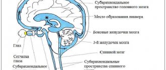
The disease occurs when excess fluid collects in the ventricles of the brain or the subarachnoid space located between the membranes.
Functions of cerebrospinal fluid:
- regulation of intracranial pressure;
- delivery of nutrients to tissues, as well as removal of harmful ones;
- preventing brain damage from any external influences.
Continuously forming in the ventricles, the fluid is excreted along with metabolic products through the circulatory and lymphatic systems. An adult produces 500 ml of it per day, and the brain contains 120-150 ml; in children - about 50 ml.
Causes and forms of hydrocephalus
As a result of the influence of certain factors, the outflow of fluid may be disrupted, and it accumulates in the brain tissue.
There are the following forms of this disease: external open, closed, mixed and secretory. The open external type appears when the absorption of cerebrospinal fluid into the bloodstream is impaired, usually as a consequence of an infection. When the internal outflow of cerebrospinal fluid is closed, a tumor, cyst, aneurysm or hematoma obstructs it. The secretory form of the disease develops with excessive formation of cerebrospinal fluid. Mixed is the most dangerous, since in this case it accumulates both in the ventricles and in the meninges.
In children, the disease can be congenital or acquired.
Causes of congenital hydrocephalus
This pathology is diagnosed during pregnancy. Most often it becomes a consequence of infections or chromosomal disorders. The reasons for this are:
- Defects and anomalies in the development of the nervous system that occur in the womb . They are formed under the influence of the bad habits of a pregnant woman and as a result of infections she suffers during pregnancy. The most dangerous for a baby are toxoplasmosis, rubella, herpes, and cytomegalovirus.
- Birth trauma (asphyxia), as well as prematurity of children.
- Genetic predisposition, chromosomal disorders.
Causes of acquired hydrocephalus
Acquired dropsy develops at a later age and can appear after just a few months of life. Most often the cause is an injury.
Sources of acquired hydrocephalus:
- Tumors of the brain and skull bones.
- Inflammatory processes in brain tissue (encephalitis, meningitis, otitis media).
- Trauma and rupture of cerebral vessels.
- Parasitic infections.
Possible complications and consequences
If a pregnant woman suffers from hydrocephalus, then there is a high probability that this pathology can be transmitted to the fetus, since it is hereditary. However, the consequences can be avoided if the problem is diagnosed in time and its timely drug treatment is started.
If dropsy appears as a result of an unhealthy lifestyle or an infectious disease, then it is necessary to urgently take measures to save the fetus, because there is a possibility of its death due to rapidly developing pathological processes. In such cases, doctors recommend terminating the pregnancy, regardless of the gestation period.
In addition to disorders of the central nervous system, hydrocephalus causes heart defects, delays in mental and physical development, lack of growth, and defects in internal systems and organs.
Types of disease
Hydrocephalus comes in different forms. The most severe is occlusive hydrocele. It is caused by blockage of the cerebrospinal fluid outflow tract with blood clots, adhesions, and neoplasms. Such hydrocephalus can lead to necrosis of certain areas of brain tissue. This form of the disease is very difficult, the prognosis is not very favorable. It progresses quickly, so the child may experience speech and intellectual impairments after treatment.
There is also a moderate form of hydrocephalus, when the brain matter decreases in volume and its place is taken by cerebrospinal fluid. It is typical for older people and later leads to dementia.
Symptoms of the disease in children
Parents of infants should be concerned if the following signs occur:
- rapid increase in skull size (the most pronounced symptom). Normally, the head grows 1.5 cm every month. The growth of the skull in the first year of a child’s life is monitored by doctors, and at the slightest deviation a diagnosis is carried out. Parents should also pay attention to this factor;
- disproportionately large head . The bones of the skull in newborn children do not fuse immediately, and a considerable volume of brain fluid prevents this, resulting in disproportion;
- thin scalp through which blood vessels are visible;
- developmental disorders (child turns over late, sits);
- swelling of the fontanel;
- the sound of a cracked pot when tapping on the skull bones;
- ophthalmological problems : bulging eyeballs, strabismus;
- frequent vomiting and regurgitation;
- paralysis of the lower limbs;
- convulsions;
- lack of emotions in a one-month-old baby;
- sleep disorders;
- the baby does not show interest in toys, does not talk, speech development suffers.
In children over two years of age whose cranial bones are aligned, the shape of the head does not change during illness. Water in a child's head causes an increase in intracranial pressure. He may complain of constant headaches, nausea and vomiting, and possible nosebleeds. Vision and coordination are impaired, muscle tone weakens, and urinary incontinence appears.
If any problems are identified, you should consult a neurologist, since this condition is dangerous for the child’s life.
Methods for diagnosing hydrocephalus
The disease is clearly visible even during intrauterine development of the fetus using ultrasound. If symptoms occur later in life, the following diagnostic methods are used:
- Ultrasound of the brain through the fontanel until it is overgrown (for newborns).
- Examination of the head using a lamp and tube.
- CT and MRI.
If after the first two methods the doctor suspects the presence of a disease, the diagnosis must be confirmed using CT or MRI. Additional methods include fundus examination, angiography and electroencephalogram. They are used to confirm complications. Angiography helps to detect a specific area with abnormalities.
Tactics for managing pregnancy with fetal hydrocephalus
The consequences of fetal hydrocephalus can be different. They depend on the size of the disorder and accompanying developmental defects. In the case when the size of the lateral ventricles does not exceed 15 mm, no other anomalies are identified and treatment is prescribed, the prognosis is relatively favorable - the newborn may not experience any abnormalities.
An unfavorable prognosis develops if the size of the ventricles exceeds 15 mm and dropsy increases rapidly, hydrocephalus is detected in the first half of pregnancy, and multiple organ lesions are noted, which are characteristic of chromosomal diseases. In this case, doctors recommend terminating the pregnancy due to fetal hydrocephalus.
Currently, intrauterine treatment of hydrocephalus is carried out. Using a puncture of the mother's anterior abdominal wall, excess fluid is pumped out from the space near the fetal brain, which puts pressure on the fetal brain. The procedure is performed 1 time. The second treatment method is intrauterine bypass surgery.
What to do if fetal hydrocephalus is detected during pregnancy? Make an appointment with a neurologist by calling the Yusupov Hospital. After the diagnosis has been established, pregnancy management tactics are developed collectively. The woman is offered the best solution to the problem.
Treatment of the disease
Hydroencephalopathy is treated conservatively or surgically. There are also supporters of traditional therapy.
Conservative treatment
It is only suitable for non-progressive disease, but this is not enough for a complete cure. Diuretics are used (Furosemide, Diacarb). These medications are also used in preparation for surgery. Patients are advised to take medications that improve brain nutrition, vitamins, homeopathy, and anti-inflammatory drugs.
Conservative methods of treatment also include diet. When the diagnosis is confirmed, patients are not recommended to consume smoked foods, fatty meats, sausages, confectionery and bakery products, as they contribute to the accumulation of fluid in the body.
Surgery
Modern treatment methods involve draining cerebrospinal fluid from the skull. For this, doctors use bypass surgery. With the help of this operation, the accumulated fluid is removed through a catheter into other cavities of the body: chest or abdominal. As the baby grows, it changes. When deciding to resort to such an operation, you must remember the possibility of infection.
Endoscopy is also used. Its advantage is that no foreign body remains in the body, and excess brain fluid is removed through an endoscope. The only drawback of the method is that it is not suitable for everyone.
The closed form of hydrocephalus requires immediate surgical removal of the cause of the deterioration in the outflow of cerebrospinal fluid, because there is a danger of dysfunction of the child’s respiratory system.
To temporarily reduce intracranial pressure, a puncture in the spine is used.
Traditional methods
The treatment regimen for hydrocephalus with folk remedies consists of taking adonis infusion three times a day. You need to drink 50 ml, diluting them in two glasses of boiling water. The course lasts 3 weeks, after which a week break is required. Then treatment is resumed.
Another folk remedy used to drain excess liquid is an infusion of parsley, prepared as follows: 2 tablespoons are poured into 0.5 liters of boiling water. You need to drink the drink throughout the day.
Parents have the right to independently decide which method to choose to treat their child, but the appropriateness of folk remedies and homeopathy is a big question among doctors.
Treatment
To treat the child, conservative therapy is used or surgery is performed.
With hydrocephalus, the child needs, first of all, to reduce intracranial pressure by reducing the production of cerebrospinal fluid or accelerating its elimination. This treatment involves the use of the drug "Diacarb". At the same time, Panangin or Asparkam is prescribed.
If intracranial pressure is high, it is necessary to use medications that ensure the removal of excess fluid from the skull. For this purpose, drugs with a diuretic effect are used: Mannitol, Glycerin, Furosemide, Lasix. At the same time, the use of drugs that stimulate the functioning of neurons is indicated - “Calcium hopantenate”, “Encephabol”, etc.
If therapy within 2 months does not produce results, then the child needs surgical intervention.
Very often, this method is the only way to prevent the progression of hydrocephalus. The method of operation will depend on the stage and form of the disease.
- The operation is performed when it is possible to remove an obstacle in the circulation of the cerebrospinal fluid (tumor, cyst, hematoma, etc.).
- If the tumor has grown into the brain tissue, the operation will be aimed at creating the ability for the cerebrospinal fluid to move. For this purpose, bypass surgery is performed.
To drain cerebrospinal fluid, silicone catheters are used, directed into the peritoneal cavity. This operation is called ventriculoperitoneal shunting. If the cerebrospinal fluid is diverted to the right atrium, the operation is called ventriculo-arterial shunting.
In endoscopic ventriculostomy, the use of an endoscope can be effective in cases of occlusive hydrocephalus.
When treating hydrocephalus, the main goal is to remove excess cerebrospinal fluid from the brain. Surgery is most often used.
But at some stages of the disease, doctors offer conservative treatment. To do this, medications are prescribed that should reduce the production of cerebrospinal fluid, for example Diacarb, diuretics, and sometimes hormonal medications.
Therapeutic treatment cannot cure the disease, it only alleviates it and provides temporary improvements.
For effective treatment, surgery is performed.
Bypass operations are most often performed. The essence of this intervention is to remove excess fluid from the brain through special silicone tubes (shunts) into the abdominal cavity, where it is absorbed.
In fact, the only way to treat hydrocephalus in newborns is surgery performed by a neurosurgeon. The main task of the specialist is to drain cerebrospinal fluid from the ventricles of the brain.
There are several surgical options for transporting cerebrospinal fluid. The most effective method is considered to be ventriculoperitoneal shunting (VPS). Using silicone catheters placed under the skin, cerebrospinal fluid from the lateral ventricle is drained into the abdominal cavity, where it is absorbed between the intestinal loops. It should be noted that VPS has saved the lives of millions of babies.
Other types of bypass are less commonly used:
- Ventriculoatrial (cerebral fluid is discharged into the right atrium);
- Lumboperitoneal (the spinal canal connects to the abdominal cavity at the lumbar level);
- Operation according to Torkildsen (cerebrospinal fluid is diverted into the occipital cistern).
The disadvantages of the latter surgical method are all kinds of postoperative complications.
Another effective method is endoscopic ventriculostomy. During the operation, the neurosurgeon inserts an endoscope through small incisions, which creates a path for the outflow of cerebrospinal fluid. Despite its great effectiveness, this method is indicated in only 10% of cases of hydrocephalus in newborns. Such a small percentage is due to certain forms of the disease.
If the surgical intervention is successful, then the development of the disease stops, the baby continues to actively develop, keeping up with his peers.
Thus, in order to avoid the severe consequences of hydrocephalus in newborns, timely diagnosis and surgical treatment are necessary. This guarantees a complete recovery and return of the child to normal life.
A positive treatment result will depend on a number of factors:
- causes of the disease;
- the amount of liquor produced;
- dynamics of increasing head size;
- the extent of the disease at the time of its discovery.
This disease can be treated with both conservative and surgical methods. The patient should be under the strict supervision of a neurosurgeon or neurologist.
If we consider the initial stage of the disease, then medications will be prescribed first. The doctor’s actions will help reduce intracranial pressure; the specialist will also prescribe hormonal drugs to increase the accumulation of excess fluid and a diuretic. Diakarb is almost always prescribed; it perfectly lowers the pressure inside the skull and is also responsible for regulating the production of cerebrospinal fluid.
Surgical treatment is almost always prescribed for advanced conditions. The operation is performed in neurosurgery.
Initially, the doctor performs a puncture to pump out excess fluid.
If there is no positive effect, they proceed to bypass surgery. Shunts are inserted into the brain vessels, through which cerebrospinal fluid drains.
The classical treatment of hydrocephalus in children consists of performing appropriate surgery with further progression of the disease. This decision is not easy for parents, but it is the only solution to the problem that has arisen and cannot be delayed. Delayed psychomotor development, an enlarged head, pain due to intracranial pressure - such symptoms of the disease do not allow parents to choose something else. Surgical intervention in this case is carried out as follows.
- The goal is to remove cerebrospinal fluid from the brain into any other free cavities of the body.
- VPS is performed - ventriculoperitoneal shunting. Through silicone catheters, cerebrospinal fluid, regulated by a special valve, flows into the abdominal cavity and is then absorbed by the intestines. All equipment is carried out under the skin and is not visible from the outside.
- VAS can be performed - ventriculoatrial shunting, when cerebrospinal fluid is drained into the right atrium.
- Sometimes doctors prescribe a Torkielsen operation, in which the cerebrospinal fluid is drained into the cistern magna.
- Another option for the operation is LPS (lumbo-peritoneal shunt), when the spinal canal has to be connected with a catheter at the lumbar level to the abdominal cavity.
- Modern medicine, using the latest developments, is increasingly using endoscopic techniques to treat hydrocephalus in newborns. It will allow you not to use a shunt system. Deep in the brain, an endoscope inserted through a small incision helps create a special bypass for the removal of cerebrospinal fluid. This operation is called endoscopic ventriculostomy, it is very effective, but can cure a limited number of babies (only 10% of the total number of patients). In other cases, a shunt system is installed.