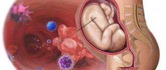
Many mothers wonder: when does the nervous system of the fetus form? Almost from the very beginning of cell laying. According to medical theories, all body systems develop unevenly in a baby. First, those systems that are most important for the future activities of the baby in the mother’s belly begin to function. The formation of the nervous system in the fetus is one of the first most important processes in the development of the body.
Already at 8-9 weeks of pregnancy, gynecologists can see the first signs of the nervous system on an echogram. The second month is marked by the baby making his first barely noticeable movements. Well, at 22-24 weeks you can accurately see a child who is sucking appendages.
Stages of neural tube formation
Nervous tissue ensures the coordinated functioning of all human organs. It consists of two types of cells:
- neurons - perform specific functions and are the main structural element of nervous tissue;
- neuroglial cells - provide protection and nutrition to neurons, provide a support function, and delimit clusters of neurons with different functions.
Nervous tissue begins to develop from the moment the embryo is formed. On the 18th day of its existence, a collection of cells called the ectoderm increases and the neural plate is formed from them. This is the rudiment of the entire future nervous system of the body. The fetal neural tube should then form - what is it?
Gradually, rollers are formed from the edges of the plate, between which there is a groove. In the anterior part, the plate is slightly expanded - this is the future brain.
The rollers move closer together over several days until they finally connect. As a result, a hollow tube is formed. Subsequently, this cavity forms the cerebral ventricles and a canal in the spinal cord. The development of the neural tube in the fetus continues during the first weeks of embryogenesis.
Some parts of the plate become unlaced and form small clusters on both sides of the tube, called a ridge. The main part will form the central nervous system. The following elements will develop from the ridge:
- peripheral nodes;
- meninges and part of glial tissue;
- nerve fiber sheath cells;
- adrenal medulla;
- skin pigment cells;
- carotid bodies and the APUD system.
At the initial stages of embryogenesis, the neural tube is represented by a continuous neuroepithelium. Gradually, it differentiates into several areas.
- The ventricular zone is formed by cylindrical cells. They are the ancestors of the main cells and macroglia.
- The subventricular zone is actively reproducing cells. It is a growth zone and almost completely disappears by the end of the embryonic period.
- The intermediate zone is cells without the ability to divide. They form the gray matter of the spinal cord.
- The marginal zone is the beginning of the white matter of the spinal cord and brain.
Embryonic development of the vertebrate nervous system
The closure of the neural tube begins in the middle of the embryo, then the process spreads to the head and tail ends of the embryo, where the openings - the anterior and posterior neuropores - remain open for some time (Fig. 7).Rice. 7. Early stages of neural tube formation using the example of human brain development (after: Nieuwenhuys R. et al., 1999). A–D – reconstruction of the appearance of the human embryo and the initial stages of the formation of the neural tube, E–H – transverse sections of the embryo at these stages of development; 1 – ectoderm, 2 – neural plate, 3 – amnion foramen, 4 – medullary plate, 5 – neural fold, 6 – neural groove, 7 – neural tube, 8 – brain rudiment, 9 – anterior neuropore, 10 – posterior neuropore, 11 – neural crest, 12 – pterygoid plate, 13 – lateral plate, 14 – basal plate, 15 – cavity of the primary cerebral ventricles, 16 – rudiments of the spinal ganglia
Even at the stage of neuropore closure, rostro-caudal differentiation of the neural tube of the embryo begins. The neural tube (believed to be under the inducing influence of the notochord) gradually sinks into the mesoderm of the embryo and, under the influence of mesodermal somites, is divided into segmental sections - neuromeres or prosomeres. Somites are located on the sides of the neural plate and are pressed into it, determining the configuration of future parts of the brain (Fig. 8).
Subsequently, the head somites fuse and form three main segments: premandibular, mandibular and hyoid. The boundary of the head segments is the region of the ear capsule, behind which from 2–3 to 10–12 trunk segments are formed (depending on the group of vertebrates). At the same time, the system of cranial nerves is formed. Each segment is innervated by certain pairs of nerves: premandibular - terminal and oculomotor nerve (III); mandibular - trigeminal (V) and trochlear (IV) nerves; hyoid – abducens (VI) and facial (VII) nerves. The two segments following the head are innervated by the glossopharyngeal (IX) and vagus (X) nerves, respectively. The rostral trunk somites in higher vertebrates are innervated by the accessory nerve system (XI), which includes a different number of roots depending on the number of trunk somites. The hypoglossal nerve (XII), which innervates the hypobranchial muscles, which develops from the anlage of the trunk segments, is similar in function to the ventral (somatomotor) roots of the spinal nerves that innervate the striated muscles of the trunk and limbs.
The anterior end of the tube at the end of the 3rd week of development, due to active processes of proliferation and migration of neurons in the brain wall, expands and forms 3 primary brain vesicles. The cranial bladder forms the primary forebrain, Prosencephalon , the middle bladder - the primary midbrain Mesencephalon , and from the third bladder the primary hindbrain Rhombencephalon develops . Next are the structures of the developing spinal cord - Medulla spinalis (Fig. 9).
Rice. 9. Development of the human brain (according to: Sade, Ford, 1976). A–B – stages of three (a) and five (b–e) brain vesicles; top view (A) and side view (B); 1–3 – primary: forebrain (1), middle (2), rhomboid (No.) brain; 4 – anlage of the spinal cord, 5 – optic cup, 6–10 – parts of the brain: terminal (6), intermediate (7), middle (8), posterior (9), medulla (10); 11 – hemispheres of the telencephalon, 20 – cerebellum, 22 – spinal cord, V–IX – cranial nerves. Arrows - bends of the neural tube (s.i. - midbrain, sh.i. - cervical, m.i. - pavement)
The spinal cord is formed from the caudal parts of the neural tube. It is a part of the central nervous system, in the structure of which the features of the embryonic stages of vertebrate brain development are most clearly preserved: the tubular nature of the structure and segmentation.
After the formation of the brain vesicles, complex processes of internal differentiation and growth begin in the nervous system. Already at the early stages of development of the embryo, the neural tube is divided over a considerable length by the boundary groove, sulcus limitans, running along the ventricular surface, into two sections: dorsal - pterygoid plate , and ventral - basal plate . The areas of the brain that develop from the pterygoid plate contain sensory nuclei, and those from the basal plate contain motor and autonomic nuclei. The rostral part of the neural tube does not contain a basal plate and is entirely derived from the pterygoid. The parts of the brain containing derivatives of both plates - middle, posterior, medulla - are often combined under the name "brain stem" .
Changes in the development of the neural tube are accompanied by the formation of several bends at the boundaries of the anlage of various parts of the brain. During the first two months of embryonic development, the main (midbrain) bend is formed when the forebrain and diencephalon bend forward and downward. Then two more (cervical and bridge) bends are formed. At the same time, the first and third primary brain vesicles are each divided into two. The stage of five brain bubbles begins. The telencephalon becomes the most rostral, then the intermediate brain (Diencephalon). After the intermediate brain comes the midbrain (Mesencephalon). The primary hindbrain vesicle is divided into the hindbrain (Metencephalon) and the medulla oblongata (Medulla oblongata). The prosencephalon includes derivatives of the first six prosomeres (neuromeres) P1–P6. The midbrain is subsequently formed from P1 structures. Prosomeres P2 and P3 develop into the thalamus and prethalamus, respectively. The telencephalon and hypothalamus develop from prosomeres P4–P6. From the more caudal segments of the neural tube (rhombomeres), the structures of the brainstem and spinal cord develop.
After the formation of the brain vesicles (5–10 weeks of development), complex processes of internal differentiation and growth of various parts of the brain and spinal cord occur in the structures of the developing nervous system.
The formation of parts of the brain is under the control of the so-called. “secondary organizers” - groups of cells that synthesize a number of morphogenetic factors, the concentration gradient of which determines the direction of migration and differentiation of different brain structures (Table 2; Fig. 9,).
Table 2. Genes and their expression products that control various processes of development of brain regions and structures (according to: Obukhov, 2008).
| Function | ||
| Dlx 1, Dlx 2, Dlx 5 | Subpallium (ganglionic eminences), diencephalon | Migration of subpallial neuroblasts, migration of neurons into the cortex from the ganglionic eminences of the anterior medullary vesicle |
| Emx 1, Emx 2 | Finite brain | Cell proliferation in the developing brain, neuroblast migration |
| Lhx 1, Lhx 2, Lhx 5 | Forebrain, cerebral cortex | Formation of subcortical and cortical (archicortex) sections of the hemispheres |
| Nkx 2.1 Nkx 2.2 | Ventral hemispheres | Proliferation and migration of neuroblasts in the striatum |
| Otx 1, Otx 2 | Forebrain, midbrain, anterior brainstem | Formation of the structure of the hemispheres, including the cerebral cortex |
| Pax 3, Pax 6 | Forebrain | Migration of neuroblasts in the dorsal hemispheres |
The development of the forebrain rudiment is controlled by a small group of cells located at the top of the neural tube and called the anterior neural ridge (ANR) and cells at the border of the second brain vesicle - zona limitans interthalamica (ZLI). The structures of the midbrain, hindbrain, medulla oblongata and the upper segments of the spinal cord are controlled by another organizer - the isthmic organizer (ISO).
During the perinatal period, the formation of the internal structure of the brain ends. Active myelination of the brain and spinal cord begins. However, these processes do not end with birth. It has been shown that for quite a long time (months and years) after birth, the maturation and differentiation of nervous structures and conductive tracts occurs. Moreover, it has now become clear that during the adult period, the formation of new populations of neurons and glial cells occurs due to the preservation of NSC populations in the brain structures.
Reasons for the formation of pathologies
What is a defect? This concept refers to any abnormal structure of tissues and organs. A neural tube defect is an anomaly in its structure, formed in the embryonic period. In a normal pregnancy, the neural plate folds join by day 23. If this does not happen within the prescribed period, the fetus develops defects.
The causes of fetal neural tube defects are quite varied:
- an increase in liquor pressure in an already formed tube;
- viral infection, especially rubella;
- genetic predisposition - a similar defect in the anamnesis of the parents, cases of children born in the family with such pathologies;
- environmental pollution - radiation, pesticides, chemical waste;
- taking antiepileptic drugs immediately before and at the beginning of pregnancy;
- prolonged fever in a woman in the early stages;
- obesity with diabetes;
- unhealthy diet with vitamin deficiencies.
If at least one of these conditions is detected, a woman is at increased risk of having a child with a neural tube defect.
Causes and consequences of abnormal development of the neural tube in the fetus
The formation of a fetal tube is a complex and multifaceted process; failure in at least one element leads to severe defects in the development of the organ. They affect the child’s brain and his spine, because the connection between the neural tube and notochord is very tight.
Defects are formed in situations where the formation of the neural tube does not occur completely or does not close. This may cause:
- Spina bifida. It is formed at the base of the column, where a hole is formed through which the spinal cord will exit.
- Anencephaly is formed in the upper part of the neural tube and leads to the immaturity of some elements of the brain and the development of the fetal skull.
- Chiari malformation is characterized by “leakage” of brain tissue into the spinal canal.
- Encephalocele manifests itself as partial protrusion of the brain and membranes through the cranial bones.
The reasons for the formation of such defects have not yet been precisely established. At some point, ontogenesis fails, which subsequently leads to disruption of the entire system. Since the tube is laid in the late gastrula and this stage is completed on the 25th day, its mechanism has not been well studied.
It is known that the likelihood of defects is influenced by hereditary factors and diseases affecting the nervous system.
It influences the formation of the problem and environmental conditions, as well as the condition of the mother. Negative aspects that increase the risk of neural tube defects include:
- radiation exposure;
- arsenic or lead poisoning;
- prolonged fever in a woman in the initial stages of pregnancy;
- lack of nutrition;
- viral infections;
- vitamin A hypervitaminosis;
- excess weight or diabetes in the mother;
- lack of folic acid in a pregnant woman;
- a woman taking certain medications.
The condition of the mother is the first factor that affects the formation of a tube in the fetus. If a woman is in an environmentally unfavorable area, where harmful substances (pesticides, exhaust gases, petroleum products, etc.) can enter the air and water, she inhales them, and together with the blood, these products enter the baby’s body.
For an adult, the risk is lower, since his body is already formed and is able to process most hazardous substances. In the body of the embryo, the defense mechanisms do not yet work and any harmful effect can disrupt the course of processes and lead to the development of pathology, including changes in the condition of the tube.
Poor nutrition also has a negative effect. A lack of nutrients leads to disruption of the processes of organ formation, as well as an excess of biologically active components such as vitamins.
Consequences of damage
Neural tube defects (NTDs) are called spinal dysraphisms. They are divided into several groups:
- spina bifida, not visually detectable;
- obvious spinal cleft in combination with spina bifida;
- splitting of the spinal column and surrounding soft tissues, leading to flattening of the spinal cord.
The first variant of the pathology is most often localized in the lumbar or sacral region. Clinically, this defect of the neural tube in the fetus does not manifest itself in any way, therefore, if it is detected, it is by chance, during radiation research methods. Externally, the skin over the area of the nonunion looks healthy, an increased number of pigment spots may be observed, and wen forms.
With extensive non-union, the following symptoms may appear over time:
- pathology of trophism and sensitivity of the skin;
- pathology of posture and improper formation of the foot;
- formation of chronic pain;
- dysfunction of the pelvic organs.
With open clefts, spinal cysts and hernias form. Depending on the level of spinal cord damage, they are divided into groups.
- Damage to the membranes, or meningocele. In this case, protrusion into the defect of the dura mater occurs. At the exit from the hernia opening, the dura mater atrophies. The covering of the hernia is a thin film. The skin over it is thinned. Inside the hernia are the meninges and cerebrospinal fluid. Clinical symptoms are usually not observed.
- Damage to the roots, or meningoradiculocele. In addition to the meninges, the roots of the spinal nerves extend into the opening of the defect. Some of the roots end blindly in the hernial sac. Depending on the number of such roots, manifestations will vary from sensory disturbances to severe paresis and paralysis.
- Brain damage, or meningomyelocele. The spinal cord along with the membranes and roots emerges into the hernial defect. The hernia is extensive, occupying several vertebrae. The condition of the child in such cases is severe, there is no motor and sensory function below the site of the lesion, and the function of the pelvic organs suffers.
- Formation of cysts, or myelocystoceles. In this case, the spinal cord in the final section expands, forming a cyst. The roots end blindly in the hernial sac, as a result of which the same symptoms are observed as in the brain form.
- Complicated forms - any of the above forms in combination with benign tumors of the hernial sac.
Complete nonfusion, or rachischisis, is in most cases a condition incompatible with life. A newborn can survive if this defect extends to no more than 5 vertebrae. This non-closure looks like a gap in the vertebral and spinal canal. The skin and muscles are atrophied; deep in the defect you can see nervous tissue with small underdeveloped blood vessels.
The vast majority of cases of this pathology are localized in the lumbosacral spine. This has biological significance. A group of Japanese researchers spent some time studying material obtained as a result of spontaneous abortions. Among the embryos, scientists chose those who had spina bifida. It was found that in almost all embryos this pathology was localized in the cervical and thoracic region. Based on these data, it was established that severe developmental pathologies, incompatible with the life of a child after birth, lead to his death even in the embryonic period.
Diagnostics
Detection of such defects is carried out in the prenatal period. Biochemical techniques and various methods of intrascopy of the fetus are used. All pregnant women are divided into several risk groups for developing neural tube defects in the fetus. What does this mean? Diagnostic measures in each group differ.
- For women at low risk, examinations by obstetricians and gynecologists are carried out once a month. After the 15th week of pregnancy, the content of fetoprotein in the woman’s blood and amniotic fluid is examined. If its level is significantly reduced, a repeat examination and ultrasound of the fetus is performed. In the third trimester, another ultrasound is performed.
- In high-risk groups, examination by an obstetrician-gynecologist is also required - already several times a month. After week 15, fetoprotein levels are repeatedly examined. Sonographic examination of the fetus is also carried out regularly. If there are difficulties in diagnosis, tomographic methods are indicated.
Ultrasound examination can be carried out in the usual way or with a three-dimensional image. This image allows you to examine the fetus from all sides and more accurately identify pathology.
Diagnostics only establishes the presence of a neural tube defect, but does not determine the degree of its severity. Therefore, the issue of maintaining or terminating a pregnancy is decided jointly by the obstetrician-gynecologist and the woman. After the presence of such a developmental defect is established, a conversation is held with the woman. All possible options for the course of pregnancy and the condition of the fetus are explained to her. The child may be born severely disabled.
Anencephaly
The content of the article
Anencephaly is the complete or partial absence of the cerebral hemispheres, and in some cases, the skull bones and soft tissues. Occurs in 1 case out of 10,000, the anomaly is accompanied by other disorders - cleft lip or hard palate, absence of the pituitary gland, spina bifida.
This happens if for some reason the anterior neuropore does not close at 3-4 weeks of pregnancy. Because of this, the frontal extensions of the neural tube, from which the cerebral hemispheres would later begin to develop, do not develop.
Instead of the “gray matter” rich in neurons, fibrous tissue is formed, in which single nerve cells, cystic formations and blood vessels are present. In 71% of cases, the fetus lacks the fronto-occipital zone and the spinal column, in 24% - the occipital lobe with the spinal column, and in 5% - the temporo-parietal zone. The baby's body does not have any abnormalities.
On ultrasound, anencephaly is diagnosed at 11-12 weeks of pregnancy, the accuracy is 96%.
The pathology is characterized by the following echo signs:
- the skull bones are not visualized;
- soft tissues of the brain are anechoic;
- in the vascular system of the brain there is a malformation - an incorrect connection of blood vessels, veins and arteries;
- maternal polyhydramnios (excess amniotic fluid).
The diagnosis of anencephaly is not limited to ultrasound examination alone. When there is a neural tube defect, the hormone alpha-fetoprotein increases in a woman’s blood. Once the diagnosis is confirmed, she is advised to stop the pregnancy, because if the newborn does not have a brain, the lungs, heart, and kidneys will fail after a while.
Ultrasound diagnosis is the primary, but not the main method of diagnosis. If neural tube pathology is detected during an ultrasound examination, the woman is prescribed amniocentesis - taking amniotic fluid from the amniotic sac under the control of a transabdominal sensor for the purpose of laboratory testing.
If a high concentration of alpha-fetoprotein and the enzyme acetylcholinesterase is detected, the pregnant woman is sent for MRI (magnetic resonance imaging). Unlike ultrasound, this method allows you to see the fetal brain in a 4D projection, magnifying the image several times.
The sooner a diagnosis is made, the sooner a woman can terminate her pregnancy. It makes no sense to preserve it with such a diagnosis, because the baby will live for a maximum of a week.
Treatment
If a woman nevertheless decides to continue her pregnancy with a reliable diagnosis of pathology of the development of the neural tube in the fetus, she is prepared in advance for surgical delivery and emergency measures to save the life of the child.
In this case, childbirth is contraindicated through the natural birth canal. A planned caesarean section is required. A few weeks before the expected due date, a woman is admitted to a hospital for pregnancy pathology. After delivery, the child is cared for by neonatologists and pediatric neurosurgeons.
The operation consists of eliminating the hernial defect, restoring the physiological structure of the spinal cord and suturing soft tissues and skin. It is preferable to perform surgical intervention on the first day after the birth of the child, since there is a high probability of infection of the nervous system through the existing hole.
Features of the nervous system in the third trimester
Mothers often ask the doctor questions about the formation of the nervous system in the fetus, at what age the baby begins to recognize sounds, and what she needs to do during this period.
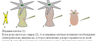
The third trimester is characterized by active growth of all organs and systems, including the growth and development of the brain and spinal cord. From the 26th week, the production of myelin protein by glial cells is activated. Until this time, the nerve impulse did not spread directly from cell to cell, but around it. Part of it was lost, which reduced the efficiency of information transfer. Myelin insulates the neuron, allowing impulses to travel directly through the cells and ensuring strong connections. Thanks to this protein, it becomes possible to store information.
After myelin appears on the surface of neurons, the child gains the ability to recognize information. At the beginning of the third trimester of gestation, the child perceives sounds in a wide range - the mother's voice, music, the voices of surrounding people, noises. He develops sound preferences and a negative attitude towards certain sounds.
The child perceives sounds of high and low frequencies negatively. For example, speaking in a raised voice causes discomfort in the fetus, which is manifested by high motor activity. The mother's calm voice, singing or pleasant music calms the baby. After birth, the baby will respond appropriately to familiar sounds.
At 31 weeks of gestation, the formation of a branched system of dendritic processes begins. The structure of a neuron includes a body, a sending process - an axon, and receiving processes - dendrites. Dendrites collect information, it is transformed in the body, and transmission is carried out by the axon.
From this moment on, it becomes advisable to enrich the environment. Expectant mothers help the child's development by creating a favorable sound background and tactile sensations.
Preventing the development of neural tube defects
Preventive measures for the development of neural tube defects in the fetus include full preparation for pregnancy. Every woman planning to become pregnant should contact a family planning center. Here, consultations are conducted by obstetricians-gynecologists, specialized specialists, and geneticists.
Doctors will calculate the risk of pathology and prescribe preventive measures. The main one is taking B vitamins and folates. A woman should take these medications several months before the expected pregnancy and during the first 12 weeks. Folate intake is especially important in the first weeks, since this is the period of formation of the fetal neural tube.
The preparations must have a certain content of folic acid. The optimal dosage is 800 mcg. The frequency and duration of administration is determined by the doctor. There are several vitamin complexes designed specifically for pregnant women.
- Vitrum prenatal is a complex drug containing all the vitamins necessary for a pregnant woman’s body. The folic acid content is 800 mcg.
- Elevit prenatal is a multivitamin complex with the addition of microelements. Used as preparation for and during pregnancy. The folic acid content is also 800 mcg.
- Natures Bounty folic acid is a natural product containing folic acid in the optimal dosage. Does not contain preservatives or dyes. Available in capsules of 100 pieces per package.
In addition to drug prevention, other methods are required that a woman should use both before and during pregnancy. What they include:
- maintaining stable emotional balance;
- being in a clean ecological environment;
- timely treatment of chronic diseases and foci of infection in the body;
- proper nutrition.
Compliance with preventive measures and proper planning of pregnancy will help to avoid pathologies in the development of the neural tube of the fetus and disability of the child.
Microcephaly
Microcephaly is a complex pathology of the brain, expressed in a decrease in the size of the organ in the fetus. The cause of underdevelopment of the brain is a violation of the division of nerve cells at the stage of formation of the neural tube of the embryo.
The pathology is provoked by several factors: in 40% it develops against the background of cytomegalovirus in the mother; there is also a hereditary form of Giacomini-Penrose-Beck disease.
Microcephaly is rare: 1 case in 5,000, and is often accompanied by other disorders of the central nervous system such as lisencephaly (impaired formation of the cerebral cortex), microgyria (small size of the cerebral convolutions), cerebellar anomaly, and underdevelopment of the spinal cord.
In terms of diagnosis, microcephaly is the most difficult defect. The ultrasound examination is based on the criterion of the ratio of the length of the femur and the circumference of the fetal head, which should not be less than 2.5. However, interpretation is complicated by the fact that the exact gestational age is not always known. False-positive and false-negative results may be due to the small size of the fetus or impaired bone growth in other pathologies.
The accuracy of ultrasound diagnostics for microcephaly is 67.4%, and in 85% of cases the diagnosis is made after 22 weeks of pregnancy. Starting from 2 weeks, the structure of the skull can be easily seen on ultrasound. With microcephaly, it has an irregular shape, the forehead is sloping, the ears are low, and the jaws are underdeveloped. There is also dilatation of the cerebral ventricles.
In addition, in 60% of cases, the fetus is diagnosed with other disorders of the central nervous system, diseases of the kidneys, heart and other internal organs.
Diagnosis of microcephaly is always complex. If a pathology is suspected, a woman's amniotic fluid is analyzed and the fetus is karyotyped. Only after a thorough examination is the woman told about the diagnosis, and she herself decides what to do next.