The structure and functions of the human nervous system are studied in 8th grade biology. The central nervous system is a complex structural network. It is divided into several sections and ensures the interaction of the body with the environment, and also connects all organs into a single whole. With its help, brain activity is formed, all internal functions are regulated and movements are controlled.
Stages of nervous system development
In evolution, the nervous system has undergone several stages of development, which became turning points in the qualitative organization of its activities.
These stages differ in the number and types of neuronal formations, synapses, signs of their functional specialization, and in the formation of groups of neurons interconnected by common functions. There are three main stages of the structural organization of the nervous system: diffuse, nodular, tubular. The diffuse nervous system is the most ancient, found in coelenterates (hydra). Such a nervous system is characterized by a multiplicity of connections between neighboring elements, which allows excitation to freely spread throughout the nervous network in all directions.
This type of nervous system provides wide interchangeability and thereby greater reliability of functioning, but these reactions are imprecise and vague.
The nodal type of nervous system is typical for worms, mollusks, and crustaceans.
It is characterized by the fact that the connections of nerve cells are organized in a certain way, excitation passes along strictly defined paths. This organization of the nervous system turns out to be more vulnerable. Damage to one node causes dysfunction of the entire organism as a whole, but its qualities are faster and more accurate.
The tubular nervous system is characteristic of chordates; it includes features of the diffuse and nodular types. The nervous system of higher animals took all the best: high reliability of the diffuse type, accuracy, locality, speed of organization of nodal type reactions.
Content
- 1 General characteristics of the nervous system 1.1 Neurons
- 1.2 Neuroglia
- 2.1 Types of nervous systems
- 2.2.6.1 Morphological division
- 2.3.1 Models 2.3.1.1 Regulatory mechanisms
- 3.1 Professional societies and journals
The leading role of the nervous system
At the first stage of the development of the world of living beings, interaction between the simplest organisms was carried out through the aquatic environment of the primitive ocean, into which the chemical substances released by them entered. The first oldest form of interaction between the cells of a multicellular organism is chemical interaction through metabolic products entering the body fluids. Such metabolic products, or metabolites, are the breakdown products of proteins, carbon dioxide, etc. This is the humoral transmission of influences, the humoral mechanism of correlation, or connections between organs.
The humoral connection is characterized by the following features:
- lack of an exact address to which a chemical substance entering the blood or other body fluids is sent;
- the chemical spreads slowly;
- the chemical acts in minute quantities and is usually quickly broken down or eliminated from the body.
Humoral connections are common to both the animal and plant worlds. At a certain stage of development of the animal world, in connection with the appearance of the nervous system, a new, nervous form of connections and regulation is formed, which qualitatively distinguishes the animal world from the plant world. The higher the development of an animal’s organism, the greater the role played by the interaction of organs through the nervous system, which is designated as reflex. In higher living organisms, the nervous system regulates humoral connections. Unlike the humoral connection, the nervous connection has a precise direction to a specific organ and even a group of cells; communication is carried out hundreds of times faster than the speed of distribution of chemicals. The transition from a humoral connection to a nervous connection was not accompanied by the destruction of the humoral connection between the cells of the body, but by the subordination of nervous connections and the emergence of neurohumoral connections.
At the next stage of development of living beings, special organs appear - glands, in which hormones are produced, formed from food substances entering the body. The main function of the nervous system is both to regulate the activity of individual organs among themselves, and in the interaction of the body as a whole with its external environment. Any impact of the external environment on the body appears, first of all, on receptors (sensory organs) and is carried out through changes caused by the external environment and the nervous system. As the nervous system develops, its highest department—the cerebral hemispheres—becomes “the manager and distributor of all the activities of the body.”
Neuroscience
Modern science of the nervous system combines many scientific disciplines: along with classical neuroanatomy, neurology and neurophysiology, molecular biology and genetics, chemistry, cybernetics and a number of other sciences make an important contribution to the study of the nervous system. This interdisciplinary approach to the study of the nervous system is reflected in the term neuroscience. In Russian-language scientific literature, the term “neurobiology” is often used as a synonym. One of the main goals of neuroscience is to understand the processes occurring both at the level of individual neurons and neural networks, the result of which are various mental processes: thinking, emotions, consciousness. In accordance with this task, the study of the nervous system is carried out at different levels of organization, from the molecular to the study of consciousness, creativity and social behavior.
Professional societies and magazines
The Society for Neuroscience (SfN, the Society for Neuroscience)[4] is the largest non-profit international organization, uniting more than 38 thousand scientists and doctors involved in the study of the brain and nervous system. The society was founded in 1969 and is headquartered in Washington. Its main goal is the exchange of scientific information between scientists. To this end, an international conference is held annually in various cities in the United States and the Journal of Neuroscience is published[5]. The society carries out educational and educational work.
The Federation of European Neuroscience Societies (FENS, the Federation of European Neuroscience Societies)[6] unites a large number of professional societies from European countries, including Russia. The Federation was founded in 1998 and is a partner of the American Society for Neuroscience (SfN). The Federation holds an international conference in different European cities every 2 years and publishes the European Journal of Neuroscience[7].
Structure of the nervous system
The nervous system is formed by nervous tissue, which consists of a huge number of neurons - a nerve cell with processes.
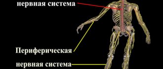
The nervous system is conventionally divided into central and peripheral.
The central nervous system includes the brain and spinal cord, and the peripheral nervous system includes the nerves that arise from them.
The brain and spinal cord are a collection of neurons. In a cross section of the brain, white and gray matter are distinguished. Gray matter consists of nerve cells, and white matter consists of nerve fibers, which are processes of nerve cells. In different parts of the central nervous system, the location of white and gray matter is different. In the spinal cord, gray matter is located inside, and white matter is outside, but in the brain (cerebral hemispheres, cerebellum), on the contrary, gray matter is outside, white matter is inside. In various parts of the brain there are separate clusters of nerve cells (gray matter) located inside the white matter - the nuclei. Clusters of nerve cells are also located outside the central nervous system. They are called nodes and belong to the peripheral nervous system.
Notes[ | ]
- Autonomic nervous system and coordination (undefined)
. biofile.ru. Retrieved June 28, 2020. - Somatic nervous system | Multidimensional and traditional medicine (undefined)
. www.ansmed.ru. Retrieved June 29, 2020. - Somatic and autonomic (autonomic) departments of the nervous system - Knowledge Hypermarket (undefined)
. Hypermarket Znan is the first in the world! Retrieved June 29, 2017. - Somatic nervous system, functions of the department that regulates (Russian), Our Nerves
(February 11, 2017). Retrieved June 28, 2020.
Reflex activity of the nervous system
The main form of activity of the nervous system is the reflex. Reflex is the body’s reaction to changes in the internal or external environment, carried out with the participation of the central nervous system in response to irritation of receptors.
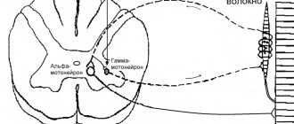
With any irritation, excitation from the receptors is transmitted along centripetal nerve fibers to the central nervous system, from where, through the interneuron along centrifugal fibers, it goes to the periphery to one or another organ, the activity of which changes. This entire path through the central nervous system to the working organ, called the reflex arc, is usually formed by three neurons: sensory, intercalary and motor. A reflex is a complex act in which a significantly larger number of neurons take part. Excitation, entering the central nervous system, spreads to many parts of the spinal cord and reaches the brain. As a result of the interaction of many neurons, the body responds to irritation.
Spinal cord
The spinal cord is a cord about 45 cm long, 1 cm in diameter, located in the spinal canal, covered with three meninges: dura, arachnoid and soft (vascular).
The spinal cord is located in the spinal canal and is a cord that at the top passes into the medulla oblongata and at the bottom ends at the level of the second lumbar vertebra. The spinal cord consists of gray matter containing nerve cells and white matter consisting of nerve fibers. Gray matter is located inside the spinal cord and is surrounded on all sides by white matter.
In a cross section, the gray matter resembles the letter H. It distinguishes the anterior and posterior horns, as well as the connecting crossbar, in the center of which there is a narrow canal of the spinal cord containing cerebrospinal fluid. In the thoracic region there are lateral horns. They contain the bodies of neurons that innervate internal organs. The white matter of the spinal cord is formed by nerve processes. Short processes connect sections of the spinal cord, and long ones make up the conductive apparatus of bilateral connections with the brain.
The spinal cord has two thickenings - cervical and lumbar, from which nerves extend to the upper and lower extremities. 31 pairs of spinal nerves arise from the spinal cord. Each nerve begins from the spinal cord with two roots - anterior and posterior. The dorsal roots are sensitive and consist of processes of centripetal neurons. Their bodies are located in the spinal ganglia. The anterior roots - motor - are processes of centrifugal neurons located in the gray matter of the spinal cord. As a result of the fusion of the anterior and posterior roots, a mixed spinal nerve is formed. The spinal cord contains centers that regulate the simplest reflex acts. The main functions of the spinal cord are reflex activity and conduction of excitation.
The human spinal cord contains reflex centers for the muscles of the upper and lower extremities, sweating and urination. The function of excitation is that impulses from the brain to all areas of the body and back pass through the spinal cord. Centrifugal impulses from organs (skin, muscles) are transmitted through ascending pathways to the brain. Along descending pathways, centrifugal impulses are transmitted from the brain to the spinal cord, then to the periphery, to the organs. When the pathways are damaged, there is a loss of sensitivity in various parts of the body, a violation of voluntary muscle contractions and the ability to move.
Evolution of the vertebrate brain
The formation of the central nervous system in the form of a neural tube first appears in chordates. In lower chordates, the neural tube is preserved throughout life; in higher chordates, vertebrates, a neural plate is formed on the dorsal side during the embryonic stage, which is immersed under the skin and folded into a tube. In the embryonic stage of development, the neural tube forms three swellings in the anterior part - three brain vesicles, from which parts of the brain develop: the anterior vesicle gives rise to the forebrain and diencephalon, the middle vesicle turns into the midbrain, the posterior vesicle forms the cerebellum and medulla oblongata. These five brain regions are characteristic of all vertebrates.
Lower vertebrates - fish and amphibians - are characterized by a predominance of the midbrain over other parts. In amphibians, the forebrain somewhat enlarges and a thin layer of nerve cells is formed in the roof of the hemispheres - the primary medullary vault, the ancient cortex. In reptiles, the forebrain increases significantly due to accumulations of nerve cells. Most of the roof of the hemispheres is occupied by the ancient cortex. For the first time in reptiles, the rudiment of a new cortex appears. The hemispheres of the forebrain creep onto other parts, as a result of which a bend is formed in the region of the diencephalon. Beginning with ancient reptiles, the cerebral hemispheres became the largest part of the brain.
The structure of the brain of birds and reptiles has much in common. On the roof of the brain is the primary cortex, the midbrain is well developed. However, in birds, compared to reptiles, the total brain mass and the relative size of the forebrain increase. The cerebellum is large and has a folded structure. In mammals, the forebrain reaches its greatest size and complexity. Most of the brain matter is made up of the neocortex, which serves as the center of higher nervous activity. The intermediate and middle parts of the brain in mammals are small. The expanding hemispheres of the forebrain cover them and crush them under themselves. Some mammals have a smooth brain without grooves or convolutions, but most mammals have grooves and convolutions in the cerebral cortex. The appearance of grooves and convolutions occurs due to the growth of the brain with limited dimensions of the skull. Further growth of the cortex leads to the appearance of folding in the form of grooves and convolutions.
Brain
If the spinal cord in all vertebrates is developed more or less equally, then the brain differs significantly in size and complexity of structure in different animals. The forebrain undergoes particularly dramatic changes during evolution. In lower vertebrates, the forebrain is poorly developed. In fish, it is represented by the olfactory lobes and nuclei of gray matter in the thickness of the brain. The intensive development of the forebrain is associated with the emergence of animals onto land. It differentiates into the diencephalon and two symmetrical hemispheres, which are called the telencephalon. Gray matter on the surface of the forebrain (cortex) first appears in reptiles, developing further in birds and especially in mammals. Truly large forebrain hemispheres become only in birds and mammals. In the latter, they cover almost all other parts of the brain.
The brain is located in the cranial cavity. It includes the brainstem and telencephalon (cerebral cortex).
The brainstem consists of the medulla oblongata, pons, midbrain and diencephalon.
The medulla oblongata is a direct continuation of the spinal cord and, expanding, passes into the hindbrain. It basically retains the shape and structure of the spinal cord. In the thickness of the medulla oblongata there are accumulations of gray matter - the nuclei of the cranial nerves. The posterior pons includes the cerebellum and the pons. The cerebellum is located above the medulla oblongata and has a complex structure. On the surface of the cerebellar hemispheres, gray matter forms the cortex, and inside the cerebellum - its nuclei. Like the spinal medulla oblongata, it performs two functions: reflex and conductive. However, the reflexes of the medulla oblongata are more complex. This is reflected in its importance in the regulation of cardiac activity, the condition of blood vessels, respiration, and sweating. The centers of all these functions are located in the medulla oblongata. Here are the centers for chewing, sucking, swallowing, saliva and gastric juice. Despite its small size (2.5–3 cm), the medulla oblongata is a vital part of the central nervous system. Damage to it can cause death due to cessation of breathing and heart activity. The conductor function of the medulla oblongata and the pons is to transmit impulses from the spinal cord to the brain and back.
In the midbrain there are primary (subcortical) centers of vision and hearing, which carry out reflexive orienting reactions to light and sound stimuli. These reactions are expressed in various movements of the torso, head and eyes towards the stimuli. The midbrain consists of the cerebral peduncles and quadrigeminalis. The midbrain regulates and distributes the tone (tension) of skeletal muscles.
The diencephalon consists of two sections - the thalamus and hypothalamus, each of which consists of a large number of nuclei of the visual thalamus and subthalamic region. Through the visual thalamus, centripetal impulses are transmitted to the cerebral cortex from all receptors of the body. Not a single centripetal impulse, no matter where it comes from, can pass to the cortex, bypassing the visual hillocks. Thus, through the diencephalon, all receptors communicate with the cerebral cortex. In the subtubercular region there are centers that influence metabolism, thermoregulation and endocrine glands.
The cerebellum is located behind the medulla oblongata. It consists of gray and white matter. However, unlike the spinal cord and brainstem, the gray matter - the cortex - is located on the surface of the cerebellum, and the white matter is located inside, under the cortex. The cerebellum coordinates movements, makes them clear and smooth, plays an important role in maintaining the balance of the body in space, and also influences muscle tone. When the cerebellum is damaged, a person experiences a decrease in muscle tone, movement disorders and changes in gait, speech slows down, etc. However, after some time, movement and muscle tone are restored due to the fact that the intact parts of the central nervous system take over the functions of the cerebellum.
The cerebral hemispheres are the largest and most developed part of the brain. In humans, they form the bulk of the brain and are covered with cortex over their entire surface. Gray matter covers the outside of the hemispheres and forms the cerebral cortex. The human cerebral cortex has a thickness of 2 to 4 mm and is composed of 6–8 layers formed by 14–16 billion cells, different in shape, size and functions. Under the cortex is a white substance. It consists of nerve fibers connecting the cortex with the lower parts of the central nervous system and the individual lobes of the hemispheres with each other.
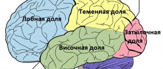
The cerebral cortex has convolutions separated by grooves, which significantly increase its surface. The three deepest grooves divide the hemispheres into lobes. In each hemisphere there are four lobes: frontal, parietal, temporal, occipital. The excitation of different receptors enters the corresponding receptive areas of the cortex, called zones, and from here they are transmitted to a specific organ, prompting it to action. The following zones are distinguished in the cortex. The auditory zone is located in the temporal lobe and receives impulses from auditory receptors.
The visual zone lies in the occipital region. Impulses from the eye receptors arrive here.
The olfactory zone is located on the inner surface of the temporal lobe and is connected to the receptors of the nasal cavity.
The sensory-motor zone is located in the frontal and parietal lobes. This zone contains the main centers of movement of the legs, torso, arms, neck, tongue and lips. This is also where the center of speech lies.
The cerebral hemispheres are the highest division of the central nervous system, controlling the functioning of all organs in mammals. The importance of the cerebral hemispheres in humans also lies in the fact that they represent the material basis of mental activity. I.P. Pavlov showed that mental activity is based on physiological processes occurring in the cerebral cortex. Thinking is associated with the activity of the entire cerebral cortex, and not just with the function of its individual areas.
| Brain department | Functions | |
| Medulla | Conductor | Connection between the spinal and overlying parts of the brain. |
| Reflex | Regulation of the activity of the respiratory, cardiovascular, digestive systems:
| |
| Pons | Conductor | Connects the cerebellar hemispheres to each other and to the cerebral cortex. |
| Cerebellum | Coordination | Coordination of voluntary movements and maintaining body position in space. Regulation of muscle tone and balance |
| Midbrain | Conductor | Approximate reflexes to visual and sound stimuli (turns of the head and torso). |
| Reflex |
| |
| Diencephalon | thalamus
hypothalamus
| |
Notes
- Due to the fact that the body of these animals always consists of the same set of cells.
- White JG, Southgate E., Thomson JN, Brenner S.
The structure of the nervous system of the nematode
Caenorhabditis elegans
//
Phil.
Trans. Royal Soc. London ,
314
, 1986. - P. 1-340. - Mednikov, 1994, p. 363.
- Society for Neuroscience -. www.sfn.org. Retrieved November 22, 2015.
- The Journal of Neuroscience. www.jneurosci.org. Retrieved November 22, 2020.
- Fens Home Page (English). FENS.org. Retrieved November 22, 2015.
- European Journal of Neuroscience.
- Harriet Cole: Drexel's Longest-Serving Employee.
Cerebral cortex
The surface of the human cerebral cortex is about 1500 cm2, which is many times greater than the inner surface of the skull. This large surface of the cortex was formed due to the development of a large number of grooves and convolutions, as a result of which most of the cortex (about 70%) is concentrated in the grooves. The largest grooves of the cerebral hemispheres are the central one, which runs across both hemispheres, and the temporal one, which separates the temporal lobe from the rest. The cerebral cortex, despite its small thickness (1.5–3 mm), has a very complex structure. It has six main layers, which differ in the structure, shape and size of neurons and connections. The cortex contains the centers of all sensory (receptor) systems, representatives of all organs and parts of the body. In this regard, centripetal nerve impulses from all internal organs or parts of the body approach the cortex, and it can control their work. Through the cerebral cortex, conditioned reflexes are closed, through which the body constantly, throughout life, very accurately adapts to the changing conditions of existence, to the environment.