Brain stem
The brain stem includes:
- medulla,
- bridge,
- midbrain.
Brain stem functions
- responsible for primitive forms of behavior,
- supports vital functions.
Medulla
The medulla oblongata (medulla oblongata) is 2.5–3 cm, located between the pons and the origin of the C1 root of the spinal cord.
Centers of the medulla oblongata
- Vital autonomic centers: respiration, vascular-motor center, digestion.
- Protective reflexes: sneezing, coughing, vomiting, blinking, sucking, chewing, swallowing.
- Centers that control the muscles of the limbs and trunk (lateral reticulospinal tract).
The medulla oblongata contains the nuclei of the IX, X, XI, XII pairs of cranial nerves, which are involved in the innervation of the head and neck.
The X pair innervates the internal organs of the thoracic and abdominal cavities.
Pons
The pons (discovered by the scientist Varolio in 1560) is located between the midbrain and medulla oblongata.
Reflex function:
- Within the bridge are located the nuclei of the V, VI, VII and VIII pairs of cranial nerves innervating the head.
- The intrinsic neurons of the pons form its reticular formation.
- Pontine RF affects the cerebral cortex, causing its activation or inhibition.
Among these neurons, a group of nuclei is localized, forming a pneumotaxic center that regulates the change of inhalation and exhalation.
Vestibular nuclei:
- ankylosing spondylitis's upper nucleus
- Roller's inferior vestibular nucleus
- medial nucleus of Schwalbe,
- lateral nucleus of Deiters.
The nuclei, together with the cerebellum, take part in maintaining the balance and tone of skeletal muscles.
From Deiters' nucleus comes the lateral vestibulospinal tract.
Excitatory effect on extensor motor neurons and inhibitory effect on flexor motor neurons.
When the vestibular apparatus is irritated, muscle tone is redistributed in such a way as to maintain balance.
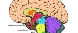
Midbrain
- quadrigemone (roof),
- peduncles of the brain
- Silviev water supply.
Reflex function
The nuclei of the midbrain perform a number of important reflex functions.
The anterior tubercles of the quadrigeminal are subcortical visual centers.
- Visual orientation reflex (eye movement and turning the head towards the light).
- Together with the autonomic nucleus of Yakubovich, III pairs (oculomotor nerve) are involved in the pupillary reflex, accommodation, convergence, gaze fixation and tracking of moving objects.
The posterior tubercles of the quadrigeminal are the primary subcortical auditory centers.
- Indicative auditory reflex (pricking of the ears in animals).
- Turning the head towards a new sound.
The quadrigeminal nuclei provide the guard reflex. A person with impairments in this area is unable to respond quickly to an unexpected stimulus.
Black substance:
- contains the pigment melanin,
- part of the extrapyramidal motor system,
- regulates the acts of swallowing and chewing,
- participates in the regulation of plastic muscle tone,
- participates in the organization of emotional behavior and fine motor skills (fine movements of the fingers),
- synthesizes dopamine, which is transported to the basal ganglia.
Red kernels:
- located at the top of each cerebral peduncle
- the color is due to the dense capillary network,
- contain iron
- associated with the cerebral cortex (descending pathways), subcortical nuclei and cerebellum.
Disruption of the connection between the red nucleus and the RF of the medulla oblongata leads to decerebrate rigidity in animals.
Decerebrate rigidity (spasticity) or contractile tone was discovered by C. Sherrington. Occurs when the brain stem is cut between the anterior and posterior colliculi . Red kernels remain above the transection site.
The animal develops a sharp increase in the tone of the extensor (limbs are extended, the head is thrown back, the back is arched, the tail is raised). The animal can be placed on its paws; it will stand on outstretched legs if the center of gravity is not disturbed.
In humans, a similar condition (opisthotonus) occurs when the midbrain is compressed due to intracerebral hematomas, tumors, cerebral edema, or trauma.
Rigidity mechanism:
The activating influence of the red nuclei and the corticospinal tract on the flexors is turned off, which increases the influence of the vestibular nuclei (Deiters nucleus) on the extensors.
The inhibitory effect of the red nucleus on the Deiters nucleus is eliminated, which is disinhibited, further enhancing the tone of the extensors.
Thus, all conditions are created for a significant increase in the tone of the extensor muscles and the formation of muscle spasticity .
13 0
Structure of the medulla oblongata
The rhomboid section includes the bulb, which is an oblong structure. The bulb originates from a thick-walled canal in the spine and enters the brain stem. Anatomically, the structure is located between the exit of the first spinal nerve and the pons (superior border).
The medulla oblongata in humans is an important part of the central nervous system. The department is located at the junction of the spinal cord and brain. The spinal cord thickens at the entrance to the foramen magnum and visually becomes like an onion. Therefore, the department received a second name - onion.
The medulla oblongata combines the structure of two structures - the spinal cord and the brain. The bulb is the connecting link for the small brain and the pons.
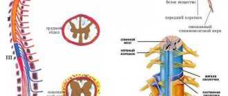
Anatomical structure and boundaries
On the surface of the medulla oblongata there is a median depression, a continuation of the spinal cord groove. Near the recess there are bundles of nerve fibers (pyramids), which pass into the spinal cords.
On the posterior surface of the oblong structure lies the dorsal part of the groove - it comes from the spinal cord. Near the recess (on the sides) lie the posterior cords - the ascending spinal tract. At the top, the cords branch in different directions and go to the cerebellum.
The structure of the bulb itself is heterogeneous, since it combines gray (neuron bodies) and white (processes) matter. The cell bodies of neurons are surrounded by numerous nuclei. They are covered on top by bundles of axons and dendrites. Long nerve fibers cross and pass into the spinal cord. Short processes extend from the long fibers through the oblongata (forming a medial loop) to the thalamus.
Short processes connect the nuclei of gray matter, which in turn continue to communicate with other nuclei. This connection connects all structures with each other and allows the brain to function as a single organ.
The anatomy of the bulb is divided into:
- External structure;
- Internal.
The special structure and functions of the medulla oblongata are inextricably linked with the control of all autonomic reactions in the body, and are also aimed at maintaining life support systems.
External structure
The outer part of the bulb is formed from pyramidal tracts - paired lobes in the form of cones, with an extension at the top and a gap. To the side of the pyramids there is an oval-shaped extension - an olive. It contains nuclei and is delimited by the anterolateral part of the sulcus (continuation of the spinal cord). The connection between the spinal cord and the brain has arcuate fibers.
The external structure of the back of the bulb has a dividing groove, which makes it visually similar to parts of the cylinder. Bundles of fibers coming from the outside have connections with the dorsal region.
On the back side of the oblong section there are ligaments that end in tubercles of the nuclei (thin and wedge-shaped). The back side is located near the rhomboid fossa (lower part), with a vascular tangle located nearby.
Scientists noted that the pyramids are structural features of the back of the brain that appeared during evolution, during the formation of the new cortex. This area has received the greatest development in humans, since it has connections with the cerebral cortex and the cranial nerves (cranial nerves). The pyramidal system allows a person to perform complex coordination movements.
Internal structure
The internal structure of the bulb is a cluster of nuclei that regulate deep systems in the body.
Anatomy of the gray matter nucleus:
- Plate of gray matter in the center;
- Reticular formation with nerve cells (reticular formation);
- ChMN (from 9 to 12 pairs);
- Vital centers (respiratory, blood circulation).
If we consider the oblong structure in a cross-section (transverse), then we can see the grooves, between which the pyramidal tracts are located. Minor elevations are olive trees.
The olive has a nucleus that is formed from gray matter and is connected to the cerebellum. This interaction regulates the vestibular apparatus and helps a person take an upright position.
The pyramidal tract and the olive are delimited by a depression. Communication with other parts of the brain occurs along an ascending path - the nerve fiber receives and transmits impulses (information).
Internal structure of the department
The ventral part of the structure includes nerve cells with complex connections - nerve fibers have weaves, between which the cells are located. The motor part contains vital centers.
Main nuclei of the medulla oblongata
The entire functional significance of the structure is formed from the nerve centers in the medulla oblongata. Control centers are paired nuclei of the cranial nerves and their own nuclei of the medulla oblongata.
CMN cores:
- Glossopharyngeal nerve (4 pairs) - Mixed nerve fibers, including motor, parasympathetic and sensory processes. The roots lie behind the olive, the processes are directed forward and extend to the opening of the skull. It has several thickenings due to nodes. From the skull, the nerve rushes down and passes between the carotid artery and the jugular vein. The nerve forms an arc and returns upward, innervating the root of the tongue. The nerve ganglia (superior and inferior) have multiple branches:
- From the lower node - the tympanic nerve (afferent and parasympathetic). The fiber runs in the tympanic cavity and forms one ganglion, from which multiple small branches extend to the mucous membrane. Thus, the nerve forms a plexus from which the petrosal nerve arises. It emerges from the skull through a rocky cleft;
- From the trunk there are 4 pairs - 5 branches depart, which in turn also have branches. Several branches arise from the pharyngeal nerve and form a plexus. Thin carotid branches enter the thickness of the glomus. The stylopharyngeal branches innervate the muscular layer. The branches running near the tonsils enter the mucous layer and then into the lymphoid tissue. The lingual branches, after entering the root of the tongue, break up into many small threads.
- The vagus nerve (10th pair) is the longest nerve cord, which, descending from the brain structures, passes through almost all organs (head, neck, abdominal and thoracic organs). The length of the nerve increased during evolution - the more distant the innervated organs were, the longer the fiber became. The nerve originates from the core of the medulla oblongata, descending, in the area of the jugular foramen it has two nodes, one of which is larger (thickens the fiber). Between the nodes lies a branch of the accessory nerve. The vagus cord is part of the vascular-nervous cervical bundle. In the thoracic cavity, parts of the nerve (left and right) occupy the surface of the aortic arch and subclavian vessel. Bending around the bronchi, large branches enter the esophagus and, after the formation of a plexus, break up into multiple branches. In the abdominal region, the fiber has many thin branches that innervate the organs;
- Accessory nerve (11th pair) - motor fibers, responsible for the ability to control muscles. The cord has two cores:
- The nucleus ambiguus (or cerebral) is the nucleus common to the 4th and 10th pairs of cranial nerves. Threads extend from the nucleus, which are the anatomical part of the motor nerve;
- The spinal nucleus is located in the posterolateral part of the anterior horn and extends to the 6th upper segment. Multiple roots are combined into one, which is directed along an ascending path to the brain structures. In the cranial cavity, thin branches are connected into a powerful trunk, which, when emerging from the foramen (jugular), is divided into an internal and external cord. The inner part is woven into the 10th pair and is its constituent unit. The outer part tends down to the trapezius muscle.
- Hypoglossal nerve (12th pair) - the nucleus is located in the medulla oblongata structure of the brain (motor), roots extend from it, which, passing through the grooves (between the olive and the pyramidal tract), are connected to the trunk. The trunk enters the canal and gives off branches to the dura mater, after which it leaves the cranial cavity along a descending path. Passing between the 10th pair of nerve fibers and the jugular vessel, it bends around the carotid artery and passes between them. The nerve forms an arc and extends to the submandibular region, into which the muscular layer of the tongue enters and splits into multiple terminal branches. Along the way to the tongue, the nerve cord has processes that connect it with branches of other nerves:
- Connection in the area of the cervical node;
- Connection in the region of the node 10 pair of cranial nerves;
- Included in the lingual branch (from the trigeminal cord);
- Weaved into the neck loop.
Cranial nerves of the brain
Also in the medulla oblongata there is part of the reticular nuclei (coeruleus), the processes going upward reach the upper layers of the cerebral cortex. The processes directed downwards rush into the spinal cord. High molecular weight granules (melanin) give the blue pigment to the nucleus.
The central olivary nucleus in the medulla oblongata has an additional plate of gray mass; together they form an intermediate coordination control center.
The nuclei of the Gaulle (thin) and Burdach (wedge-shaped) bundles originate from the spinal ganglia and have a system of ascending fibers. The fibers form two bundles, which are separated in the middle by a layer of nervous tissue. Closer to the middle (thin bundle) there are long conductive strands - they come from the lower extremities.
The lateral bands (wedge-shaped fasciculus) are shorter and begin in the upper limbs. Both bundles end their path in the nuclei of the bulb (the first and second path), but their axons extend to the parietal lobe of the cortex, where they end their direction (the third).
From the cochlear nuclei, processes are directed to the nuclei of the trapezoid body. The axons form a bundle of fibers called the auditory lemniscus. These cords rush to the subcortical hearing centers.