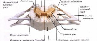
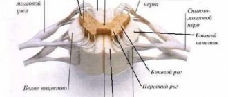
Function of the posterior and anterior roots.
The experiments of physiologists Bell and Magendie established that the posterior and anterior roots contain nerves that carry impulses of different functions. The transection of the anterior roots of the spinal cord on one side that they performed led to complete paralysis of the limb on the same side, while sensitivity was completely preserved. Transection of the dorsal roots led to loss of sensitivity in the absence of any disturbances in movements. These observations served as the basis for formulating the position that the dorsal roots are sensitive and contain centripetal fibers, and the anterior roots are motor, containing centrifugal fibers.
Rice. CROSS SECTION OF THE SPINAL CORD. 1 - posterior root; 2 - anterior roots; 3 - anterior horn; 4 - lateral horn, 5 - posterior horn.
The essence of the spinal conduction mission
Pathways are special neural fibers that transmit signals of a certain kind to various brain centers. In medical practice, it is customary to differentiate three groups of the above fibers.
Spinal tracts
- Associative. They are intended to connect gray matter cells from dissimilar segments to form, directly near the gray matter, special bundles of their own (meaning anterior, lateral, posterior).
- Commissural. The function of these fibers is to connect the gray matter from both hemispheres, as well as similar and equidistant nerve centers of both halves of the brain to correlate and coordinate their work.
- Projection. These fibers connect the overlying and underlying brain regions. They are responsible for projecting pictures of the surrounding world onto the cerebral cortex, like on a scoreboard or television screen.
Projection fibers differ depending on the direction of the urges sent to the ascending and descending pathways. The following three groups of ascending pathways are responsible for the delivery of signals to the brain that appear as a result of the influence of various factors and environmental phenomena on the human body.
- Exteroceptive - deliver impulses from two types of receptors.
- Impulses delivered by exteroceptors. This refers to temperature, tactile and pain signals.
- Impulses of the senses: the ability to see, hear, smell and taste.
- Proprioceptive - responsible for impulses coming from the organs of movement and muscles.
- Interoceptive - intended to conduct impulses sent by internal organs.
Along descending pathways, signals pass from the subcortical centers and the cortex itself to the nuclei of the brain, as well as to the motor nuclei of the spinal horns located in front. The descending tracts include several fiber systems.
- The corticospinal cord is responsible for the mission of movement.
- The tectospinal tract, otherwise called the tectospinal tract, is a projection of the descending nervous system.
- The vestibular-spinal cord is responsible for the proper coherence in the work of the vestibular apparatus.
- The reticular-spinal cord, otherwise called the reticular-spinal tract, ensures the proper level of muscle tissue tone.
In addition, the pathways of the brain and spinal cord are also differentiated according to the tasks performed.
- Motor pathways responsible for the reflex response. Their task is to transmit “pointers” from the brain to the spinal cord and further to the muscles. Thanks to the coordinated work of these paths, the proper level of coordination of movement is ensured.
- Sensory pathways help in recognizing pain, temperature and its changes, and tactile sensations.
Nerve fibers are the guarantors of the inextricable relationship between the brain and the spinal cord, and through it, with all organ systems. The rapid transmission of appropriate signals ensures the consistency of all body movements, eliminating significant efforts made by the person himself. The pathways form bundles of nerve cells.
Types of conductive paths by direction
The ascending pathways of the spinal cord recognize urges received from various life-supporting organs of a person, with their subsequent provision to the “center”.
Ascending and descending tracts connect the spinal horns with the cerebral cortex
Descending pathways send “instructions” immediately to certain internal organs, various glands, and muscles. Signals and impulses in this case are transmitted through the spinal neural connection.
Fast and accurate data transfer is ensured thanks to the double course of the spinal tracks.
White matter
White matter - substantia alba, envelops the gray matter, is formed by a collection of nerve fibers, which also come in three types:
- front;
- rear;
- lateral.
Moreover, all the roots have a different direction, and some of them are connected directly to the brain and central nervous system (hereinafter simply the CNS). And if the reflex function of the spinal cord is to transmit signals from the motor neurons of the gray matter, then the task of the white matter neurons is the prompt delivery of impulses from the muscles and joints to the medulla oblongata. Thus, the transmission of all commands along the entire spinal cord is realized.
Here are the paths through which all information regarding sensitivity and pain is transmitted. Only before entering the cerebral cortex, the information first reaches the diencephalon, and only then rushes further to its destination.
Localization of paths as they move
The ascending and descending tracts connect the spinal horns with the cerebral cortex. The spinal tracts are nerve bundles and tissues that pass through the corresponding parts of the brain. In this case, impulses can be transmitted only in one direction. The location of the spinal tracts is clearly demonstrated by the diagram in the video above.
Ascending spinal tracts and their characteristics
The bodies of the first nerve cells, acting as transmitters of various types of spinal sensitivity, lie in the corresponding brain nodes. The cellular axons of these nodes enter the spinal cord. Among them there are a couple of groups.
The medial group moves towards the posterior cord. At this point, each existing fiber is divided into a pair of branches. They are called ascending and descending. A certain number of the above branches, when moving up and down, form bundles in various spinal segments and points.
The lateral group moves to the edge, and then to the posterior column of gray matter to contact the cells of the posterior genus.
The ascending tracts of the spinal cord, otherwise called centrifugal or afferent, with their characteristics and direction of movement are described in detail in Table No. 1.
| No. | View of the ascending path | Characteristics | |
| 1 | Posterior spinocerebellar | The task of this direct cerebellar pathway is to conduct impulses to the cerebellum from muscle receptors. The spinal ganglion is the home of the first neurons. The refuge of the second neurons is the entire surface of the spinal cord in the thoracic nucleus. These neurons move towards the outside. Having reached the posterolateral spinal cord, they turn upward and follow close to the lateral spinal cord. Then they go to the cortex of the cerebellar vermis. | |
| 2 | Anterior spinocerebellar | This tract is also designed to carry impulses to the cerebellum from muscle receptors. The spinal ganglion is the home of the first neurons. And the medial nucleus of the intermediate region is the habitat of the bodies of the second neurons. Their fibers are sent to the lateral cords of both sides. Having reached the anterior outer sections of the cords, the fibers will be located above the posterior spinocerebellar tract. Turning upward, crossing the bridge and crossing, the fibers reach the cerebellar vermis, which completes this path. | |
| 3 | Spino-olive | Let this ascending conduction begin in the cells of the dorsal horns. After crossing, the axons of these cells move upward along the spinal surface. The final destination of the spino-olive tract is, accordingly, the olive nuclei. Through the above tract, data from muscle and skin receptors enters the brain. | |
| 4 | Anterior spinothalamic | Responsible for transmitting signals regarding tactile sensitivity. | The spinal ganglia are the area where the cell bodies of the first neurons are located. The path of the second neurons runs to the opposite side towards the cords. The fibers of these pathways bypass the medulla oblongata, pons and cerebral peduncles, subsequently reaching the thalamus. The third neurons lie precisely in the thalamus, following directly to the cerebral cortex. |
| 5 | Lateral spinothalamic | Carries out signals regarding temperature and pain sensations. | |
| 6 | Spinoreticular | The elements of this tract are fibers from both spinothalamic tracts. | These two pathways run through the lateral spinal cords, ending in the plate of the mesencephalic roof. |
| 7 | Dorsal-tegmental | ||
| 8 | Thin Bun | This bundle transmits “instructions” directed by the lower parts of the human torso along with its lower limbs below the 4th thoracic segment. Having reached the medulla oblongata, the bundle begins to contact its own nuclear cells. | The muscles provide “instructions” to both bundles. The first neurons of the above tracks lie in certain spinal ganglia. They move to the nuclei of the medulla oblongata. The two tubercles are the second neurons of the corresponding bundles. Their axons reach the opposite side when moving. There they form a sensory chiasm, and then move to the thalamus, already being an integral part of the medial loop. The fibers of these bundles come into direct contact with thalamic cells. The processes of these neurons are sent directly to the brain. |
| 9 | Wedge-shaped bundle | It is formed from fibers that initiate movement in the cells of the spinal ganglia and end in the sphenoid tubercle. | |
Conducting function of the spinal cord
Basic reflexes of the spinal cord
• Stretch reflexes (myotatic)
basic extensor - posture reflexes, pushing (jumping, running) reflexes
• Flexion jerk reflexes
• Rhythmic reflexes (
carding, walking)
• Positional reflexes (
cervical tonic reflexes of inclination and position)
• Autonomic reflexes
Extensor reflexes of the limbs
The knee extensor reflex is carried out due to the contraction of the quadriceps femoris muscle, the level of closure is L2-L4.
Flexion reflexes of the limbs:
Achilles reflex - proprioceptive, expressed in plantar flexion of the foot as a result of contraction of the triceps muscle of the leg, occurs when the Achilles tendon is struck with a hammer; the reflex arc closes at the level of the sacral segments - S1-S2.
Plantar reflex - cutaneous, expressed in flexion of the foot and toes when the sole is irritated by strokes; the reflex arc closes at the S1-S2 level.
Closing function of the spinal cord
ü The presence in the spinal cord of nerve centers that regulate the functioning of internal organs;
— Centers affecting the muscles of the eye – C8-T2;
— Centers regulating salivation (T2-T5), sweating, cardiac activity (T1-T5),
- secretory-motor reflexes of the stomach (T6-T9) and intestines, respiratory (reflexes that expand the lumen of the bronchi), thermoregulation, kidney function (T5-L3).
— Parasympathetic centers that empty the rectum, evacuate urine from the bladder, as well as spinal genital centers (S2-S4).
Conducting function of the spinal cord
The connection of the spinal cord with the overlying parts of the central nervous system (brain stem, cerebellum and cerebral hemispheres) is carried out through ascending and descending pathways. The pathways are located in the lateral, posterior and anterior columns of the spinal cord.
Ascending (sensitive) pathways carry information received from receptors of peripheral organs to sensitivity centers (thalamus, cerebral cortex);
Descending (motor) pathways conduct impulses from the cerebral cortex and other higher-lying sections to the neurons of the spinal cord, and from them to the performing organs (skeletal muscles, glands, internal organs);
Age characteristics
of the spinal cord concern both its topography and structure.
In the 2nd half of the prenatal period, the growth of the spinal cord lags behind the growth of the spinal column. During childhood, spinal cord growth continues to lag. The length of the spinal cord increases 2.7 times over the entire period of growth, mainly due to the thoracic segments. The mass of the spinal cord increases 6-7 times. The gray and white matter of the spinal cord grow unevenly, the volume of gray matter increases 5 times, and the volume of white matter increases 14 times.
The completeness of the structure is already observed in the fetus before birth. This is explained by the increased purposeful activity of the newborn's limbs. An increase in the size of spinal cord neurons is observed in children during school years. Consequently, the motor ability of a primary school child largely depends on the spinal cord.
Development of motor skills in children.
In the first days after birth, motor activity in children is little pronounced. They are in a state of some inhibition. If the child is kept in comfortable conditions, then motor reactions are associated only with stimulation of the food center.
By the time of birth, the child has completely completed the development of the spinal and medulla oblongata, globus pallidus, red nucleus, paleocerebellum and all pathways except the pyramidal one.
Many newborn children exhibit postural reflexes: when the head is turned to the side, the arms and legs are extended on the side where the face is facing, and the limbs are flexed on the other side. More often, this redistribution of tone occurs only in the upper limbs.
• A newborn has a large number of unconditioned reflexes: sucking and swallowing reflexes, urination and defecation reflexes, Hering and Breuer respiratory reflexes are well developed. At this age, an orienting reflex appears: in a darkened room, the child constantly turns his eyes towards the bright light. Posotonic reflexes are well expressed in newborns, controlled by the medulla oblongata and consisting in the fact that when the head is turned to the side, the arm on this side extends, and on the opposite side it bends and rises.
• In healthy newborns and infants, knee and Achilles reflexes and Robinson's grasping reflex are easily manifested (if the surface of the child's palm is stroked, the fingers clench). With line irritation of the skin of the plantar surface of the foot, the Babinski reflex is observed, most often in the form of extension of the big toe and flexion of the rest.
• The reflexes of newborns, which are used by pediatricians to study the functions of the central nervous system, also include the proboscis reflex - stretching the lips forward when a finger touches them; Moro reflex - when hitting the table on which the child is lying, he spreads his arms and then brings them together; crawling reflex - when pressing with the palm of your hand on the feet of a child lying on his stomach, repulsion and a crawling-type movement occurs. These reflexes make it possible to assess the degree of development of the newborn’s central nervous system.
Additional Information.
Spinal cord
• the first half of the intrauterine period occupies the entire length of the spinal canal,
• subsequently its growth lags behind the growth of the spinal column,
• in a newborn, the lower end of the spinal cord is at the level of the 3rd lumbar vertebra.
• In childhood, the growth of the spinal cord continues to lag behind the growth of the spinal column,
• in adults it ends at the level of the 1st lumbar vertebra, and in some cases at the level of the 2nd lumbar or 12th thoracic vertebrae.
• The following indicators allow you to compare the length of the spinal cord with the length of the body:
in a newborn, the length of the spinal cord is 30%, in a 1-year-old child - about 27%,
in children from 3 to 5 years old - 21%,
from 7 years of age -22
in an adult - 26% (A.K. Koveshnikova, 1958).
• A comparison of these percentage data suggests that with body growth, the ratio of the length of the spinal cord to the length of the body first decreases, then in an adult it increases slightly again. These relationships change during the formation of the child’s body. %
• The length of the spinal cord of a newborn is on average 14.1 cm and increases 3-4 times over the entire postnatal period.
• There is a close relationship between the length of the spinal cord and the body: these two quantities increase in direct proportion.
• The thoracic region increases the most, the lumbar region increases the slowest.
• The average weight of the spinal cord in a newborn is 3.2 g, i.e. 0.1% body weight (0.04% body weight in adults) and 1% brain weight (2% in adults).
Until 6-10 months after birth, his weight doubles, by 3 years it increases 3 times, after which weight gain occurs more slowly.
• Cervical and lumbar thickenings begin to contour after 3 years of life. There may be more grooves on the surface of the spinal cord than in an adult. There are permanent furrows, which deepen with age, and non-permanent ones, which subsequently disappear.
• The white matter of the spinal cord in newborns occupies twice the surface area compared to the surface of the gray matter. The growth of gray and white matter occurs unevenly. The volume of gray matter increases 5 times, white matter 14 times.
• The central canal of the spinal cord is relatively wider than in an adult.
• The sensory tracts and anterior pyramidal tract are well myelinated. The rest begin to myelinate from 4-6 months and this process ends in a child aged 3 to 7 years.
• The average weight of the spinal cord in a newborn is 3.2 g, i.e. 0.1% body weight (0.04% body weight in adults) and 1% brain weight (2% in adults). Until 6-10 months after birth, his weight doubles, by 3 years it increases 3 times, after which weight gain occurs more slowly.
• During development, the relationship between the weight of the brain and spinal cord changes. In a newborn, the spinal cord is equal to 1% of the weight of the brain, in a one-year-old child - 1.2%, in a 2-year-old child - 1.3%, in an adult - 2%. Relative to body weight, the spinal cord varies from 0.09% in a newborn to 0.04% in an adult.
• Intensive growth of the spinal cord in postnatal ontogenesis is inextricably linked with the formation of spinal reflexes, the development of pathways and the cortical ends of the analyzers. In ontogenesis, as in phylogeny, old pathways of the spinal cord are formed earlier and later - new pathways and corresponding cellular formations.
• During development, the configuration of the spinal cord changes, cervical and lumbar thickenings appear, associated with the growth of the rudiments of the upper and lower extremities. The spinal cord in these thickenings is most massive, and the cervical thickening develops faster than the lumbar thickening, which must be associated with the earlier development of the upper limb.
• The most intensive process of formation of cervical and lumbar thickening occurs in the first years of a child’s life.
• According to N.P. Gundobin, the spinal cord grows slowly in thickness. By the age of 12, its thickness doubles and subsequently remains almost the same.
• According to F.I. Walker (1938), the lumbar and sacral sections of the spinal cord grow more slowly than the thoracic.
• In a newborn, the length of the spinal cord is on average 14-16 cm, and in an adult - 42-45 cm.
• During development, the configuration of the spinal cord changes, cervical and lumbar thickenings appear, associated with the growth of the rudiments of the upper and lower extremities. The spinal cord in these thickenings is the most massive, and the cervical thickening develops faster than the lumbar one, which must be associated with the earlier development of the upper limb - 16 cm, and in an adult - 42-45 cm. • .
• Cervical and lumbar thickenings begin to contour after 3 years of life. There may be more grooves on the surface of the spinal cord than in an adult.
• There are permanent grooves, which deepen with age, and non-permanent ones, which subsequently disappear. • .
• The white matter of the spinal cord in newborns occupies twice the surface area compared to the surface of the gray matter.
• Gray and white matter grow unevenly.
• The volume of gray matter increases 5 times, white matter - 14 times.
• The central canal of the spinal cord is relatively wider than in an adult.
• The sensory tracts and anterior pyramidal tract are well myelinated.
• The rest begin to myelinate from 4-6 months and this process ends in a child aged 3 to 7 years.
• In both prenatal and postnatal ontogenesis, the cells of the dorsal horns of the spinal cord are distinguished by a variety of shapes and are smaller in size than the cells of the anterior horns. According to A.I. Molochek (1936), the fibrillar structure in the motor cells of the anterior horns is detected later (in the fifth month of the embryonic period) than in other cells of the spinal cord, where it appears earlier (in the third month). •
• By the end of the embryonic period, the nerve cells of the spinal cord are already in a developed state in terms of shape, size, position of the nucleus and the content of Nissl bodies in them.
• The initially wide lumen of the neural tube transforms into a narrow central canal during postnatal development in school-aged children and adults. However, in general, the macro-microscopic structure of the spinal cord in the early stages of postnatal ontogenesis is almost the same as in an adult.
• The spinal cord, its cellular and fibrous structure develops earlier than other parts of the nervous system, which is in full accordance with the earlier formation of spinal reflex mechanisms.
Descending pathways
All descending tracts of the spinal cord with their detailed characteristics and course of movement are clearly demonstrated in Table No. 2.
| No. | Descending Path View | Characteristics | |
| 1 | Lateral corticospinal, also called lateral corticospinal or main crossed pyramidal. | This pathway includes a considerable proportion of fibers of the pyramidal system. The lateral tract is localized in the lateral funiculus. As they travel, the fibers gradually become thinner. Lateral fibers conduct signals that cause conscious actions in humans. | Lateral fibers conduct signals that cause conscious actions in humans. |
| 2 | Anterior corticospinal, otherwise called corticospinal, and also straight or uncrossed pyramidal. | This path lies in the anterior spinal cord. Like the lateral pyramidal tract, the direct pyramidal tract includes cellular axons of the motor hemisphere, although they are located ipsilaterally. Initially, these axons descend towards their “own” segment. After this, as part of the anterior spinal commissure, they are transported to the opposite side, ending in the mononeurons of the anterior horn. | |
| 3 | Red-spinal or rubrospinal. | Beginning in the red nucleus of the spinal cord, this tract subsequently descends to the motor nerve cells of the anterior horns. This pathway is responsible for transmitting unconscious motor signals. | |
| 4 | Tectospinal, otherwise called tectospinal. | It is localized in the anterior funiculus near the anterior pyramidal tract. This tract starts on the roof of the midbrain. The mononeurons of the anterior horns are its final point. The tectospinal tract provides reflex protective actions in response to visual and auditory stimuli. | |
| 5 | vestibulospinal, otherwise called vestibulospinal. | This path is localized in the anterior spinal cord. The vestibular nuclei of the pons are its beginning, and the anterior spinal horns are its end. The balance of the human body is ensured precisely through the transmission of impulses from the vestibulospinal tract. | |
| 6 | Reticulospinal or reticulospinal. | This pathway ensures the transmission of excitatory signals from the reticular formation to the spinal nerve cells. | |
To understand the neurophysiology of the human spinal cord pathways, you will need to briefly familiarize yourself with the structure of the spine. In its structure, the spinal cord is a bit like a cylinder, covered with muscle tissue on all sides. The pathways control the functioning of the internal organs, as well as all organ systems and functions performed by the body. Injuries, various injuries, and other ailments of the spinal cord can somehow reduce conductivity. By the way, conduction may even stop completely due to the death of neurons. Complete loss of conduction of spinal signals is characterized by paralysis, manifested in a complete lack of sensation in the limbs. This is very fraught with problems with internal organs that are responsible for damage to the communication of nerve cells. Thus, injuries and other ailments of the lower spinal cord are often characterized by urinary incontinence and even spontaneous defecation. Drug treatment will consist of prescribing medications that prevent the death of brain cells, as well as additionally increasing blood flow to the damaged spinal areas. Electrical impulses may be prescribed as an additional treatment to stimulate the functioning of neurons and also help maintain muscle tone.
Surgical operations to restore spinal conduction are performed in specialized spinal clinics.
Also, if necessary, the attending physician may prescribe the use of the following folk remedies.
Apitherapy
- Apitherapy. Bee stings effectively restore the conductivity of efferent tracts. Thus, the poisons of these insects, penetrating into damaged areas, provide them with additional blood flow. If the cause of spinal pathology is radiculitis, a growing hernia and other similar ailments, apitherapy will be an excellent addition to traditional treatment.
- Herbal medicine. Medications are prescribed to normalize blood circulation and improve metabolism.
- Hirudotherapy. Thanks to treatment with leeches, it becomes possible to eliminate congestion - inevitable attributes of vertebral pathologies.
The resulting degenerative changes almost immediately lead to disruption of conduction and reflex activity. Dying neurons are quite difficult to restore. The disease can often develop rapidly, significantly impairing conductivity. Therefore, it is advisable to consult a doctor for medical help when the first signs of pathology are detected.
Characteristic
The spinal cord lies in the canal of the spinal column. In an adult, the length of the cord is about 45 cm. The upper segment of the spinal cord borders the medulla of the brain, which ensures the continuity of the pathways. The basis of the cord is gray matter, which when cut looks in the shape of a butterfly. Gray matter is surrounded by white fibers, which form pathways - ascending and descending.
The spinal cord contains tracts of white matter that support sensory functions and complex motor activities. There are ascending (afferent) and descending (efferent) pathways within the spinal cord. In the first case, we are talking about the transmission of impulses from all parts of the body to the head part of the central nervous system, in the second - about the direction of impulses from the cortical structures of the head part of the central nervous system to parts of the body and limbs.
The medulla receives impulses emanating from exteroreceptors (perceive environmental stimuli) located on the surface of the skin, proprioceptors (sensitive receptors), and partially visceroreceptors (located in blood vessels and internal organs). The ascending sensory pathways within the spinal cord perform a conducting function. They convey:
- Proprioceptive sense (receive impulses from elements of the musculoskeletal system - muscles, tendons, joint capsules).
- Extraceptive feeling (receive impulses from skin receptors).
- Intraceptive sense (perceive impulses from elements of the circulatory system and internal organs).
The fibers that carry the proprioceptive sense are divided into conscious and unconscious sensory pathways. In the first case, impulses arrive in the cortical parts of the hemispheres, in the second - in the cerebellum, where balance reactions and complex motor activity that require fine coordination are modulated.
The nerve endings of the spinal cord innervate the skeletal muscles with the exception of the muscles of the head, which are innervated by the cranial nerves. The pathways pass in the posterior, lateral, and anterior cords, formed from white matter, where nerve fibers are grouped within the spinal cord, transmitting impulses of various types to certain parts of the central nervous system.