The central nervous system has delimited sections that interact with each other by transmitting and receiving impulses through thin conductors. Nerve fibers unite all structures into a single mechanism that controls the internal functioning of the body. Medulla oblongata - the functions of the department are associated with maintaining the vital functions of the body.
Division of the medulla oblongata
Characteristic
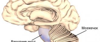
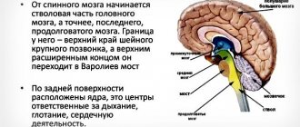
Oblongata is a cone-shaped section of the brain about 25 mm long, which is located between the dorsal region and the pons (ventral localization - on the anterior side), where it ends. The upper edge borders the posterior part of the brain. The location of the medulla oblongata immediately behind the spinal cord determines the structural, morphological and functional similarity of the designated areas.
The topography of the department suggests the presence of an outer layer of white matter. The gray matter within the structure is grouped into nuclei. On the dorsal (posterior) side, the edge of the medulla oblongata borders the cavity of the 4th ventricle and the cerebellum, which is part of the hindbrain. In the anatomy of the medulla oblongata, there are 4 surfaces - 2 lateral, dorsal (posterior) and ventral (anterior). The superficial area is riddled with grooves similar to the surface of the spinal cord.
On the ventral side there are paired elements of the medulla oblongata - pyramids, presented in the form of rollers. The fibers that form the pyramids intersect in the lower part of the brain region and become part of the cords formed from white matter that lie within the boundaries of the spinal cord. The chiasm represents the boundary between the oblongata and dorsal regions.
The border coincides with the area of the foramen magnum. Laterally (to the side, further from the median plane) from the pyramid lies a groove that separates the pyramid and the olive, which is part of the medulla oblongata. Olives are oval-shaped elevations located on the surface of the brain. Branches of the XII pair of nerves extend from the groove. Behind the olive lies another groove, from which nerve endings IX (glossopharyngeal), X (vagus), XI (accessory) pairs branch.
The dorsal surface is equipped with structural formations belonging to the medulla oblongata - longitudinal groove, bundles consisting of nerve fibers (thin, wedge-shaped). The thin bundle, also known as the Gaulle bundle, is located medially (closer to the median plane), in the upper segment it forms a convexity of the thin nucleus. The wedge-shaped bundle is located laterally and is the basis for a similar convexity.
The nuclei, together with the lateral cords, form the basis of the inferior peduncle, which belongs to the cerebellum and extends upward. The legs in the lower part are the boundaries of a triangular-shaped area, which is called the diamond-shaped fossa. A transverse section within the medulla oblongata, passing at the level of the olive, shows a number of structures consisting of gray matter.
These are olive kernels, a section of reticular formation, the fibers of which intertwine kernels of small diameter. The olivary nuclei of the medulla oblongata in humans are located in a layer called interolive and consists of white matter, where fibrous branches extending from the nuclei (thin, wedge-shaped) are concentrated.
The fibers are grouped into a medial loop, which is part of the structure of the pathway that transmits impulses from proprioceptors (sensitive receptors located in muscles, joint capsules, ligaments, and skin). If a transverse incision is made above the level of the olives, the lower peduncles belonging to the cerebellum are visible.
In the anterior section from the legs lie the fibers of the conductive tracts - spinocerebellar, red nuclear spinal cord. The fiber structures of the pyramids are visible from the side, and the fibers extending from the longitudinal bundle are visible from behind and above. At the back and sides there are ascending pathways along which nerve impulses enter the brainstem, cerebellum and the terminal part of the brain (hemispheres).
Where is the medulla oblongata located?
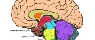
In a mammal, the brain consists of 2 parts - the brain and the spinal cord; the brain consists of 5 sections, one of which is the medulla oblongata. To understand where the medulla oblongata is located, you need to imagine that it is an anatomical continuation of the spinal cord. The location of the medulla oblongata is between the spinal and midbrain. Externally, the medulla oblongata looks like an onion compressed from behind.
Structure
The structure of the medulla oblongata determines its role and functions. This brain region contains the respiratory and vasomotor centers, so it supports the vital functions of the body. The medulla oblongata continues the spinal cord; this brain structure is located in the most posterior zone of the trunk. During embryonic development, this area is formed from the brain bladder located at the back.
The department consists of gray and white medulla. Nerve centers - nuclei within the medulla oblongata, are formed by gray matter. The fibers connecting the spinal cord and brain are formed from white matter. The structure of the medulla oblongata in the brain suggests the presence of a lower segment of the fourth ventricle, which in humans extends from the aqueduct of Sylvius to the valve.
Considering the internal structure of the medulla oblongata, it is worth noting the pyramids, which are located on the ventral (front) side and are delimited by the median sulcus. The basis of the pyramids is the fiber structures of the corticospinal tract (pathways of the pyramidal system). At the junction of the medulla oblongata and the spinal cord, partial crossing of the fibers of the pathways occurs (crossover).
On the side of the pyramid there is an olive tree. The posterior region of the oblongata is divided by a groove. To the left and right of the sulcus are bundles of nerve fibers that continue the spinal cord. The Gaulle bundle, like the Burdach bundle, ends in the form of convexities - nuclei, bearing the same names (Gaull, Burdach).
In the medulla oblongata there are nuclei of 4 types - cranial nerves (centers where the first synaptic connections are formed during the transmission of impulses from cranial nerves), sensory, reticular formation, autonomic nervous system. There are types of cranial nerve nuclei (pairs - VIII-vestibular-cochlear, IX-glossopharyngeal, X-vagus, XI-accessory, XII-hyoid):
- Sensitive (sensory). They perceive information coming from exteroceptors (receptors that respond to environmental stimuli).
- Motor. Nerve branches extending from the nucleus innervate the skeletal muscles.
- Mixed. Nerve fibers extend from the nucleus, including sensory (sensitive) and motor branches, as well as conducting components of the autonomic system. The nerve branches of the autonomic system innervate areas of the internal organs.
The main function of the sensory nuclei grouped in the medulla oblongata is the primary (intermediate) processing of information coming from sensory structures lying below. Sensitive nuclei include olives, Gaulle's nuclei, Burdach's nuclei, Bekhterev's nucleus, and Deiters' nucleus. The red nuclei located in the midbrain have a constant regulatory (inhibitory) effect on the Deiters nucleus.
For example, when brain structures are transected below the red nucleus, decerebrate rigidity develops (increased tone of the extensor muscles and relative relaxation of the flexor muscles), which leads to extensor-type hypertonicity. Characteristics of the nuclei VIII, IX, X, XI, XII pairs of cranial nerves are presented in the table:
| Kernel view | Type of nerve fibers | Innervation zone |
| Hypoglossal nerve | Motor | Musculature of the tongue |
| Accessory nerve | Motor | Neck muscles |
| Vagus nerve | Mixed | The vegetative component is the organs located in the chest and abdominal cavity (heart, stomach, digestive tract organs). The motor component is the muscles of the soft palate, the muscles of the pharynx and larynx. Sensitive component - perceives impulses coming from receptors located in the area of internal organs (liver, heart, lungs) |
| Glossopharyngeal nerve | Mixed | The vegetative component is the parotid salivary glands. The motor component is the pharyngeal muscles, the muscle tissue of the oral cavity. Sensitive component - perceives stimuli from taste buds located in the tongue, from chemoreceptors (react to chemical stimuli) of the aorta. |
| vestibulocochlear nerve | Sensitive | Perceives stimuli from the vestibuloreceptors (determine the speed and change in direction of movement when the body moves in space) of the inner ear, as well as from the auditory receptors located in the middle ear. |
The reticular formation is a section of the brain stem, consisting of clusters of neurons and interconnected nuclei, partially located in the medulla oblongata. This structure controls the level of excitability of different areas of the central nervous system. In the medulla oblongata, the reticular formation contains 3 nuclei (giant cell, small cell, reticular lateral localization), which regulate the degree of behavioral arousal and level of consciousness.
Medulla oblongata and pons - lectures on PostNauka
VIDEO When the second month of embryonic development begins, the neural tube in its anterior part begins to produce swellings, which are called medullary vesicles.
These brain bubbles gradually form our brain. At first, three brain bladders are formed - they are called the primary hindbrain, the primary midbrain and the primary forebrain. Then a more subtle division of these vesicles into parts begins, and the primary hindbrain is ultimately divided into three structures: the medulla oblongata, the pons and the cerebellum. The medulla oblongata and the pons are zones that line the central part of our brain, and they form what is called the brainstem.
The stem structures are a direct continuation of the spinal cord, that is, the neural tube becomes wider and thicker, and the medulla oblongata and pons appear. The midbrain also belongs to the stem structures, sometimes they even include the diencephalon, that is, those unpaired structures that largely make up our central nervous system.
In addition to the stem structures, the brain includes paired structures - the cerebral hemispheres and the cerebellum, since the cerebellum also consists of two hemispheres. Anatomists contrast the trunk and on this trunk two crowns - the cerebral hemispheres and the cerebellar hemispheres.
The medulla oblongata and pons are the areas closest to the spinal cord, which means they handle the most ancient and basic functions of our nervous system. To briefly characterize what they do, we can say that they are engaged in vital functions, that is, functions without which it is generally impossible to exist - the body dies almost immediately.
Of course, the most vital function is respiratory; it is in the medulla oblongata and the pons that our respiratory center is located, and here are also the control centers for the heart, vascular tone, sleep and wakefulness centers, and a number of other very important clusters of nerve cells.
If we begin to look at the medulla oblongata and the pons at the anatomical level, then we see that something that was in the spinal cord gradually turns into stem structures, and, for example, the anterior, lateral and posterior horns of the gray matter of the spinal cord turn into cranial nuclei nerves. The intermediate nucleus, which was in the spinal cord, turns into the reticular formation, that is, the central integrative zone of the medulla oblongata and the pons, which calculates different signal flows and makes decisions on the launch of certain reactions.
Books
First, a few words about cranial nerves. The spinal cord connects to our body through 31 pairs of spinal nerves, and the brain through 12 pairs of cranial nerves. All these 12 pairs are very individual.
In the spinal cord, the spinal nerves are quite stereotypical, they are simply designated by numbers from 1 to 31, but in the brain, each nerve has its own character, its own functions and deserves not only a number, but also its own name. The medulla oblongata and the pons are richest in cranial nerve nuclei.
Cranial nerves from the 5th to the 12th begin precisely in the structures of the medulla oblongata and the pons.
The first pair of cranial nerves, the olfactory nerves, are connected to the cerebral hemispheres. The second pair is associated with the diencephalon. The third and fourth pairs are the midbrain pairs, and the 5th to 12th nerves are the nerves of the medulla oblongata and pons. Nerves 5 to 8 are connected to the pons, and nerves 9 to 12 are the nerves that exit the medulla oblongata.
The trigeminal nerve, the fifth, is so called because it consists of three branches: one branch comes from the frontal zone, the second from the upper jaw, and the third from the lower jaw. The trigeminal nerve is associated with skin, pain and muscle sensitivity of the head.
For example, when our teeth hurt, it is the trigeminal nerve. The sixth nerve, the abducens nerve, is part of the oculomotor system. This system is based primarily in the midbrain.
The seventh nerve, the facial nerve, is so called because its main function is to control the facial muscles: the muscles of the forehead, cheeks, and lips.
In addition, the facial nerve also controls, for example, part of the salivary glands, it controls the lacrimal glands, that is, we cry when the fibers of the facial nerve are activated. The facial nerve is associated with taste sensitivity from the front of the tongue, and it is very rich in its functions.
The eighth nerve is called the vestibulo-auditory nerve, it enters the stem structures at the border of the medulla oblongata and the pons and comes from our inner ear. The very name “vestibulo-auditory” suggests that some of the fibers conduct information from the organ of balance, and some from the organ of hearing, that is, from our cochlea.
The ninth nerve is called the glossopharyngeal nerve. (The name of each nerve tells you what the nerve does.) For the glossopharyngeal nerve, the division of functions is approximately as follows: the first function is taste sensitivity, the glossopharyngeal nerve, together with the facial nerve, provides taste sensitivity.
The glossopharyngeal nerve belongs to the back of the tongue; it is involved in swallowing and controls the muscles of the pharynx, the initial part of our digestive tract. This, of course, is also a very important function, and it should work immediately when the child is born.
In addition, the glossopharyngeal nerve controls part of the salivary glands, that is, it again works together with the facial nerve. These two nerves form a tandem and carry out many functions together.
The tenth nerve is called the vagus nerve. Each nerve, of course, has a Latin name, the vagus nerve is especially known - nervus vagus, which is why they often write “vagal influences”. The vagus nerve is the most important parasympathetic nerve in our body. It is associated with the management of the organs of the chest and abdominal cavities.
Most of our internal organs experience both sympathetic and parasympathetic influences at the same time. The lion's share of parasympathetic influences is the vagus nerve.
Parasympathetic neurons are located mainly in the brain, in the region of the medulla oblongata and pons, in contrast to sympathetic neurons, which are located mainly in the spinal cord, in the lateral horns of the thoracic segments.
The vagus nerve is called the vagus because at one time, when anatomists described it, they were amazed at how widely it branches throughout our body - it seemed to get lost.
It descends along the esophagus, then passes through the chest cavity and branches, giving its fibers to the bronchi and heart, then passes through a special opening in the diaphragm along with the esophagus, goes along the wall of the stomach and then branches very widely in the abdominal cavity. That is, this is a huge nerve that controls many of our functions.
In addition to being parasympathetic, there are also motor and sensory fibers. Sensory fibers are needed to read information from internal organs - for example, the degree of distension of the intestines or stomach. One of the very famous reflexes - when a person hiccups - is associated with excessive distension of the stomach and the reaction of the vagus nerve to the fact that the stomach is full of something. The motor fibers that make up the vagus nerve are associated with the larynx, that is, with the control of the vagus ligaments. It turns out that we speak largely due to the work of the vagus nerve.
What remains are simple nerves - the 11th and 12th, the first of which is called the accessory, and the second - the hypoglossal. Both of these nerves are purely motor, they control muscles. The accessory nerve is the nerve that controls head movements. There is a muscle called the sternocleidomastoid, and it ensures head rotation.
Another 11th nerve controls part of the fibers of the trapezius muscle. When we shrug our shoulders, this muscle contracts. The hypoglossal nerve, the 12th, controls the muscles of the tongue. Pay attention to how difficult it is to control the tongue: the fibers of the 12th, 7th, 9th, and also the 5th nerve, the trigeminal, converge in the tongue.
For example, touching the tongue and temperature of food are functions of the trigeminal nerve.
In addition to the nuclei of the cranial nerves, the reticular formation occupies a huge place in the medulla oblongata and the pons. It is here that nerve cells are located that collect sensory signals, trigger reactions, and take into account the influence of, say, the higher parts, the cerebral cortex, the hypothalamus. The most important place here is occupied by the respiratory center.
Each of our inhalations and exhalations starts from the medulla oblongata and the pons. Here are nerve cells, the so-called respiratory pacemakers, otherwise known as respiratory pacemakers. In a sleeping person, they emit electrical impulses approximately once every five seconds, and these impulses trigger inhalation.
At the same time, for inhalation to take place, the signal must first go to the spinal cord, then be transmitted to the intercostal muscles and the diaphragm. Inhalation begins, the lungs stretch - information appears that they have stretched and the volume of the chest has increased, and this information ultimately stops inhalation and starts exhalation.
We inhaled, exhaled - a pause, then the pacemaker works again, and a new breath, and then a new exhalation. This system is automatic, that is, there is no need for voluntary control, no need to think every time that it is time to inhale, although, since this is a movement, the possibility of voluntary control exists, of course, and we can change the rhythm of our breathing.
This is very important in order to speak, because our speech mainly occurs on the exhale. Fine work with the respiratory centers is a very important function.
The respiratory center must take into account a lot of additional signals: the concentration of oxygen and carbon dioxide in the blood, body temperature, and so on. When the concentration of carbon dioxide increases and oxygen decreases, pacemakers begin to work more often and breathing becomes more frequent.
Next to the respiratory center is the so-called vasomotor center, which controls the functioning of the heart and the tone of the blood vessels. When there is more carbon dioxide in the blood and less oxygen, the heart begins to beat faster.
These two functions—breathing control and heart control—are coupled because they are most relevant when there has been physical activity, and there is an urgent need to pump additional oxygen into the body and remove accumulated carbon dioxide.
In addition, the centers of innate eating behavior are located here, that is, the medulla oblongata and the pons interface taste signals and signals that are associated with sucking, swallowing, salivation, secretion of gastric juice, that is, innate food reflexes. They are terribly important, they must work right away. The baby was born, and these reflexes are already functioning, otherwise how will he eat and receive energy and “building materials” in order for his body to grow?
The reticular formation of the pons is characterized by the presence of wakefulness centers. Here are nerve cells, the axons of which branch very widely throughout the central nervous system and set its tone. The reticular nuclei of the medulla oblongata are characterized by the presence of sleep centers.
In addition to the nuclei of the cranial nerves and reticular formations in the medulla oblongata and the pons, there are also nuclei that perform completely separate functions. For example, the locus coeruleus is a zone that is located in the front upper part of the bridge.
Here are neurons that produce norepinephrine as a transmitter, and norepinephrine provides mental support for stress, increasing the level of wakefulness, changing learning, and changing pain sensitivity.
This is a very important function, including those related to training. When we learn under stress, these are also locus coeruleus reactions.
There are other centers - for example, the inferior olive, which works together with the cerebellum and is involved in motor learning.
There are so-called tubercles of the thin and cuneate fasciculi, which are involved in the transmission of skin and muscle sensitivity from the spinal cord to the thalamus and cerebral hemispheres.
Due to the fact that almost all neurons of the medulla oblongata and pons have innately assigned functions, any damage in this area is very dangerous and practically cannot be compensated.
Fortunately, firstly, evolution has fine-tuned these zones very well, they work very reliably, and secondly, the medulla oblongata and the pons are hidden in the very depths of our head, and their damage occurs very rarely. If they occur, then, of course, many of the most important functions of our body are disrupted or the brain generally falls into a comatose state.
Source: https://postnauka.ru/video/72361
Functions
The medulla oblongata in humans is responsible for conduction and reflex function, which indicates the versatility of the tasks performed. The physiology (functional activity) of the medulla oblongata is based on communication with the peripheral parts through sensory and motor fibers. Sensitive fibers carry impulses from receptors located in the skin of the head, in the mucous membranes of the organs of vision, nose and mouth.
Nerve impulses are transmitted from the hearing organs, including from the vestibular apparatus, which ensures postural stability and balance. The reflex function performed by the medulla oblongata covers most of the body's life support systems.
In short, the vital centers of the medulla oblongata support the reflex activity of the body within the systems - respiratory, cardiovascular, digestive. Basic reflexes, which are a reflection of the activity of the structures of the medulla oblongata:
- Protective. Lacrimation, coughing and sneezing reactions, blinking, gagging.
- Food. Swallowing, sucking reflex in infants, secretory activity of the digestive glands.
- Cardiovascular. Control of contractile activity of the heart and tone of the vascular wall.
- Vestibular. Maintaining a stable posture in conditions of gravity and other external influences.
- Respiratory. Ventilation of the lungs in automatic mode (regulation of the contractile activity of the intercostal muscles - internal and external), determination of such breathing characteristics as the frequency and depth of inspiration.
In the medulla oblongata there are pathways along which nerve impulses travel from the brain to the spinal cord and back, which determines the nature of its conduction function. The pathways passing through the medulla oblongata bilaterally connect the cortical structures, intermediate, middle, spinal sections and the cerebellum, which is part of the hindbrain.
Age-related features of the medulla oblongata
If you compare the features of the medulla oblongata of a child and an adult, you can notice some differences in the degree of myelination of the tracts and nuclei, since the organ changes as a person grows older. The development of this part of the brain continues until the age of seven, by which time the nuclei of the vagus nerve are finally formed. Another feature of the medulla oblongata is that in this section the crossing of nerve fibers occurs, due to which the opposite sides of the brain are responsible for the sides of the body.

