The cerebellum is connected through pathways to almost all parts of the central nervous system. Its functions were studied in animals in which the cerebellum was completely or partially removed.
Removal or damage to the cerebellum affects the movements and position of the animal’s body. When only half of the cerebellum is removed, the animal's limbs on the side of the operation are stretched, the animal cannot stand and falls on the same side. After a few days, all phenomena largely subside, and the animal can get up, walk and perform quite complex movements. Only some awkwardness of movements on the operated side remains.
Rice. Loss of muscle tone due to damage to the cerebellum.
If you remove the entire cerebellum, very deep and serious disorders occur. In the first days such an animal cannot get up and make any movements. After a few days, movements are partially restored.
However, in order to stand, the animal spreads its paws wide, and when walking it makes many unnecessary and uncertain movements.
The changes that occur in the movements of the animal as a result of removal of the cerebellum are reduced to the following four groups.
General information about the organ
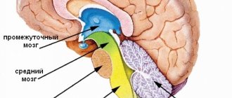
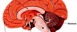
The posterior part of the brain is occupied by the cerebellum. It is located at the base of the occipital and temporal parts above the medulla oblongata and the pons. The main brain and cerebellum are separated by a deep fissure, where a small outgrowth of the telencephalon, called the tentorium, is located.
On the outside, the cerebellum has folds and deep winding grooves. In appearance, it is similar to a head of cabbage: in the middle there is a white stalk, and leaves extend from it.
The volume of the cerebellum is 130–190 g, which is 10% of the total volume of the brain. It contains more than 50% of all neurons. Transverse length – 9-10 cm, front and back – 3-4 cm.
It is a brain center whose main task is to maintain balance and muscle activity, as well as maintain coordination of movements and maintain a certain body position. It controls conditioned reflexes and participates in the functioning of the senses.
The cerebellum, also called the small brain, is located above the pons and the medulla oblongata. Outwardly it looks like other brain structures with characteristic convolutions, depressions, and folds on the surface. The condition of the organ and the presence of disturbances in its functioning can be clearly seen when performing fast movements - playing a musical keyboard instrument, typing using a typewriter or laptop keyboard, running.
The organ controls the consistency, clarity, and measuredness of actions performed by different parts of the body. It does not cause muscle contractions, but determines the sequence of movements and the time frame for their execution. Information about the desired sequence of actions enters the cerebellum from other parts of the brain and parts of the body.
Feedback information allows you to evaluate the consistency of planned and executed movements. The cerebellum adjusts the motor activity program taking into account deviations. Correction occurs by increasing or decreasing the activation of individual muscles. The organ, in cooperation with the cortical structures of the cerebrum, plans a new movement in advance. Irritation of this part of the brain provokes dilation of the pupils and an increase in blood pressure.
Serious damage to the cerebellar fibers leads to malfunctions in the body:
- Astasia. Impaired ability to hold the body in a standing position.
- Atony. Abnormally decreased tone of skeletal muscles and muscle fibers of internal organs.
- Asthenia. A state of powerlessness, general weakness.
- Ataxia. Impaired fine motor functions, lack of coordination of movements.
Autonomic functions are impaired when the structures that make up the cerebellum are damaged. The mechanisms of natural regulation of breathing, blood circulation, and maintaining normal body temperature are slowed down. Damage to all or part of the cerebellum does not provoke the development of paralysis and has virtually no effect on cognitive abilities.
A person experiences difficulties when it comes to learning and remembering new movements. To understand the questions of what the cerebellum is needed for and what it is responsible for, it is worth remembering that the organ is the control center for gross and fine motor skills. It coordinates complex and simple movements on a subconscious level without involving thought processes and involving the work of consciousness.
How to protect your brain and avoid cerebellar damage
Maintaining overall brain health is the best way to avoid damage to the cerebellum.
Reducing the risk of stroke, traumatic brain injury, and exposure to poisons may help prevent some forms of ataxia.
- Quitting smoking: Smoking increases the risk of stroke by thickening the blood and increasing blood pressure.
- Limit alcohol consumption: Large amounts of alcohol can damage the cerebellum. Alcohol also increases blood pressure, which increases the risk of stroke.
- Regular physical activity has a beneficial effect on the functioning of the heart and blood vessels and reduces the risk of stroke.
- Head protection: Wearing seat belts, helmets, and recording safety hazards at home reduces the risk of traumatic brain injury.
- Take measures to prevent falls. Parents should also ensure that children do not have access to balconies or fire escapes.
- Avoid contact with lead: Construction companies no longer use lead, but older homes may have lead pipes and paint. People should keep their homes free of dust that may contain lead and prevent children from playing in the soil.
Regular counseling and management can help limit the physical limitations of genetic ataxia.
J. Keith Fisher, MD This article represents the opinions of our medical experts. All content is strictly informational and should not be construed as medical advice.
Structure of the cerebellum
The structure of the cerebellum is complex. The organ consists of many parts - lobes of the hemispheres, vermis, cortex, nuclei and legs, which perform certain functions and connect the cerebellum with parts of the brain. The anatomy of the cerebellum is represented by two hemispheres, lying on either side of the vermis. The cerebellar cortex is formed from three areas:
- Archicerebellum. Otherwise called the flocculonodular lobe.
- Paleocerebellum. Bilaterally interacts with the receptors of the spinal cord and the sensorimotor area of the cortex covering the cerebral hemispheres.
- Neocerebellum. Bilaterally interacts with cortical neurons that make up the cerebral hemispheres, receptors of the visual and auditory apparatus.
The cortex consists of an inner (granular), outer (molecular) and middle layer formed from Purkinje cells. The layered structure of the organ indicates integration processes that reflect the active interaction of different parts of the brain. The layers of the cerebellum provide its connection with different parts of the central nervous system.
The cerebellum is connected to the parts of the brain through the peduncles. The fiber composition of the peduncles located in the cerebellum includes the nuclei of the vestibular nerve, as well as nerve bundles - the gentle and sphenoid. The lower legs connect the organ with a portion of the medulla oblongata. The middle legs in the form of a spacious, protruding part of the cerebellum are located laterally and pass into the pons.
The structure and functions of the cerebellum cannot be imagined without the vermis, which connects both hemispheres of the organ together. The vermiform structure is composed of white matter nerve fibers. Pathological changes in the tissues of the worm cause ataxia.
Slices
The lobes of the hemisphere located in the cerebellum are delimited by grooves. The worm and hemispheres contain 8 lobules. The anterior, as well as the flocculonodular lobes, located in the cerebellum, are connected by working functions with the brain of the spinal column and the vestibular apparatus. The cerebellar hemispheres receive impulses from muscle and joint tissue receptors. On the front side, a small formation is adjacent to the hemisphere - a shred.
The nuclei located in the cerebellum control the axial and proximal muscles of the limbs. The idea of movement appears in the associative cortical structures of the brain, then is transmitted through afferent pathways towards the elements of the musculoskeletal system. The program of planned movement is formed in the dentate nucleus and simultaneously in the cerebellar hemispheres, then redirected to the motor centers of the cortex using the thalamic nuclei.
Functions
When looking at the cerebellum, functions are also something we need to talk about. Here it is worth clarifying that this organ is not associated with the body’s receptors. It has contact exclusively with the central nervous system. Multiple sensory pathways are directed to it, which carry impulses from muscles, ligaments, tendons, and vestibular nuclei. The cerebellum itself can send impulses to all parts of the central nervous system.
Anatomy of the cerebellum
The outer layer of the worm is gray matter, and the inner layer is white. His job includes controlling the posture of the body, maintaining muscle activity and maintaining a balanced state. Problems in its functioning lead to walking disorders and the inability to stand normally.
Slices
The lobules of this organ are grouped into separate sections of convolutions and separated by large grooves. They continuously cover the hemispheres and the worm. One lobe of the worm is in contact with the lobes of the hemispheres on both sides. Collectively, they are parts of the small brain, divided into several types: upper, posterior and lower.
This body has another division into parts:
- anterior, including the tongue, the central lobule, and the apex;
- back: it includes a slope, a leaf, a tubercle, a sleeve;
- The flocculo-nodular one contains a nodule on the vermis and a zone on the hemisphere.
According to the structure, this organ is divided into three types:
- Old (archicerebellum), including a nodule and a hub on the worm. These parts control the respiratory muscles and the muscles of the groin area. The sleeve is involved in the process of controlling the muscles of the body.
- The ancient one (palerecebellum) includes the uvula, central lobule, apex and clivus. With their help, the head moves, the eyeballs, tongue, pharynx, chewing muscles and facial muscles are well coordinated. The stingray is responsible for the movement of the neck muscles.
- New (neocerebellum), including the leaf, tubercle and pyramid of the vermis. The leaf and tubercle are responsible for the movements of the limbs on both sides. The superior and inferior semilunar lobules control that the limbs above and below do not move synchronously. To control the movements of the arms, the control centers are located in the superior semilunar lobule, and for the legs - in the lower lobule.
Each part of the small brain is responsible for specific motor functions. Malfunctions in their operation are manifested in the following:
- a person is not able to maintain balance with problems in the old cerebellum;
- problems with muscle movements in the neck and torso indicate dysfunction of the ancient cerebellum;
- If problems arise with the muscles of the arms or legs, then there may be problems in the new cerebellum.
Within this organ there are several types of nuclei. Their composition is represented by gray matter. Thanks to their work, signals from the brain also arrive to the body. The following types are distinguished:
- cortical nucleus: located in the deepest part of the organ. With its help, a person can make precise movements. Formed from a wedge-shaped structure of gray matter. Its cells reach the red nuclei of the midbrain and several nuclei of the thalamus, which affect certain parts of the brain. The signal to them comes from nerve impulses of the cerebellum from its intermediate zone;
- dentate nucleus: occupies the lower part of the white matter. Is the largest. Has a wave-like shape. Thanks to its functioning, a person is able to plan and control his actions. With its help, the muscles of the skeleton move, a person feels space and is capable of thinking. Signals are transmitted to it by nerve impulses from the cerebellum and hemisphere, which are located on the sides;
- tent core: its composition is represented by gray matter. Nerve impulses from the cerebellum send commands to it. It includes two zones: rostral and caudal. The rostral has a relationship with the control of the vestibular apparatus, and the caudal is responsible for the movements of the eyeballs.
- globular nucleus: located in the deep zone of the cerebellum. It consists of small and large neurons.
The nuclei are located in the zone of the cortex from where signals come to them. The tent cores are located in the middle. They take information from the worm. Spherical and cork-shaped nuclei are located on the side. The signal comes to them from the side of the intermediate zone. The dentate nucleus is located in the very lateral part. Data comes to it from the left or right hemisphere. Also, the inferior olive of the medulla oblongata provides them with information.
The cerebellum is supplied with blood by several arteries:
- anterior lower: the anterior zone of the lower part of the organ receives blood;
- superior: nourishes the upper region of the organ. In the upper zone it divides into the pia mater, which has a connection with the anterior and posterior inferior arteries.
- posterior inferior: divided into middle and lateral parts as it approaches the inferior artery. The medial branch goes in the opposite direction to the depression in the middle of the hemispheres. The branch located laterally supplies blood to the lower region, where it interacts anteriorly with the inferior and superior arteries.
Problems with cerebellar dysfunction
The small brain contacts only the nervous system. It has connections with pathways that carry signals from muscle tissue, ligaments, and tendons. The organ itself transmits signals to all parts of the central nervous system. It plays a primary role as a comparison mechanism when a decision about any action is made in the motor part of the cortex. It receives information about the likely results of this movement, which is stored there.
To study this organ, scientists conducted experiments on animals. They removed their cerebellum. Scientists described the consequences of this method with several symptoms:
- Astasia: an animal without an organ spreads its legs wide and sways to the sides.
- Atony: impairment of muscle function during flexion and extension.
- Asthenia: inability to control your movements.
- Ataxia: sudden movements.
After some time, the animal's movements become smooth.
Researchers have also found that in the absence of the cerebellum, problems in the functioning of the nervous system are disrupted, blood vessels change, and the functioning of the digestive system changes.
Based on the above, the following tasks of the small brain should be highlighted:
- Make movements coordinated.
- Adjust muscle tone.
- Maintain balance.
Symptoms of disorders of the cerebellum depend on the causes of their occurrence, among which are:
- Defective development from birth.
- Disorders transmitted by inheritance.
- Acquired dysfunctions (alcoholism, vitamin E deficiency, etc.).
- In children, lesions are often caused by brain tumors, which are usually located in the middle part of the cerebellum. Sometimes, in rare cases, a child may acquire a cerebellar disorder after suffering a viral illness.
There are two methods for studying small brain problems:
- Analysis of human gait and movements, study of muscle tone. The gait and shape of a person’s feet are examined based on their footprints: paper is placed on metal coated with paint.
- Using the same research methods that are used to study the main brain: radiography, echoencelography, etc.
Among the symptoms of malfunction of the cerebellum are:
- Impaired coordination of movements.
- Fatigue sets in quickly, and after light physical work the body requires rest.
- Decreased and weak muscle tone.
- No ability to move smoothly. All body movements are sharp. You cannot contract the muscles for a long time.
- A quick change of movements is not possible for a person. Before his shift, he thinks about it.
- Impaired precision of movements.
- Presence of tremors.
- The appearance of pendulum-like reflexes.
- Increased intracranial pressure. Most often, it occurs in connection with tumors and injuries of this organ.
- Speech impairment: words are spoken slowly.
Treatment of cerebellar disorders only partially corrects them and is supportive.
The physiology of the cerebellum is mainly related to motor activity and regulation of the autonomic system. Increased heart rate and breathing during sports activities are triggered by the cerebellum’s reaction to increased physical activity. As a result, blood supply to tissues, their saturation with oxygen and nutrients improves. The organ plays a crucial role in maintaining balance. Functions of the cerebellum include:
- consistency, synchronization of movements;
- maintaining balance and a given posture;
- ensuring the accuracy of targeted movements with timing and taking into account clearly marked coordinates;
- interaction of agonist and antagonist muscles;
- regulation of the activity of the autonomic system.
The organ takes part in memorizing motor information. Its activities are associated with the correct execution of physical techniques, poses and exercises in sports.
The influence of the cerebellum on autonomic functions
The small brain is endowed with the ability to influence the functioning of all body systems. Damage to the cerebellum leads to a decrease in blood pressure and the development of bradypnea. The tone of the respiratory muscles on the affected side will be decreased, while on the opposite side there will be an increase.
The tone of the intestinal muscle fibers changes, it will be reduced, and this causes a disruption in the removal of the contents of the intestines and stomach. There is a suppression of the absorption of nutrients. Metabolic processes accelerate, the level of glucose in the blood increases, which persists for a long period of time compared to the norm. Appetite worsens, the patient loses weight, and the process of tissue regeneration is suppressed.
In drawing a conclusion, I would like to focus on the fact that the cerebellum, despite its small size, plays a very important role in the functioning of the entire organism. Its defeat can cause severe disorders; in the absence of adequate therapy, this is fraught with irreversible consequences.
Symptoms of cerebellar damage
Pathological processes are often accompanied by dysmetria. Limbs cannot move smoothly to a given position due to incorrect force and incorrect direction of movement. Another sign of clinical damage is dysdiadochokinesis, which is expressed in awkward, angular execution of movements that alternate rapidly.
An attempt to reproduce a complex movement leads to fragmentation of the action into separate stages. As a result, the movement is performed intermittently. If the patient tries to touch an object, an intention tremor of the limb occurs. The amplitude of the jitter increases as the distance to the target decreases. Patients with cerebellar dysfunction when walking move their legs too far apart, lose balance and often fall. Words and sentences are spoken at a slow pace, indistinctly. The speech takes on a chanting character.
Hypotonia is a decrease in muscle tone, manifested in combination with a decrease in muscle strength. Muscle resistance to passive movements decreases. Palpation reveals increased muscle softness. A pendulum-shaped knee reflex appears. When the doctor hits the knee tendon below the cap with a special hammer, the leg swings for a while due to weakened muscle tone. Normally, long-term vibrations of the limb do not occur. Other symptoms:
- Dizziness, nausea, headache. The conditions develop due to a breakdown in the functional connection between the vestibular apparatus and the cerebellum.
- Increased fatigue due to physical and mental stress.
- Malfunctions in the functioning of the cardiovascular system.
- Dysfunction of the gland that produces gastric juice.
- Neurological abnormalities.
The location of the cerebellum in the posterior cranial fossa determines the characteristics of symptoms in some diseases. For example, when a tumor forms, the disease in the first stages manifests itself as symptoms of damage to the brain stem or signs of blocking the drainage of cerebrospinal fluid.
Diseases
Diseases that affect the cerebellum - cyst, malignant tumor, malformations and anomalies, abscess, olivopontocerebellar degenerative processes, cerebellar ataxia caused by hereditary factors, known as Pierre Marie's ataxia. Pathologies of the cerebellum cause disorders in the functioning of the musculoskeletal system. Violations affect:
- coordination of movements (dynamic ataxia);
- maintaining balance (static ataxia);
- muscle tone.
The defeat of a specific lobe is reflected in symptoms. For example, damage to the tissue of the flocculo-nodular area leads to atony of the muscles running along the spinal column.
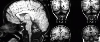
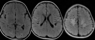
Diagnosis of pathologies
Instrumental diagnostics are carried out using ultrasound, MRI and CT methods. Other research methods are cerebrospinal fluid puncture and vascular Dopplerography. To identify disturbances in the functioning of the organ, vestibulometry is performed - a series of neurological tests:
- Romberg's test. The patient is in a standing position, the feet are moved together, the eyes are closed, the upper limbs are extended forward, then spread to the sides.
- Romberg test with complication. The body position is the same as in the usual Romberg test. The only difference is that the feet are on the same line. In this case, the right foot is located in front of the left foot.
- Walking along a straight line. The test is performed with eyes open, then with eyes closed.
- Unterberger's test. The patient walks in one place with his eyes closed, raising his knees high. The angle of deviation from the initial position after taking 50 steps normally does not exceed 30°.
- Finger test. An attempt to touch your own nose with your index finger with your eyes closed ends in a miss or trembling of the finger.
A blood test will show the presence of specific markers of inflammation or stroke. A lumbar puncture is performed if the neurologist suspects an infectious disease occurring in the cerebrospinal fluid.
The cerebellum is an organ that regulates the functions of the musculoskeletal system. Failures in the functioning of this part of the brain are accompanied by multiple disorders of motor activity. Organ dysfunction impairs quality of life. With significant damage to the cerebellar tissue, a person is unable to hold the body in the desired position, walk smoothly, or make precise movements with the arms and legs.
What is it needed for?
What is the cerebellum of the brain responsible for? First of all, it regulates gait and other actions with stereotypical movements, keeps the body in balance and in the desired position. In addition, this department is necessary for regulating the tone of flexors, extensors, and other antagonist muscles.
The functions of the cerebellum of the human brain include the regulation of speech due to the coordinated control of the muscles of the tongue and lips, fine motor skills (handwriting).
In case of injuries, hemorrhagic and ischemic strokes, inflammatory processes, multiple sclerosis, tumors, the cortex or nerve fibers can be damaged. The pathways are affected, and adequate transmission of nerve impulses to the motor neurons of the spinal cord does not occur.