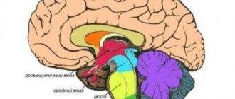
| Cranial nerves | |
| Diagram of the brain, brain stem and cranial nerves | |
| Latin name | nervus cranialis (plural: nervi craniales) |
| Catalogs |
|
Cranial nerves
(lat. nervi craniales) - twelve pairs of nerves extending from the brain stem. They are designated by Roman numerals in the order in which they are located, each of them has its own name.
In Russian-language sources, the term “cranial nerves” is often used. According to the anatomical terminology adopted in Sao Paulo in 1997, the term is designated as lat. nervi craniales - cranial nerves
[1]. In the 6th edition of Sinelnikov’s atlas of human anatomy, monographs devoted to human anatomy, the term is unified under the international anatomical classification. At the same time, the frequency of use of the combination “cranial nerves” is evidenced by the first phrase of the corresponding article in the Great Soviet Encyclopedia:
| cranial nerves, more correctly cranial[2] |
Content
- 1 List of nerves
- 2 Development of cranial nerves in embryogenesis
- 3 Where cranial nerves exit the brain
- 4 Cranial nerve nuclei
- 5 Functions of cranial nerves 5.1 Olfactory nerve
- 5.2 Optic nerve
- 5.3 Oculomotor nerve
- 5.4 Trochlear nerve
- 5.5 Trigeminal nerve
- 5.6 Abducens nerve
- 5.7 Facial nerve
- 5.8 Vestibulocochlear nerve
- 5.9 Glossopharyngeal nerve
- 5.10 Vagus nerve
- 5.11 Accessory nerve
- 5.12 Hypoglossal nerve
Conducting pathways of pain and temperature sensitivity (Fig. 85b).
The cell body of the first neuron is located in the spinal ganglion.
The peripheral process (dendrite) is directed into the skin, where it ends with a receptor. The central process (axon) as part of the dorsal root enters the spinal cord. Some of the fibers enter the gray matter of the dorsal horn, where the nerve impulse switches to the cells of the gelatinous substance (II neuron). The other part of the fibers, without entering the gray matter, is directed to the posterior cord, and, as part of the thin and wedge-shaped fascicles, reaches the nuclei of the same name in the medulla oblongata (II neuron). The processes of the second neurons move to the opposite side, i.e. undergoes a crossover, and as part of the anterior spinothalamic tract (from the spinal cord) and the bulbothalamic tract (from the medulla oblongata) as part of the medial loop they reach the lateral nucleus of the thalamus (III neuron).
https://www.youtube.com/watch?v=7EKgsbu4EhM
The processes of thalamic cells as part of the thalamo-cortical tract reach the cortex, where the IV neuron (cortical end of the analyzer) is located in the postcentral gyrus and superior parietal lobule.
a b
a – pathway of tactile sensitivity: 1 – spinal cord; 2.3 – anterior spinothalamic tract; 4 – lateral nuclei of the thalamus (III neuron); 5 – thalamocortical tract; 6 – nerve cells of the postcentral gyrus (IV neuron); 7 – thalamus; 8 – midbrain; 9 – bridge; 10 – thin and wedge-shaped nucleus (II neuron); 11 – medulla oblongata; 12 – spinal node (I neuron); 13 – gelatinous substance (II neuron).
b – pathway of pain and temperature sensitivity: 1 – spinal cord; 2 – lateral spinothalamic tract; 3 – medial loop; 4 – thalamus; 5 – thalamocortical tract; 6 – nerve cells of the postcentral gyrus (IV neuron); 7 – cerebral cortex; 8 – thalamus; 9 – midbrain; 10 – bridge; 11 – medulla oblongata; 12 – own nucleus (II neuron); 13 – spinal node (I neuron)
I neuron – cells of the spinal ganglion. The peripheral process goes into the skin, the central one enters the spinal cord, where the second neuron is located in the nucleus proper. The processes of these cells move to the opposite side, undergoing decussation, and as part of the lateral spinothalamic tract they join the medial lemniscus and reach the thalamus (III neuron). IV neuron - in the cortex of the postcentral gyrus.
3. Pathways for the sense of stereognosis (recognition of objects by touch).
I neuron – cells of the spinal ganglion. The peripheral process is directed into the skin, the central one enters the spinal cord and, as part of the thin and wedge-shaped fascicles, reaches the nuclei of the same name in the medulla oblongata (II neuron). The processes of these nuclei undergo decussation (decussation of the medial lemniscus) and reach the thalamus (III neuron). IV neuron – in the superior parietal lobule.
Conducting pathways of the motor analyzer
The cell body of the first neuron is located in the spinal ganglion. The peripheral process passes as part of the spinal nerve and ends with a nerve ending in the muscle or joint capsule (proprioceptor). The central process enters the spinal cord and, as part of the thin and wedge-shaped fascicles, reaches the nuclei of the same name in the medulla oblongata (II neuron).
Proprioceptive pathways to the cerebellum
List of nerves
- I pair - olfactory nerve (lat. nervus olfactorius)
- II pair - optic nerve (lat. nervus opticus)
- III pair - oculomotor nerve (lat. nervus oculomotorius)
- IV pair - trochlear nerve (lat. nervus trochlearis)
- V pair - trigeminal nerve (lat. nervus trigeminus)
- VI pair - abducens nerve (lat. nervus abducens)
- VII pair - facial nerve (lat. nervus facialis)
- VIII pair - vestibulocochlear nerve (lat. nervus vestibulocochlearis)
- IX pair - glossopharyngeal nerve (lat. nervus glossopharyngeus)
- X pair - vagus nerve (lat. nervus vagus)
- XI pair - accessory nerve (lat. nervus accessorius)
- XII pair - hypoglossal nerve (lat. nervus hypoglossus)
Development of cranial nerves in embryogenesis
The olfactory and optic nerves develop from protrusions of the anterior medullary bladder and consist of axons of neurons that are located in the mucous membrane of the nasal cavity (the organ of smell) or in the retina of the eye. The remaining sensory nerves are formed by the eviction of young nerve cells from the developing brain, the processes of which form sensory nerves or sensory (afferent) fibers of mixed nerves. Motor cranial nerves are formed from motor (efferent) nerve fibers, which are processes of cells of the motor nuclei located in the brain stem. The formation of cranial nerves in phylogenesis is associated with the development of visceral arches and their derivatives, sensory organs and reduction of somites in the head region.
Cranial nerves - classification
There are 2 grouping options, by starting points and functionality. In the first method, the existing pairs of cranial nerves are classified into 4 types:
- above the trunk zone – I, II;
- from the central department – III, IV;
- from Varoliev Bridge – V-VIII;
- from the oblong section (bulb zone) – IX-XII.
According to functional “responsibilities”, there are the following cranial nerves, a table with clarification below:
- sensory – sensitivity;
- activator – movements;
- mixed.
Where cranial nerves exit the brain
It is impossible to say about the first (olfactory) nerve that it “comes out” of the brain, since it carries only afferent (sensitive) information. The olfactory nerve is the processes of the olfactory cells of the nasal mucosa, collected in olfactory filaments. The olfactory filaments reach the olfactory bulb through the openings of the cribriform plate of the ethmoid bone.
It is also impossible to say about the second (optic) nerve that it “comes out” of the brain, for the same reason. It originates from the optic nerve head, located at the posterior pole of the eye. The optic nerve passes into the cranial cavity through the optic canal formed by the lesser wing of the sphenoid bone. In the cranial cavity, the optic nerves of both eyes form a cross (chiasm), and only part of the fibers cross. Further, the fiber paths are called the “optic tract”.
The third (oculomotor) nerve exits from the side of the trunk next to the interpeduncular fossa (fossa interpeduncularis).
The IV (trochlear) nerve is the only one emerging from the dorsal (“dorsal”) side of the trunk, from the upper edge of the rhomboid fossa, bending and emerging to the ventral side from under the cerebral peduncles.
The V (trigeminal) nerve exits the ventral side of the pons.
Nerves VI to VIII also exit on the ventral side of the brain stem between the medulla oblongata and the pons, from the edges to the center in a row, with VII and VIII lying close to each other at the “corner” of the medulla oblongata, and VI (abducens) at the level of the anterolateral sulcus .
Nerves IX to XII emerge from the medulla oblongata on the ventral side. The XI (accessory) nerve is somewhat special - it combines, in addition to the head part, some roots of the spinal cord. Nerves IX to XI emerge from the lateral surface of the medulla oblongata, from bottom to top in a row.
The XII (hypoglossal) nerve emerges from the anterolateral groove (lat. sulcus ventrolateralis).
Types of nerves and pathologies
There are several types of nerves:
- motor;
- mixed;
- sensitive.
The neurology of motor cranial nerves, both sensory and mixed, has pronounced manifestations that specialists can easily diagnose. In addition to isolated damage to individual nerves, those that simultaneously belong to different groups can also be affected. Thanks to knowledge of their location and functions, it is possible not only to understand which nerve is damaged, but also to localize the affected area.
https://www.youtube.com/watch?v=7EKgsbu4EhM
Carotid and verbal angiography reveal good values. But more detailed information can be obtained using computed tomography. With it you can see individual nerve trunks and identify tumors and other changes in the auditory, optic and other nerves.
It became possible to study the trigeminal and auditory nerves thanks to the method of cortical somatosensory potentials. Also in this case, audiography and nystagmography are used.
The development of electromyography has expanded the ability to obtain more detailed information about cranial nerves. Now you can study, for example, the reflex blink response, spontaneous muscle activity during facial expressions and chewing, muscles of the tongue, palate, and so on.
Let us dwell in more detail on each of the pairs of these nerves. There are a total of 12 pairs of cranial nerves. A table containing all of them is indicated at the end of the article. For now, let's look at each of the pairs separately.
1. Olfactory nerve - has no nuclei, olfactory cells are located in the mucous membrane of the olfactory region of the nasal cavity. Contains visceral sensory fibers.
The exit from the brain is from the olfactory bulb.
The exit from the skull is from the cribriform plate of the ethmoid bone.
The nerve is a collection of 15-20 thin nerve filaments, which are the central processes of the olfactory cells. They pass through openings in the ethmoid bone and then end in the olfactory bulb, which continues into the olfactory tract and triangle.
The optic nerve has no nuclei; ganglion neurocytes are located in the retina of the eyeball. Contains somatic sensory fibers.
Exit from the brain - optic chiasm at the base of the brain
Exit from the skull - optic canal
Moving away from the posterior pole of the eyeball, the nerve leaves the orbit through the optic canal and, entering the cranial cavity along with the same nerve on the other side, forms the optic chiasm, lying in the optic groove of the sphenoid bone. The continuation of the visual pathway beyond the chiasm is the optic tract, ending in the lateral geniculate body and in the superior colliculus of the roof of the midbrain.
3. Oculomotor nerve - has 2 nuclei: autonomic and motor, located in the tegmentum of the midbrain (at the level of the superior colliculi). Contains efferent (motor) fibers to most of the external muscles of the eyeball and parasympathetic fibers to the internal eye muscles (ciliary muscles and muscles that constrict the pupil).
- The exit from the brain is from the medial sulcus of the cerebral peduncle/interpeduncular fossa/from the oculomotor sulcus.
- The oculomotor nerve leaves the brain along the medial edge of the cerebral peduncle, then goes to the superior orbital fissure, through which it enters the orbit.
- Entering the orbit it divides into 2 branches:
- A) The superior branch - to the superior rectus muscle of the eyeball and to the muscle that lifts the upper eyelid.
B) Inferior branch - to the inferior and medial rectus muscles of the eyeball and the inferior oblique muscle of the eyeball. From the lower branch, a nerve root extends to the ciliary ganglion, carrying parasympathetic fibers to the ciliary muscle and the muscle that constricts the pupil.
4. Trochlear nerve – has 1 motor nucleus, located in the tegmentum of the midbrain (at the level of the inferior colliculi). Contains only efferent (motor) fibers.
- The exit from the brain is from under the lower colliculi/on the sides of the frenulum of the superior medullary velum.
- The exit from the skull is the superior orbital fissure.
- Leaving the brain, it bends laterally around the cerebral peduncle and enters the orbit through the superior orbital fissure, where it innervates the superior oblique muscle of the eyeball.
5. Trigeminal nerve - has 4 nuclei: 3 sensory and 1 motor nucleus. Located in the tegmentum of the midbrain, tegmentum of the pons, tegmentum of the medulla oblongata. Contains afferent (sensory) fibers and efferent (motor) fibers.
- The exit from the brain is at the pons and middle cerebellar peduncle.
- The exit from the skull is the ophthalmic nerve - the superior orbital fissure, the maxillary nerve - the round foramen, the mandibular nerve - the foramen ovale.
- Branches of the trigeminal nerve:
- 1. The ophthalmic nerve enters the orbital cavity through the superior orbital fissure, but before entering it it divides into 3 more branches:
- a) The frontal nerve, runs directly anteriorly under the roof of the orbit through the supraorbital notch (or foramen) into the skin of the forehead, here it is called the supraorbital nerve, giving branches along the way to the skin of the upper eyelid and medial corner of the eye.
b) The lacrimal nerve goes to the lacrimal gland and, after passing through it, ends in the skin and conjunctiva of the lateral corner of the eye. Before entering the lacrimal gland, it connects with the zygomatic nerve (from the second branch of the trigeminal nerve). Through this anastomosis, the lacrimal nerve receives secretory fibers for the lacrimal gland and also supplies it with sensory fibers.
c) Nasociliary nerve, innervates the anterior part of the nasal cavity (anterior and posterior ethmoidal nerves), the eyeball (long ciliary nerves), the skin of the medial corner of the eye, the conjunctiva and the lacrimal sac (subtrochlear nerve).
Cranial nerve nuclei
| Character of the core | Nerve | Character of innervation | Innervation of the organ | Function |
| The magnocellular nucleus of the oculomotor nerve (paired) | III pair | motor | the levator superioris muscle, the superior, inferior and medial rectus muscles of the eye, the inferior oblique muscle of the eye | eye movement, raising the upper eyelid |
| Small cell nucleus of the oculomotor nerve (syn. Yakubovich nucleus), paired | III pair | parasympathetic | muscle constrictor pupil (lat. m.sphincter pupillae), ciliary muscle (lat. m.ciliaris) | constriction of the pupil and accommodation of the eye |
| Small cell unpaired nucleus of the oculomotor nerve (syn. nucleus of Perlia) | III pair | motor, convergence | medial rectus oculi muscle | simultaneous approach to the median plane (convergence) |
| Trochlear nerve nucleus | IV pair | motor | superior oblique muscle (lat. m. obliquus superior) | moving the eye to the side and down |
| The nucleus of the spinal tract of the trigeminal nerve (lat. nucleus tractus spinalis n.trigemini) | V pair | sensitive | face | superficial (pain and tactile) sensitivity |
| Core of deep sensitivity of the trigeminal nerve (lat. nucleus sensorius principalis n.trigemini) | V pair | sensitive | face | deep (proprioceptive) sensitivity |
| Motor nucleus of the trigeminal nerve (lat. nucleus motorius (masticatorius) n.trigemini) | V pair | motor | masticatory muscles (masseter, temporalis, lateral and medial pterygoid, mylohyoid muscles, anterior belly of the digastric muscle and muscle that tightens the soft palate) | chewing |
| The nucleus of the abducens nerve (lat. nucleus n.abducentis) | VI pair | motor | lateral rectus muscle (lat. m.rectus lateralis) | abduction of the eyeball to the side |
| The nucleus of the facial nerve (lat. nucleus n.facialis) | VII pair | motor | facial muscles | facial expressions |
| Core of the solitary tract (lat. nucleus tractus solitarius) | VII,IX,X pairs | sensitive | tongue (taste buds) | taste |
| Superior salivatory nucleus (lat. nucleus salivatorius superior) | VII pair | parasympathetic | lacrimal gland, submandibular and sublingual salivary glands | tearing, salivation |
| Anterior and posterior cochlear nuclei (lat. nuclei cochleares anterior et posterior) | VIII pair | sensitive | inner ear (auditory receptors) | hearing |
| Vestibular nuclei (superior, lateral, medial and inferior) (lat. nuclei vestibulares) | VIII pair | sensitive | inner ear (vestibular receptors) | vestibular apparatus |
| Double nucleus (lat. nucleus ambiguus) | IX, X and XI pairs | motor | muscles of the soft palate, pharynx and larynx | chewing, voice, articulation |
| Lower salivatory nucleus (lat. nucleus salivatorius inferior) | IX pair | parasympathetic | parotid gland | salivation |
| Sensitive nucleus of the glossopharyngeal and vagus nerves (lat. nucleus alae cinereae) | IX and X pairs | sensitive | oral cavity, middle and inner ear | overall sensitivity of these areas |
| Posterior nucleus of the vagus nerve (lat. nucleus dorsalis n.vagi) | X pair | parasympathetic | heart muscle, smooth muscles of the lungs, bronchi, stomach and intestines | heart rate, secretion of endocrine glands of the gastrointestinal tract, bronchial smooth muscle tone |
| Accessory nerve nucleus (lat. nucleus n.accessorii) | XI pair | motor | trapezoid and sternocleidomastoid (lat. m.sternocleidomastoideus) | turning the head, lifting the shoulder, scapula and acromial part of the clavicle upward (“shrug”), pulling the shoulder girdle back and bringing the scapula to the spine |
| The nucleus of the hypoglossal nerve (lat. nucleus n.hypoglossi) | XII pair | motor | muscles of the tongue and orbicularis oris | tongue movement, swallowing. |
Damage to cranial nerves
Impaired functionality of individual couples is diagnosed based on specific symptoms. Motor cranial nerves with lesions cease to carry out the corresponding tasks, as well as mixed ones, which is manifested by paralysis and inflammatory processes. A change in the conductivity of the remaining pairs affects sensitivity, and sensory perception worsens. Often, damage to the cranial nerves occurs in a complex manner, especially if a person receives head injuries in the area of the trunk and jugular fossa.
Possible signs of damage to different couples:
- deterioration of hearing, smell, vision, taste and tactile sensitivity;
- drooping eyelid;
- severe pain in the face area;
- paresis and paralysis;
- strabismus;
- problems with swallowing and breathing;
- dizziness;
- loss of the ability to navigate in space and maintain balance;
- deterioration of the cardiovascular system;
- atrophy of the muscles of the neck and shoulder girdle;
- speech disorders;
- digestive pathologies and others.
Functions of cranial nerves
Olfactory nerve
Main article: Olfactory nerve
Olfactory nerve
(
olfactory nerves
) (lat. nervi olfactorii) - the first of the cranial nerves, responsible for olfactory sensitivity.
Optic nerve
Main article: Optic nerve
Optic nerve
(lat. Nervus opticus) - the second pair of cranial nerves, through which visual stimuli perceived by the sensitive cells of the retina are transmitted to the brain.
Oculomotor nerve
Main article: Oculomotor nerve
Oculomotor nerve
(lat. nervus oculomotorius) - III pair of cranial nerves, responsible for the movement of the eyeball, raising the eyelid, and the reaction of the pupils to light.
Trochlear nerve
Main article: Trochlear nerve
Trochlear nerve
(lat. nervus trochlearis) - IV pair of cranial nerves, which innervates the superior oblique muscle (lat. m. obliquus superior), which turns the eyeball outward and downward.
Trigeminal nerve
Main article: Trigeminal nerve
V (trigeminal) nerve
(lat. nervus trigeminus) is mixed. Its three branches (ramus ophthalmicus - V1, ramus maxillaris - V2, ramus mandibularis - V3) through the Gasserian ganglion (ganglion trigeminale) carry information from the upper, middle and lower thirds of the face, respectively. Each branch carries information from the muscles, skin and pain receptors of each third of the face. In the Gaserian node, information is sorted by type, and information from the muscles of the entire face goes to the sensitive nucleus of the trigeminal nerve, located mostly in the midbrain (partially enters the pons); cutaneous information from the entire face goes to the “main nucleus” (nucleus pontinus nervi trigemini), located in the pons; and pain sensitivity is in the nucleus spinalis nervi trigemini, coming from the bridge through the medulla oblongata to the spinal cord.
The trigeminal nerve also belongs to the motor nucleus (lat. nucleus motorius nervi trigemini), which lies in the bridge and is responsible for the innervation of the masticatory muscles.
Abducens nerve
Main article: Abducens nerve
Abducens nerve
(lat. nervus abducens) - VI pair of cranial nerves, which innervates the lateral rectus muscle (lat. m. rectus lateralis) and is responsible for abduction of the eyeball.
Facial nerve
Main article: Facial nerve
Facial nerve
(lat. nervus facialis), the seventh (VII) of the twelve cranial nerves, exits the brain between the pons and the medulla oblongata. The facial nerve innervates the facial muscles. Also included in the facial nerve is the intermediate nerve, which is responsible for the innervation of the lacrimal gland, the stapedius muscle and the taste sensitivity of the two anterior thirds of the tongue.
vestibulocochlear nerve
Main article: Vestibulocochlear nerve
vestibulocochlear nerve
(lat. nervus vestibulocochlearis) is a nerve of special sensitivity responsible for the transmission of auditory impulses and impulses emanating from the vestibular part of the inner ear.
Glossopharyngeal nerve
Main article: Glossopharyngeal nerve
Glossopharyngeal nerve
(lat. nervus glossopharyngeus) - IX pair of cranial nerves. Is mixed. Provides:
- motor innervation of the stylopharyngeus muscle (lat. m. stylopharyngeus), levator pharynx
- innervation of the parotid gland (lat. glandula parotidea), providing its secretory function
- general sensitivity of the pharynx, tonsils, soft palate, Eustachian tube, tympanic cavity
- taste sensitivity of the posterior third of the tongue.
Nervus vagus
Main article: Vagus nerve
Nervus vagus
(lat. nervus vagus) - X pair of cranial nerves. Is mixed. Provides:
- motor innervation of the muscles of the soft palate, pharynx, larynx, as well as striated muscles of the esophagus
- parasympathetic innervation of the smooth muscles of the lungs, esophagus, stomach and intestines (up to the splenic flexure of the colon), as well as the muscles of the heart. Also affects the secretion of the glands of the stomach and pancreas
- sensitive innervation of the mucous membrane of the lower part of the pharynx and larynx, the skin behind the ear and part of the external auditory canal, the eardrum and the dura mater of the posterior cranial fossa.
The dorsal nucleus of the vagus nerve, nucleus dorsalis nervi vagi, is located in the medulla oblongata lateral to the nucleus of the hypoglossal nerve.
Accessory nerve
Main article: Accessory nerve
Accessory nerve
(lat. nervus accessorius) - XI pair of cranial nerves. Contains motor nerve fibers that innervate the muscles responsible for turning the head, raising the shoulder and adducting the scapula to the spine.
Hypoglossal nerve
Main article: Hypoglossal nerve
Hypoglossal nerve
(lat. nervus hypoglossus) - XII pair of cranial nerves. Responsible for the movement of the tongue.
Cranial nerves - innervation
Each pair has branches consisting of fibers that connect certain organs and muscles to the nuclei. The cranial nerves innervate not only structures located in the head, but also more distant formations. Some reach the abdominal muscles and even the intestines. Below is how cranial nerves are innervated; the table reflects all organic relationships. Additionally, the nuclei responsible for neural transport of impulses are indicated.
History of nerve names
| Nerve | Etymology of the name | First named | The scientist who gave the name | Reason for the name |
| Terminal nerve (lat. nervus terminalis) | from lat. terminalis - extreme | 1905 | Albert William Losey | The nerve was first called the accessory olfactory nerve, but due to the lack of understanding of its functions, its name was changed to terminal, due to its proximity to the terminal plate of the brain |
| Olfactory nerve (lat. nervus olfactorius) | classical lat. olfacere - I sniff, postclassical olfactorius (two suffixes -tor (a suffix to form a noun from a specific verb) and -ius (indicates belonging to a function)) | 1651 | Thomas Bartolin | The nerve got its name due to its connection with the function of smell. |
| Optic nerve (lat. nervus opticus) | from ancient Greek ὀπτικός (optikos) | not known for sure; Galen points out that some of his contemporaries called the optic nerve | ???? | The nerve is so named because it is involved in the function of vision. |
| Oculomotor nerve (lat. nervus oculomotorius) | post-classical Latin word, combined from two Latin words: oculus - eye and motore - I move; two suffixes have also been added: -tor and -ius | 1783 | Johann Pfeffinger | Named for its function (innervates the muscles of the eyeball and thus moves them) |
| Trochlear nerve (lat. nervus trochlearis) | from lat. trochlea - block | 1670 | William Molins | The nerve is so named because it innervates the superior oblique muscle, the tendon of which makes a bend that resembles a pulley. |
| Trigeminal nerve (lat. nervus trigeminus) | from lat. trigeminus - trigeminal | 1732 | Jacob Winslov | It got its name because of its shape: the main trunk, which emerges from the cerebellopontine angle, is divided into three massive branches |
| Abducens nerve (lat. nervus abducens) | from lat. abducere - to take away, with the addition of the suffix -ens , characteristic of imperfect participles | 1778 | Samuel Thomas Soemmering | The nerve got its name because of the function it provides, namely, to move the eye outward |
| Facial nerve (lat. nervus facialis) | from lat. faciei - face; postclassical facialis - that which relates to the face | 1778 | Samuel Thomas Soemmering | The nerve received its name through the innervation of the facial muscles |
| Intermediate nerve (lat. nervus intermedius) part of the facial nerve | from lat. intermedius - intermediate | 1778 | Heinrich August Wriesberg | Due to the close proximity of the facial and vestibulocochlear nerves, they were long considered one nerve; in this case, the intermediate nerve was considered as a connecting branch between them, that is, an intermediate |
| vestibular-cochlear nerve (lat. nervus vestibulocochlearis) | from lat. vestibulum - vestibule; from lat. cochlea - curl, twist and suffix -aris | 1961 | Collegium when viewing PNA | The name comes from the two anatomical structures with which the nerve connects in the inner ear |
| Glossopharyngeal nerve (lat. nervus glossopharyngeus) | from ancient Greek γλῶσσα (glossa) - language and from ancient Greek. φάρυγξ (pharynx) - pharynx, throat | 1753 | Albrecht von Haller | The name comes from the fact that anatomists who examined the nerve described that it is woven into the pharynx and the root of the tongue |
| Vagus nerve (lat. nervus vagus) | from lat. vagus - wandering, vagrant, wandering | 1651 | Thomas Bartolin | The nerve got its name because of its length and extensive branching in the human body. |
| Accessory nerve (lat. nervus accessorius) | from the post-classical Latin word accesorius - additional | 1666 | Thomas Willis | Due to its close location to the vagus nerve and branches to it, it was considered as an “addendum” to the modern X pair |
| Hypoglossal nerve (lat. nervus hypoglossus) | from ancient Greek γλῶσσα (glossa) - language and with the addition of the prefix hypo- - sub- | 1732 | Jacob Winslov | Characterizes the relationship to the function of the tongue and anatomical placement |
| Notes:[3][4] | ||||
ChMN functionality
I – olfactory nerve. Consists of receptors, which are thin processes that thicken towards the end. There are special hairs on the ends of the processes that capture odors. II is the visual nerve. It runs through the entire eye, ending in the vision canal. At the exit from it, the nerves cross, after which they continue their movement to the central part of the brain.
The visual nerve delivers signals received from the outside world to the necessary sections of the brain. VIII - vestibulocochlear nerve. Belongs to the sensory type. Consists of 2 components, different in functionality. The first conducts impulses emanating from the vestibule of the inner ear, and the second transmits hearing impulses that emanate from the cochlea. In addition, the vestibular component is involved in regulating the position of the body, arms, legs and head and, in general, coordinates movements.
Motor group
III – oculomotor nerve.
These are processes of nuclei. Runs from the midbrain to the orbit. Its function is to engage the muscles of the eyelashes, which carry out accommodation, and the muscle that constricts the pupil.
IV - trochlear nerve.
It is of the motor type, located in the orbit, entering there through a gap from above (on the side of the previous nerve). It ends at the eyeball, or more precisely its superior muscle, which it supplies with nerve cells.
VI – abducens nerve.
Like the block one, it is motor. It is formed by processes. It is located in the eye, where it penetrates from above, and provides nerve cells to the external eye muscle.
XI – accessory nerve.
Representative of the motor type. Dual-core. The nuclei are located in the spinal cord and medulla oblongata.
XII – hypoglossal nerve.
Type - motor. Nucleus in the medulla oblongata. Provides nerve cells to the muscles and muscles of the tongue and some parts of the neck.
Mixed group
V – trigeminal.
Leader in thickness. It got its name because it has several branches: ophthalmic, mandibular and maxillary.
VII – facial nerve.
It has a front and an intermediate component. The facial nerve forms 3 branches and provides normal movement of the facial muscles.
IX – glossopharyngeal nerve.
Belongs to the mixed type. Consists of three types of fibers.
X – vagus nerve.
Another representative of the mixed type. Its length exceeds that of the others. Consists of three types of fibers. One branch is the depressor nerve, ending in the aortic arch, regulating blood pressure. The remaining branches, which have a higher susceptibility, provide nerve cells to the membrane of the brain and the skin of the ears.
The vagus nerve can be divided (conditionally) into 4 parts: the head region, the neck region, the chest region and the abdominal region. The branches extending from the head go to the brain and are called meningeal. And those that suit the ears are ear-friendly. The pharyngeal branches come from the neck, and the cardiac branches and thoracic branches depart from the chest, respectively. The branches directed to the plexus of the esophagus are called esophageal.
Notes
- Terminologia anatomica: international anatomical terminology (English). Federative Committee on Anatomical Terminology (1997). Retrieved November 15, 2010.
- cranial nerves. Great Soviet Encyclopedia. Retrieved November 15, 2010. Archived February 29, 2012.
- Simon F, Marečková-Štolcová E, Páč L. (2011). "On the terminology of cranial nerves". Annals of Anatomy 193
(5): 447– 452. DOI:10.1016/j.aanat.2011.04.012. PMID 21724380. (English) - Matthew C. Davis, Christoph J. Griessenauer (2014). "The naming of the cranial nerves: A historical review". Clinical Anatomy 27
(1): 14-19. DOI:10.1002/ca.22345. PMID 24323823. (English)