Parts of the brain
During intrauterine development, a complex brain was formed from an ordinary neural tube. This happened due to the bulging of five brain vesicles, which gave rise to the corresponding parts of the brain:
- telencephalon, or forebrain, from which the cerebral cortex, basal ganglia, and anterior part of the hypothalamus were formed;
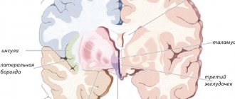
- diencephalon, or diencephalon, which gave rise to the thalamus, epithalamus, and posterior part of the hypothalamus;
- mesencephalon, or midbrain, from which the quadrigeminal peduncle and cerebral peduncles subsequently formed;
- the metencephalon, or hindbrain, which gave rise to the cerebellum and pons;
- myelencephalon, or medulla oblongata.
Later in the article we will talk in more detail about the telencephalon, or forebrain. After all, the relief of the cerebral cortex belongs specifically to this part of the central nervous system.
Structure of the cortex
Thanks to the presence of the cortex, a person is able to experience emotions, navigate himself and the surrounding space. What is noteworthy is that the structure of the bark is unique. The grooves and convolutions of the cerebral cortex of one person have a different shape and size than that of another. But the general plan of the building is the same.
What is the difference between the sulci and convolutions of the brain? Fissures are depressions in the cerebral cortex that look like slits. They are the ones who divide the bark into shares. There are four lobes of the cerebral hemispheres:
- frontal;
- parietal;
- temporal;
- occipital
Gyri are convex areas of the cortex that are located between the furrows.
Embryology
First at 3 months. During embryonic development, the lateral (Sylvian) fossa appears. Its bottom is formed by a slowly growing bark, which later forms an island. Rapidly growing neighboring areas of the cortex cover it and form folds—tires. The line of their contact forms the lateral (Sylvian) fissure. At 5-6 months.
In the cerebral hemispheres, the superficial (cortical) mantle part [cerebral mantle (pallium)] is distinguished with the grooves and convolutions located on it. Based on phylogenetic development, the brain cloak is divided into ancient (paleopallium), old (archipallium) and new (neopallium). So-called
Formation of the cortex in embryogenesis
Embryogenesis is the intrauterine development of the fetus from conception to birth. First, uneven depressions form on the cerebral cortex, which give rise to furrows. The primary grooves are formed first. This occurs around the 10th week of intrauterine development. After this, secondary and tertiary depressions are formed.
The deepest groove is the lateral one; it is one of the first to form. It is followed in depth by the central one, which separates the motor (motor) and sensory (sensitive) zones of the cerebral cortex.
Most of the cortical relief develops from 24 to 38 weeks of gestation, and some of it continues to develop after the baby is born.
Fissures and convolutions: formation and functions
The structure of the human brain distinguishes it from other mammals, and for this reason may explain its unique mental abilities compared to other animals. The number of folds in the cortex may correlate with some specific cognitive, sensory, and motor abilities. Although there is no clear explanation of how the unique division of the human brain into sulci and convolutions occurs.
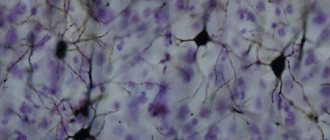
Today there is progress in understanding the extremely complex processes in the brain, the cortex of which is built with so many grooves and convolutions. Even though all cells have the same DNA, different neural stem cells are formed. It is their work with various properties that creates the basic structure of the brain, consisting of neurons and glial cells.
Brain growth occurs through two types of stem cells—neural stem cells and neural progenitors. Both of these forms form neurons, which become permanent in the brain, as well as intermediate cells that create the building material for building the brain. Four different types of stem cells determine the structure of the cortex.
During early embryonic development, expansion of the rostral domain of the neural tube leads to the appearance of two telencephalic vesicles. The dorsal half of these vesicles is molecularly defined as the primordium of the cerebral cortex. At this stage, the cortical primordium consists exclusively of a monolayer of neuroepithelial progenitor cells.
- basal-directed movement during G1 phase;
- basal position during S phase;
- apically directed movement during G2 phase;
- mitosis on the apical surface.
The cycling movement is known as interkinetic nuclear migration and is completely asynchronous between neuroepithelial cells, giving the neuroepithelium a pseudostratified appearance. Cells undergo only symmetrical self-aggressive divisions, with each division generating two daughter cells, hence exponentially increasing their number.
Because they are the fundamental progenitor cells of the cerebral cortex, the size of their association determines the number of derived neurogenic progenitor cells and the final number of cortical neurons, and therefore it has a fundamental influence on the size of the mature cerebral cortex. An increase in quantity leads to an expansion of surface area and the formation of neuroepithelium.
Immediately before the onset of neurogenesis, neuroepithelial progenitor cells begin to lose tight junctions and acquire features typical of glial cells (including expression of brain lipid-binding protein, vimentin, and Pax6), thereby becoming apical radial glial cells (ARGCs).
However, they gradually begin to divide asymmetrically to generate one similar cell plus another cell. These new cells accumulate in the basal part of the cortical primordium, while the cell bodies of the ARGC remain on the apical side, forming the ventricular zone (VZ). With the accumulation of cells above the GC, the ARGK process is prolonged, remaining attached to the basal plate, and is now called radial glia.
Asymmetric ARGK divisions generate one ARGK plus one neuron or one intermediate progenitor cell. Intermediate progenitor cells (secondary progenitor cells without apical-basal polarity) do not undergo interkinetic nuclear migration, divide in a layer located in the ventricular zone, the subventricular zone (SVZ), and all express a transcription factor (Tbr2).
However, due to the fact that each neuron itself consumes during mitosis, their relative quantity compared to ARGK is quite low. Intermediate progenitor cells in the cerebral cortex generate the majority of cortical excitatory neurons. As neurogenesis progresses, the need for ARGK expansion/renewal decreases and the production of neurons increases.
In addition to the expanded ventricular zone, the subventricular zone thickens, populated in abundance by basal precursors, especially in the later stages of neurogenesis. As a result, the SVZ splits into internal and external parts. The outer portion contains a wide variety of progenitor cell types with high developmental potential, which is a key factor for the expansion and formation of the cortex.
The brain is the most advanced, and therefore one of the most difficult to study, part of the human body. And its most highly organized component is the cerebral cortex. More details about the anatomy of this formation, the structure of the grooves and convolutions of the brain later in the article.
During intrauterine development, a complex brain was formed from an ordinary neural tube. This happened due to the bulging of five brain vesicles, which gave rise to the corresponding parts of the brain:
- telencephalon, or forebrain, from which the cerebral cortex, basal ganglia, and anterior part of the hypothalamus were formed;
- diencephalon, or diencephalon, which gave rise to the thalamus, epithalamus, and posterior part of the hypothalamus;
- mesencephalon, or midbrain, from which the quadrigeminal peduncle and cerebral peduncles subsequently formed;
- the metencephalon, or hindbrain, which gave rise to the cerebellum and pons;
- myelencephalon, or medulla oblongata.
Later in the article we will talk in more detail about the telencephalon, or forebrain. After all, the relief of the cerebral cortex belongs specifically to this part of the central nervous system.
Structure of the cortex
Thanks to the presence of the cortex, a person is able to experience emotions, navigate himself and the surrounding space. What is noteworthy is that the structure of the bark is unique. The grooves and convolutions of the cerebral cortex of one person have a different shape and size than that of another. But the general plan of the building is the same.
What is the difference between the sulci and convolutions of the brain? Fissures are depressions in the cerebral cortex that look like slits. They are the ones who divide the bark into shares. There are four lobes of the cerebral hemispheres:
- frontal;
- parietal;
- temporal;
- occipital
Gyri are convex areas of the cortex that are located between the furrows.
Embryogenesis is the intrauterine development of the fetus from conception to birth. First, uneven depressions form on the cerebral cortex, which give rise to furrows. The primary grooves are formed first. This occurs around the 10th week of intrauterine development. After this, secondary and tertiary depressions are formed.
The deepest groove is the lateral one; it is one of the first to form. It is followed in depth by the central one, which separates the motor (motor) and sensory (sensitive) zones of the cerebral cortex.
Most of the cortical relief develops from 24 to 38 weeks of gestation, and some of it continues to develop after the baby is born.
The grooves are classified according to the function they perform. The following types are distinguished:
- primary formed - the deepest in the brain, they divide the cortex into separate lobes;
- secondary - more superficial, they perform the function of forming convolutions of the cerebral cortex;
- additional, or tertiary - the most superficial of all types, their function is to provide an individual relief of the bark, increasing its surface.
- Although the shape and size of some of the sulci and convolutions of the cerebral hemispheres differ from individual to individual, their number is normally unchanged. Every person, regardless of age and gender, has the following grooves:
- Sylvian fissure - separates the frontal lobe from the temporal lobe;
- lateral sulcus - separates the temporal, parietal and frontal lobes, and is also one of the deepest in the brain;
- Roland's fissure - separates the frontal lobe of the brain from the parietal lobe;
- parieto-occipital sulcus - separates the occipital region from the parietal;
- cingulate sulcus - located on the medial surface of the brain;
- circular - is the boundary for the insular part on the basal surface of the cerebral hemispheres;
- The hippocampal sulcus is a continuation of the cingulate sulcus.
Main convolutions
The relief of the cerebral cortex is very complex. It consists of numerous convolutions of different shapes and sizes. But we can highlight the most important of them, which perform the most important functions. The main convolutions of the brain are presented below:
- angular gyrus - located in the parietal lobe, is involved in recognizing objects through vision and hearing;
- Broca's center - the posterior part of the inferior frontal gyrus on the left (in right-handers) or on the right (in left-handers), which is necessary for correct speech reproduction;
- Wernicke's center - located in the posterior part of the superior temporal gyrus on the left or right (similar to Broca's area), is involved in the understanding of oral and written speech;
- cingulate gyrus - located on the medial part of the brain, takes part in the formation of emotions;
- hippocampal gyrus - located in the temporal region of the brain, on its inner surface, necessary for normal memorization;
- fusiform gyrus - located in the temporal and occipital regions of the cerebral cortex, is involved in face recognition;
- lingual gyrus - located in the occipital lobe, plays an important role in processing information coming from the retina;
- precentral gyrus - located in the frontal lobe in front of the central sulcus, necessary for processing sensitive information entering the brain;
- postcentral gyrus - located in the parietal lobe behind the central sulcus, necessary for voluntary movements.
The anatomy of the cerebral convolutions and sulci is best studied in sections. Let's start with the outer surface. It is on the outer surface of the brain that the deepest groove is located - the lateral one.
- The frontal lobe is separated from the parietal lobe by a deep central groove, sulcus centralis.
- It begins on the medial surface of the hemisphere, passes to its superolateral surface, runs along it slightly obliquely, from back to front, and usually does not reach the lateral sulcus of the brain.
Main grooves
Although the shape and size of some of the sulci and convolutions of the cerebral hemispheres differ from individual to individual, their number is normally unchanged. Every person, regardless of age and gender, has the following grooves:
- Sylvian fissure - separates the frontal lobe from the temporal lobe;
- lateral sulcus - separates the temporal, parietal and frontal lobes, and is also one of the deepest in the brain;
- Roland's fissure - separates the frontal lobe of the brain from the parietal lobe;
- parieto-occipital sulcus - separates the occipital region from the parietal;
- cingulate sulcus - located on the medial surface of the brain;
- circular - is the boundary for the insular part on the basal surface of the cerebral hemispheres;
- The hippocampal sulcus is a continuation of the cingulate sulcus.
Additional images[edit | edit code]
- Position of the central sulcus (marked in red)
- Position of brain structures in the head. The central sulcus separates the parietal lobe (in yellow) from the frontal lobe (in blue)
- Lateral surface of the left hemisphere. The central sulcus is marked with the number 6
- Lateral surface of the right hemisphere. Central sulcus - in the center, marked in red
- Medial surface of the hemispheres
- Play media file
Video with dissection of the telencephalon, demonstrating the central sulcus of the left hemisphere
Main convolutions
The relief of the cerebral cortex is very complex. It consists of numerous convolutions of different shapes and sizes. But we can highlight the most important of them, which perform the most important functions. The main convolutions of the brain are presented below:
- angular gyrus - located in the parietal lobe, is involved in recognizing objects through vision and hearing;
- Broca's center - the posterior part of the inferior frontal gyrus on the left (in right-handers) or on the right (in left-handers), which is necessary for correct speech reproduction;
- Wernicke's center - located in the posterior part of the superior temporal gyrus on the left or right (similar to Broca's area), is involved in the understanding of oral and written speech;
- cingulate gyrus - located on the medial part of the brain, takes part in the formation of emotions;
- hippocampal gyrus - located in the temporal region of the brain, on its inner surface, necessary for normal memorization;
- fusiform gyrus - located in the temporal and occipital regions of the cerebral cortex, is involved in face recognition;
- lingual gyrus - located in the occipital lobe, plays an important role in processing information coming from the retina;
- precentral gyrus - located in the frontal lobe in front of the central sulcus, necessary for processing sensitive information entering the brain;
- postcentral gyrus - located in the parietal lobe behind the central sulcus, necessary for voluntary movements.
Anatomical structure of the lobes of the brain
In the structure of any organ of the human body that has undergone development in evolutionary dynamics, one can trace the expediency of nature, which provided and transformed some organs.
The furrows and convolutions of the hemispheres are present in the structure of the human brain and in the brain structures of some mammals. Their formation was dictated by the need to fit a large surface under the cranial vault, which practically did not change in size.
The transition of humans to upright walking and its development led to the need to form a larger volume of brain structures.
But an increase in the bony protective arch would lead to disproportion, weighting, due to which the head could not be at the top of the body.
And there was an objective need for this, since it is in it that the main catchers and analyzers of information coming from outside are located.
Thanks only to the incoming significant volume of information flow, a person would not be able to achieve his current, dominant and exceptional position - the surrounding reality required the formation of sensations, practical experience, the ability to analyze and compare already accumulated knowledge and relate it to a new or similar situation.
Speech appeared thanks to the accumulation of gnosis and praxis, and its formation led to abstract thinking - this ability, the performance of complex movements and their chains, higher mental functions, all this is the result of transformations that nature has made, increasing the surface area by forming folds and depressions and accommodating a large volume in a limited space.
65-70% of the cortex is located in the newly formed grooves; if not for the packaging carried out by nature in a similar way, a person would have at his disposal only a third of the existing volume, which is located on the surface of both hemispheres.
Studies have shown that the pattern of gyri and sulcus varies from person to person. All people and some animals have deep and large folds. The answer to the question - how many convolutions a person has in the brain - will not be exact, the number is variable, the formation of new ones or the absence of familiar ones is so varied that there are special atlases that contain examples of the individual structure of the telencephalon.
The main differences between the sulcus and the gyrus are not so much in the functions performed, but in the location of these formations relative to each other. But they are inextricably linked because they are formed at the gyrification stage during intrauterine development from the frontal (or primary anterior) medullary vesicle.
Gyri are elevations above the surface of the brain, while sulci are localized in the intergyral space and delimit these projections. The undoubted connecting role of white matter with its specific component - a bundle of axons (conductors of nerve impulses) covered with special myelin.
The differentiation of GM grooves by type, embryogenesis and functionality implies differentiation into primary, secondary and tertiary. The latter are not taken into account when studying the anatomical structure, since they are deeply individual for each person. They are quite small and have no names.
Toponyms and eponyms are found only in the primary and secondary grooves, but the primary ones differ not only in location (they are present both inside the lobes and as an interlobar unifying and delimiting depression).
Types and location
Primary grooves are the main and delimiting grooves located between the two hemispheres or main parts of the brain.
Their formation occurs as early as the 10th week of gestation; it is embedded in the program according to which the brain vesicles work:
- The Sylvian fissure, which separates the temporal from the frontal and parietal, is now more often called the lateral fissure; together with the central and parieto-occipital fissure, they delimit the superolateral surface into lobes.
- The frontal lobe is located in front of the central lobe, and the parietal lobe is behind it; it separates them from each other, just like the central lobe. It initially received an eponymous name, you can find the term “Rolandova”.
- The cingulate serves as a boundary for all the main segments localized in the olfactory brain.
- The parieto-occipital divides those lobes from whose names its own lobe is derived (parietal and occipital).
The medial surface is simply dotted with deep permanent grooves - here are the hippocampal, cingulate, collateral, geographically close to the occipital lobe, and not far from it - the calcarine. But the first one that anatomists remember when mentioning the medial surface is the groove of the corpus callosum, because between it and the cingulate, which forms the branches, the cingulate gyrus is located. Although the hippocampal and collateral are also a kind of boundary for the gyrus of the temporal cortex.
The lower surface of the cortex contains the olfactory, orbital, rectus and inferior temporal sulci. They are part of different sections, because the lower surface belongs to the temporal, frontal and occipital lobes.
In this brain segment there is only one gyrus that deserves attention - the lingual (in another transcription - lingual, gyrus lingualis), which is separated by the calcarine and collateral sulci.
The presence of grooves is invariably associated with the presence of convolutions formed by the transformation of brain tissue to save space:
- The frontal lobe has as many as 6 convolutions, four of them are on the outer surface, and two are on the inner surface. This amount is easily explained by the size of the LD, which occupies more than a quarter of the entire human brain. The straight and orbital gyri are internal formations, and the vertical precentral, superior, inferior and middle gyri are external. The precentral (in other sources - precentral) is formed by the sulcus of the same name and the central one, the place of dislocation of the middle one is, naturally, between the upper and lower frontal sulcus, the lower one is located between the corresponding frontal and Sylvian. The posterior third of the middle frontal gyrus contains the writing center, and the inferior frontal gyrus contains the motor center.
- The boundaries of the parietal region are Rolandova (from the frontal), Silvius (at the temporal), and parieto-occipital. It contains the postcentral, limited by the fissure of the same name and the central, angular, near the superior temporal sulcus, two horizontally located lobules (inferior and superior parietal), and supramarginal, next to the Sylvian fissure.
- On the basal surface of the temporal lobe there are only two, but very important, convolutions: the hippocampus and the lateral occipitotemporal. The dislocation in the internal space of the occipital lobe of 2 formations: the gyrus lingualis and the parieto-occipital gyrus, is due to the presence of the calcarine groove, which separates them from each other.
The cerebral convolutions and cerebral sulci are difficult to consider separately. Their deep and lasting connections are due to centuries-old changes and transformations. Nature worked, creating the basic architectonics of the most important structural formation, adjusting its constituent elements as closely as possible to existing needs.
We can consider them not as separate parts, but as part of a single whole, peculiar keys that the central nervous system presses if a person changes his type of activity, performs some actions, starts, finishes or continues what he has learned in the process of long-term improvement .
Each hemisphere is divided into four lobes: frontal, parietal, temporal and occipital. Most brain functions rely on different regions throughout the brain working together, but each lobe performs the bulk of relatively specific functions.
The frontal lobe is located in the most anterior region of the cerebral cortex, separated from the parietal lobe by the central sulcus, and from the temporal lobe by the lateral sulcus. The region typically contains the most important executive functions for a person, including emotion regulation, planning, reasoning, and problem solving.
The parietal lobe is responsible for the integration of sensory information, including contact, temperature, pressure, and pain. Because of the processing that occurs in the parietal lobe, it is possible to distinguish between the touch of two objects at nearby points (rather than as a single object). This process is called two-point.
The temporal lobe also contains areas involved in sensory processing, especially important for hearing, language recognition, and memory formation. The primary auditory cortex receives audio information through the ears and secondary areas and processes the data so that a person understands what he hears (words, laughing, crying, etc.).
Outside surface
The anatomy of the cerebral convolutions and sulci is best studied in sections. Let's start with the outer surface. It is on the outer surface of the brain that the deepest groove is located - the lateral one. It begins in the basal (lower) part of the cerebral hemispheres and moves to the outer surface. Here it branches into three more recesses: the ascending and anterior horizontal, which are shorter, and the posterior horizontal, which is much longer. The last branch has an upward direction. It is further divided into two parts: descending and ascending.
The bottom of the lateral groove is called the insula. It then continues as the transverse gyrus. The insula is divided into anterior and posterior lobes. These two formations are separated from each other by a central groove.
Parietal lobe
The boundaries of this part of the brain are outlined by the following grooves:
- central;
- parieto-occipital;
- transverse occipital;
- central.
Behind the central sulcus is the postcentral gyrus of the brain. At the back it is bounded by a groove with the appropriate name - postcentral. In some literary publications, the latter is further divided into two parts: upper and lower.
The parietal lobe, using the interparietal sulcus, is divided into two regions, or lobules: superior and inferior. The latter contains the supramarginal and angular gyri of the cerebral hemispheres.
In the postcentral, or posterior central, gyrus there are centers that receive sensory (sensitive) information. It is worth noting that the projection of different parts of the body in the posterior central gyrus is unevenly located. So, most of this formation is occupied by the face and hand - the lower and middle third, respectively. The last third is occupied by projections of the torso and legs.
Praxis centers are located in the lower part of the parietal lobe. It implies the development of automatic movements throughout life. This includes, for example, walking, writing, tying shoelaces, etc.
The furrows and convolutions of the parietal and occipital lobes of the cerebral hemispheres.
Semester.
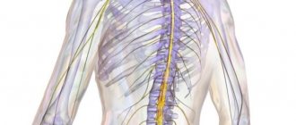
Spinal cord, structure.
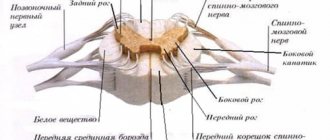
The average length is 43 cm, a flattened cord of cylindrical shape, located in the spinal canal, passes into the brain at the level of the foramen magnum . The lower border of the 1-2 lumbar vertebrae, here it narrows and is called the cone brain; then goes to the terminal thread. Below the 2nd vertebral column, a spinal puncture can be performed, since the spinal cord is not presented here in the form of a cord, but consists of thin nerve fibers-roots that form a “cauda equina”. The spinal cord has 2 thickenings - cervical and lumbosacral , associated with the innervation of the limbs, i.e. there are more nerve cells and fibers here. The anterior median fissure and posterior median sulcus divide the spinal cord into symmetrical left and right parts. There are also 2 anterior and 2 posterior lateral grooves - this is the place of origin of the anterior (motor) and posterior (sensitive) incoming roots. The grooves divide each half of the spinal cord, its white matter, anterior, lateral and posterior funiculi . The cords contain the conductive tracts of the spinal cord. The dorsal roots have a spinal ganglion - these are the bodies of the first sensory neurons. The anterior and posterior roots fuse to form the spinal nerves. There are 31-33 pairs of them, they emerge from the intervertebral foramina. The section of the spinal cord corresponding to 2 pairs of roots or one pair of spinal nerves is called a spinal cord segment. There are 31-33 segments in total: 8 cervical, 12 thoracic, 5 lumbar, 5 sacral and 1-3 coccygeal. The length of the spinal cord is less than the length of the spinal column, so the segments lie above the corresponding numbers of the vertebrae, starting from the lower cervical region. The lumbar vertebrae are at the level of 10-11 thoracic, and the sacral and coccygeal vertebrae are at the 12th thoracic and 1-2 lumbar vertebrae.
The structure of the gray matter of the spinal cord. Sheaths of the spinal cord.
The gray matter of the spinal cord forms columns throughout its entire length - anterior, posterior and lateral. The lateral one is present only at the level from the 8th cervical to the 2nd lumbar segments. In a cross section, the gray matter has the appearance of horns. Wider front, narrower rear and side. Hence the characteristic appearance of the “gray butterfly”. In the anterior horns there are large cells of motor neurons, forming 5 nuclei, two lateral, two medial and central. In the dorsal horns, smaller cells of sensory neurons form their own nucleus; Clark's pectoral nucleus is located at the base of the posterior horn The lateral horns contain the lateral intermediate substance. These are the centers of the sympathetic nervous system . The posterior horn is adjacent to the border, spongy zones and gelatinous substance. At the level of the cervical segments, between the lateral and posterior horns, the reticular formation is located in the white matter. In the center of the gray matter there is a central canal, which ends at the bottom with the terminal ventricle, and at the top communicates with the 4th ventricle of the brain. The center of the canal contains spinal cerebrospinal fluid; around the canal there is gray matter in the center. After 30 years, the canal becomes overgrown. The spinal cord is surrounded by three membranes. The external dura materspinalis, between it and the periosteum of the spinal canal there is an epidural space with fatty tissue and the internal venous plexus. Inward from the hard shell there is a thin arachnoid membrane - the arachnoid, between which there is a narrow slit-like subdural space. The third pia mater spinalis fuses with the spinal cord and is pierced by blood vessels. Between it and the arachnoid membrane there is a subarachnoid space filled with subarachnoid cerebrospinal fluid - 120-140 ml. The subarachnoid space of the spinal cord continues into the subarachnoid space of the brain.
Finite brain. Furrows and convolutions of the frontal and temporal lobe.
The brain is divided into 5 sections: telencephalon, diencephalon, midbrain, hindbrain and medulla oblongata.
The telencephalon , the telencephalon, consists of two hemispheres of the cerebrum, separated by a longitudinal fissure and connected to each other by the corpus callosum, anterior and posterior commissures and the commissure of the fornix. The telencephalon cavities are the right and left lateral ventricles in the corresponding hemispheres. Each hemisphere consists of gray and white matter. Gray matter covers the outside of the hemisphere - this is the cerebral cortex or mantle. Another part of the gray matter in the form of clusters is located deep in the hemispheres in the white matter - these are the basal ganglia. The white matter of the hemispheres is the corpus callosum, fornix, commissures and internal capsule. Each hemisphere has lateral, medial and inferior surfaces and consists of the frontal parietal, temporal and occipital lobes, separated by sulci and having prominent poles. The relief of each lobe is formed by grooves and convolutions - the elevations of the brain between the grooves. The frontal lobe is separated from the parietal central (Rolandic) fissure and from the temporal lobe by the lateral (Sylvian) fissure . In the frontal lobe, there are precentral sulcus , superior and middle frontal sulcus and, respectively, precentral gyrus , and superior, middle and inferior frontal gyri . In the inferior frontal gyrus, three parts are distinguished: orbital, triangular and tegmental , formed by the anterior and ascending branches of the sulci from the lateral sulcus. The tegmental part covers the insula, which lies deep in the lateral sulcus. The temporal lobe has superior and inferior temporal sulci, and, respectively, superior, middle and inferior temporal gyri . On the medial surface there is a hippocampal sulcus, separating the parahippocampal gyrus and the medial and lateral occipitotemporal gyri, separated by the sulcus of the same name.
The furrows and convolutions of the parietal and occipital lobes of the cerebral hemispheres.
The parietal lobe is separated from the central frontal lobe (Rolandic fissure), from the occipital lobe by the parieto-occipital beard, and from the lateral temporal lobe (Sylvian fissure). The parietal lobe is divided into the postcentral sulcus and the postcentral gyrus . The superior and inferior parietal lobules are separated by the intraparietal sulcus. Below are the supramarginal and angular gyri . There is a paracentral lobule on the medial surface .
The occipital lobe does not have noticeable grooves or convolutions on the lateral surface. the calcarine groove is clearly visible , as well as two sections of the occipital and parietal lobes - the cuneus and the precuneus . Below the calcarine groove on the occipital lobe, the lingual gyrus .
Frontal lobe
The frontal part of the cerebral hemispheres is located in front of all other structures of the brain. Posteriorly, this area is limited from the parietal lobe by the central sulcus, and laterally, by the lateral sulcus, from the temporal region.
In front of the central sulcus is the precentral gyrus of the brain. The latter, in turn, is limited from other formations of the frontal lobe cortex by means of the precentral recess.
The precentral gyrus, together with the adjacent posterior parts of the frontal lobe, plays an important role. These structures are necessary for the implementation of voluntary movements, that is, those that are under the control of consciousness. In the fifth layer of the cortex of the precentral gyrus there are giant motor neurons, which are called pyramidal cells, or Betz cells. These neurons have a very long process (axon), the endings of which reach the corresponding segment of the spinal cord. This pathway is called the corticospinal pathway.
The relief of the frontal region of the brain is formed by three large convolutions:
- superior frontal;
- average;
- bottom.
These formations are delimited from one another by the superior and inferior frontal grooves.
In the posterior part of the superior frontal gyrus there is an extrapyramidal center, which is also involved in movements. This system is historically more ancient than the pyramidal one. It is necessary for accuracy and smoothness of movements, for automatic correction of motor acts that are already normal for humans.
In the posterior part of the inferior frontal gyrus there is Broca's motor center, which was already mentioned earlier in the article.
Additional images
- Position of the central sulcus (marked in red)
- Position of brain structures in the head. The central sulcus separates the parietal lobe (in yellow) from the frontal lobe (in blue)
- Lateral surface of the left hemisphere. The central sulcus is marked with the number 6
- Lateral surface of the right hemisphere. Central sulcus - in the center, marked in red
- Medial surface of the hemispheres
- Play media file
Video with dissection of the telencephalon, demonstrating the central sulcus of the left hemisphere
Occipital lobe
The boundaries of the occipital region of the brain are outlined by the following formations: it is separated from the parietal lobe by the parieto-occipital recess, from below the occipital part smoothly flows into the basal surface of the brain.
It is in this area of the brain that the most unstable structures are located. But the posterior occipital gyrus of the brain is present in almost all individuals. Moving closer to the parietal region, transitional gyri are formed from it.
On the inner surface of this area there is a calcarine groove. It separates three convolutions from each other:
- wedge;
- lingular gyrus;
- occipitotemporal gyrus.
There are also polar grooves that have a vertical direction.
The function of the most posterior lobe of the brain is the perception and processing of visual information. It is noteworthy that the projection of the upper half of the retina of the eyeball is in the wedge, but it perceives the lower part of the visual field. And the lower half of the retina, which receives light from the upper visual field, is projected in the region of the lingual gyrus.
Development of sulci and convolutions of the brain
The hemispheres of the brain remain free from furrows for a very short time: already in the third
a significant number of them appear in the month of uterine life. During this period of development, the hemispheres still represent bubbles in the full sense, that is, they have thin walls and a relatively voluminous cavity. The grooves that appear on them represent real folds of the walls of these bubbles, so that each groove on the outer surface corresponds to an elevation on the inner surface - a roller. Of the furrows that appear in the third month of uterine life, some subsequently (namely in the fifth month) disappear; others from among these early grooves remain forever, and most of them retain in the adult the same character of the folds of the wall of the hemisphere, that is, they form protrusions or shafts protruding into the cavity of the ventricle - a property that the grooves that appear later do not have. Furrows that exist temporarily, between the 3rd and 5th months, cannot interest us*, and therefore we will not go into a description of them. The primary remaining grooves (sulci primitivi. permanentes Kölliker) are few in number, these are: 1) fiss. Sylvii, formed due to the fact that the hemisphere takes a horseshoe shape - as if bent in two, 2) fiss. parieto-occipitalis, 3) fiss. calcarina, 4) fiss. hippocampi.
* (It has been suggested that these grooves are an artificial product - the result of shrinkage of the delicate embryonic brain under the influence of alcohol, in which drugs are usually preserved. Recently, Cunningham has become convinced of their natural origin and permanence.
)
At the end of the fifth month, after the disappearance of temporary primary fissures, the outer surface of the hemisphere appears completely smooth again (all remaining fissures, with the exception of the Sylvian fissures, are concentrated on the inner surface). At the 6th month, a second series of remaining grooves appears, and of these the first is the Rolandic, then the precentral inferior, sometimes the superior frontal; further, on the parietal lobe, the interparietal groove always appears in the form of two segments (anterior and posterior), which corresponds to the different constancy of these parts (see above); the first temporal and occipitotemporal (absolutely constant of the four temporal sulci) appear on the temporal lobe; on the inner side of the hemisphere at this time fiss is added to the already existing ones. calloso-marginalis. This month, the Sylvian fissure begins to take on the shape that it has in an adult: the insula (Reilii) grows from its bottom in the form of a triangular blunt pyramid; the banks of the Sylvian fissure clearly begin to grow onto the island from above and in front in separate capes, from which the ascending branch fiss is designated. Sylvii.
At the 7th month of uterine life, all other so-called typical furrows (i.e., the first and second categories) appear, in the form of irregular or torn into sections of depressions.
By the end of the 7th and 8th month, some furrows of the third category begin to develop in the form of round or slightly oblong dimples, but in very small numbers, and the furrows of the second category take on more definite shapes, approaching the shape that they have in adult. Thus, the groove pattern of an 8-month-old fetal infant, as Ecker pointed out, can serve as a blueprint for the adult groove pattern - it consists almost exclusively of elements that are more permanent and simplified in form.
At the 9th month, the length of the furrows increases, their outline is somewhat complicated by the appearance of branches. The number of grooves of the third category increases, however, slightly. As a result, at the moment of birth, the pattern of grooves appears to be already formed to a certain extent, but clearly not completed precisely because of the smaller number of bends and branches of the main grooves, as well as the lack of grooves of the third category. Kölliker describes this drawing as already fully developed; but we, on the basis of numerous observations, argue that this is unfair: when comparing the pattern of grooves on a number of brain specimens of different ages, starting from the moment of birth to the 5th week after birth, one can always clearly observe a gradual complication of the pattern due to an increase in the number of grooves of the third category. At the 5th week, the pattern of the convolutions is indeed fully developed and, in various cases, represents all those individual changes that are observed in adults. The brain at this age seems even richer in convolutions than the brain of an adult, because the child’s small hemisphere has the same number of grooves as an adult’s. However, this period of development of furrows is the shortest and is characteristic only of very well-fed children. Extremely often among those children whose corpses are brought to the anatomical theater, there appears to be a slower development of the pattern of grooves: it turns out to be unfinished even in three-month-old children who are normally but poorly developed.
As for the reasons for the development of furrows and convolutions, nothing is known about them. There are only a few hypotheses, more or less probable. Thus, Reichert thinks that the reason for the development of grooves is pressure from the vessels of the pia mater, and Seitz attributes it to the influence of the vessels on the degree of nutrition of various parts of the cerebral cortex. The nature of the pattern of the grooves itself, which is not at all similar to the well-known picture of the branching of blood vessels, speaks against this assumption. Further, observation of the adult brain, and especially the embryonic brain, directly shows that the arteries very often run across the convolutions, leaving only shallow impressions on them, such as are seen, for example, on the inner surface of the skull bones. The main and most obvious proof of the injustice of this assumption is provided by the surface of the cerebellum, on which it is very easy to observe the discrepancy between the tree-like branching vessels and the straight and parallel grooves.
Another opinion, the most common, is that the reason for the formation of grooves is the slower growth of the skull compared to the growth of the brain, which causes the surface of the brain to form folds. This opinion is supported by the fact that in animals with a dolichocephalic skull, longitudinal grooves predominate, while in those with a brachycephalic skull, predominantly transverse folds are found. Kölliker attaches importance to this reason, but only for the formation of primary furrows, which subsequently disappear. The formation of permanent grooves cannot be explained by this influence, if only because during their development, that is, in the second half of uterine life, the brain does not form a skull, but lies freely, surrounded by a large amount of serous fluid. Of equal importance is the fact that the grooves finally develop at a time when the process of ossification of the skull is still far from complete, and, therefore, the skull can present little resistance to pressure from the brain.
The most likely explanation for the development of the grooves is that they are the result of a partial and non-simultaneous growth of various parts of the mass of the hemisphere, moreover, in connection with the gradual and consistent development of the cortical centers*.
* (Schnopfhagen (Die Entstehung d. Windungen des Grosshirns, Leipzig, 1891) considers the cause of this uneven growth of the surface of the brain to be the growth of the fibers of the projection system and the corpus callosum, mainly in the radial direction.
)