Subcortical visual centers and optic radiance
In the lateral geniculate bodies , which are subcortical visual centers, the bulk of the axons of retinal ganglion cells end and nerve impulses are switched to the next visual neurons, called subcortical or central. Each of the subcortical visual centers receives nerve impulses coming from the homolateral halves of the retinas of both eyes. In addition, information also comes to the lateral geniculate body from the visual cortex (feedback). It is also assumed that there are associative connections between the subcortical visual centers and the reticular formation of the brain stem, which contributes to the stimulation of attention and general activity (arousal).
The lateral geniculate body consists of six layers . Each of them has several (usually six) layers of nerve cells. It has been established that in the six-layer subcortical visual center the arrangement of neurons maintains a certain orderliness and topographic-anatomical relationships characteristic of the retina. The alternating layers of the lateral geniculate body receive visual impulses only from the homolateral (right or left) halves of the retina corresponding to the side of the location of the geniculate body of one or the other eye. There is no absolute sequence in the alternation of layers.
Thus, in the left geniculate body, the projections of the right halves of the retinas are located in the following order (from the superficial layer to the deep): left eye, right, left, right, right, left. There is no explanation yet for the incomplete sequence of alternating projections of the homogeneous halves of the retinas of the right and left eyes. The listed projections of the halves of the retinas in the layers of the lateral geniculate body are located exactly one below the other.
The experiment proved that the cells of the lateral geniculate body respond to visual impulses reaching them in approximately the same way as retinal ganglion cells respond to visual impulses coming to them from photoreceptors. At the same time, the central visual neurons of the geniculate bodies and the corresponding retinal ganglion cells, which can be called peripheral visual neurons, have a similar structure of receptive fields with the on- and off-centers of visual neurons and give similar bioelectric responses, depending on the intensity and color of light impulses.
It has also been established that neighboring retinal ganglion cells and central visual neurons of the subcortical visual center are located among themselves in an identical sequence. It is assumed that some neurons of the lateral geniculate body have short axons that provide local interneuronal synaptic connections, which suggests their interaction leading to a possible preliminary analysis and synthesis of visual information entering the subcortical centers. However, there is currently no consensus on the role of the external geniculate bodies in the processing of visual information. D. Hubel in 1990 suggested that, apparently, no significant transformations of visual impulses coming from the retina occur in it. At the same time, J.G. Nicolet, A.R. Martin, B.J. Wallas and P.A. Fuchs (2003) recognize that geniculate neurons are involved “in providing the first steps in the analysis of visual scenes: identifying lines and shapes based on retinal input...”
The axons of the neurons of the lateral geniculate body, emerging from the six layers of the lateral geniculate body, unite into a single bundle and participate in the formation of the posterior limb of the internal capsule, and then form the next part of the visual pathways, the optic radiance, which has a significant extent.
Visual radiance
The axons of optic neurons located in the lateral geniculate body are part of the white matter of the cerebral hemispheres. At the same time, they first form a compact bundle that participates in the formation of the posterior leg of the internal capsule, more precisely its sublenticular part (pars sublenticularis), and subsequently form optic radiation (radiatio optici), or Graziole bundles . After passing through the so-called isthmus of the temporal lobe of the brain, the optic radiance expands and takes the form of a wide ribbon. This peculiarity of the organization of this part of the optic radiance leads to the fact that its damage is often partial, due to its considerable width and the non-compact arrangement of the nerve fibers included in its composition. In this regard, total damage to the optic radiance occurs only with a fairly common pathological process.
The nerve fibers that make up the optic radiation are involved in the formation of white matter in the temporal, parietal and occipital lobes. In the temporal lobe, near the outer wall of the inferior horn of the lateral ventricle, most of the fibers of the inferior part of the optic radiation first pass forward to the pole of the temporal lobe. Then these fibers, forming Meyer's loop , turn back and pass through the white matter of the temporal and occipital lobes.
As a result, they reach the cortex of the lingual gyrus (gyrus linqualis), which forms the lower “lip” of the calcarine groove (sulcus calcarinus), located on the medial surface of the occipital lobe.
The upper part of the visual radiance is straighter and therefore shorter than the lower one. It passes through the white matter of the parietal and occipital lobes of the hemisphere and ends by coming into contact with cortical cells located in the upper lip of the calcarine sulcus, formed by a gyrus known as the cuneus. The cortex of the medial surface of the occipital lobe, surrounding the calcarine sulcus and extending into its depth, constitutes the primary projection visual field , occupying cytoarchitectonic area 17, according to Brodmann.
It should be recalled that the visual pathways conduct visual impulses throughout their entire length, being located in a strict retinotopic order and at the same time maintaining the topographic-anatomical relationships characteristic of the retina.
"Renes" - combining the best experience in audiology.
Our hearing center in Moscow offers its clients the opportunity to undergo diagnostics and buy Widex and Phonak equipment - digital hearing aids, the prices of which will be the most favorable. At the same time, the quality of the equipment remains impeccable. The sale of hearing aids we carry out in Moscow also includes:
- competent consultations with specialists;
- assistance in using equipment;
- a whole range of measures for successful rehabilitation.
The RENES company offers a full range of services in the field of hearing care - a thorough examination of the patient using the most modern medical equipment, selection of a hearing aid, production of in-ear and intra-canal hearing aids and individual earmolds, repair and service, sale of various accessories for people with hearing impairments.
The flexible pricing policy of RENES makes it affordable for every client to purchase even a fairly expensive device.
In addition, from us you can purchase methodological literature, collections of exercises for working with children whose sound perception function is impaired, and receive free consultation from a specialist.
Modern technologies have made correction devices compact, aesthetic and almost invisible, allowing to significantly improve the level of sound perception.
In addition to Phonak and Widex hearing aids, you can purchase hearing aids from Oticon, Bernafon, Siemens or Tondi, the prices of which will pleasantly surprise you. A distinctive feature of such devices is their ability to operate effectively in almost any climatic conditions.
Our employees are specialists of the highest class - they have extensive work experience, regularly undergo retraining abroad (Germany, Denmark, Switzerland) and are always ready to assist every client.
We try to find a personalized approach to clients of any age, take into account their needs and capabilities, find the right solutions and effective techniques for everyone.
We have been working since 1992 and do it at a high professional level. We value our clients and are always ready to help them.
To the question of how to choose the right hearing aid, you can clearly answer that this must be done in a specialized hearing restoration center, by visiting an audiologist. But a patient with hearing loss should remember some important points himself.
General information
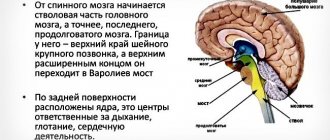
The diencephalon is located under the corpus callosum and consists of: the thalamus (thalamic brain) and the hypothalamus.
The thalamus (aka: visual thalamus, sensitivity collector, body informant) is a section of the diencephalon located in its upper part, above the brain stem. Sensory signals and impulses from various parts of the body and from all receptors (except smell) flow here. Here they are processed, the organ evaluates how important the incoming impulses are for a person and sends the information further to the central nervous system (central nervous system) or to the cerebral cortex. This painstaking and vital process occurs thanks to the components of the thalamus - 120 multifunctional nuclei that are responsible for receiving signals, impulses and sending processed information to the corresponding section of the cerebral cortex.
Thanks to its complex structure, the “visual thalamus” is capable of not only receiving and processing signals, but also analyzing them.
Ready information about the state of the body and its problems reaches the cerebral cortex, which, in turn, develops a strategy for solving and eliminating the problem, a strategy for further actions and behavior.
Inside or outside?
If the aesthetics of the device comes first, and you want it to be completely unnoticeable, then when you come to the hearing and speech center, you should ask for an in-ear device that is completely unnoticeable.
But devices of this type have some disadvantages, for example, their batteries are smaller in size, which means they waste their charge faster. If you prefer more massive behind-the-ear devices, you can ask the audiology and hearing care center to choose a hearing aid that will match your skin tone and will not stand out too much.
For those patients who suffer from inflammation of the middle ear, in-ear devices are contraindicated, and the hearing correction center will definitely be told about this.
Advantages of Renes
Ours is one of the best hearing centers in Moscow. You can purchase various types and models of hearing aids from us, some of which are budget, while others are recognized as elite products. At the same time, competent and experienced specialists who work at the Renes Hearing Center will help you choose a device not only in accordance with your audiogram, but also with all additional requirements and wishes. We will try to help you understand what type of devices will be most effective and convenient for you, suitable for your lifestyle and profession.
Price matters
If you want the sounds reproduced by the device to be “live”, then you should not skimp on them. In particular, everything that costs less than 15,000 rubles in hearing aid centers is not equipped with a good noise suppression system, so it is better not to choose such devices, but to spend money on more expensive ones.
After all, you will have to wear them for a very long time, and their quality will directly affect the quality of your perception of the world. As a rule, in modern audiology centers the price of high-quality hearing aids ranges from 20,000 rubles and above.
Sounds become even clearer if the device has two adaptive microphones. Due to the fact that devices can have such a large number of features, among which the patient can choose according to his capabilities, when he comes to the hearing care center, it is worth clarifying the technical features of the devices that are offered there. If the institution is self-respecting, then the center’s experienced specialists will definitely answer all your questions. Today, different Moscow hearing care centers offer very different products, and, on the one hand, this provides the patient with a very large choice, but at the same time it significantly complicates it, making the help of knowledgeable people necessary.
Functions of the thalamus
The “sensitivity collector” receives, filters, processes, integrates and sends information to the brain that comes from all receptors (except smell). We can say that in its centers the formation of perception, sensation, and understanding occurs, after which the processed information or signal enters the cerebral cortex.
The main functions of the body are:
- processing of information received from all organs (receptors of vision, hearing, taste and touch) senses (except smell);
- managing emotional reactions;
- regulation of involuntary motor activity and muscle tone;
- maintaining a certain level of activity and excitability of the brain, which is necessary for the perception of information, signals, impulses and irritations coming from the outside, from the environment;
- responsible for the intensity and feeling of pain.
As we have already said, each lobe of the thalamus consists of 120 nuclei, which, based on functionality, can be divided into 4 main groups:
- reticular;
- lateral (lateral);
- medial (middle);
- associative.
Reticular group of nuclei (responsible for balance) – responsible for ensuring balance when walking and balance in the body.
The lateral group (vision center) is responsible for visual perception, receives and transmits impulses to the parietal, occipital part of the cerebral cortex - the visual zone.
The medial group (hearing center) is responsible for auditory perception, receives and transmits impulses to the temporal part of the cortex - the auditory zone.
Associative group (tactile sensations) - receives and transmits tactile information to the cerebral cortex, that is, signals emanating from receptors of the skin and mucous membranes: pain, itching, shock, touch, irritation, etc.
Also, from a functional point of view, nuclei can be divided into: specific and nonspecific.
Specific nuclei receive signals from all receptors (except smell). They provide a person’s emotional reaction and are responsible for the occurrence of pain.
Specific kernels, in turn, are:
- external - receive impulses from the corresponding receptors and send information to specific areas of the cortex. Through these impulses feelings and sensations arise;
- internal - do not have direct connections with receptors. They receive information already processed by the relay cores. From them, impulses go to the cerebral cortex to the associative zones. Thanks to these impulses, primitive sensations arise and the relationship between sensory areas and the cerebral cortex is ensured.
Nonspecific nuclei support the general activity of the cerebral cortex, sending nonspecific impulses and stimulating brain activity. Having no direct connection with the cortex, the nonspecific nuclei of the thalamus transmit their signals to subcortical structures.

