PET-CT
What does it diagnose?
PET/CT for brain diseases
Registration for PET/CT
PET/CT of the brain is a diagnostic method that allows one to assess the condition of various parts of the brain at the cellular level and track deviations in tissue metabolism. PET/CT is sometimes called functional tomography, since it can be used to study not only the anatomical structure of the area under study, but also the functional activity of tissues.
Interesting! As a rule, in modern medical centers when studying the brain using PET/CT, they use the latest radiopharmaceutical - 11c-methionine. But in some clinics (especially domestic ones) they still do diagnostics with labeled glucose, although its indicativeness and accuracy are an order of magnitude lower.
We will talk more about the difference in radiopharmaceutical data below.
In the arsenal of modern neurologists
In neurology, PET is used to study metabolic processes in nerve cells of the brain in various diseases.
The advantage of the technique is the ability to evaluate functional changes at the cellular level, which are recognized already at the initial stages of the development of the disease without the presence of obvious clinical manifestations, and not an anatomical image of the organ under study, as during MRI. Indeed, in such cases, neuroimaging methods that study tissue structure do not help in identifying pathology.
There is no need to worry about high radiation exposure, because the administration of the maximum dose of the drug is equal to the radiation dose that the patient receives during a chest x-ray in two projections.
Positron emission tomography in neurology is performed for the following purposes:
- In case of cerebrovascular pathology or ischemic brain damage, the doctor determines the area of cells that can recover with adequate therapy. This makes it possible to recognize new changes and those already existing in patients with recurrent stroke.
- In epilepsy, it becomes possible to accurately determine epileptogenic foci, which is extremely important in focal forms of the disease, and to assess the degree of metabolic disturbance in them. PET scan of the brain helps to decide on the indications for surgery, choose the optimal method and make an approximate prognosis of the outcome of such treatment for epilepsy.
- Alzheimer's disease is defined by a clear focal decrease in metabolic processes in the neocortical associative areas of the cerebral cortex - posterior cingulate, temporoparietal and frontal multimodal, while more severe pathological changes most often develop in the dominant hemisphere. Dementia caused by vascular pathology is accompanied by damage to the frontal lobes with the cingulate and superior frontal gyrus. In this case, “spotty” areas of decreased metabolic processes in the white matter and cortex are often identified, and the cerebellum and subcortical structures are often affected. Frontotemporal pathology is characterized by abnormalities in the frontal, anterior and medial parts of the temporal cortex. Dementia with Lewy bodies is accompanied by bilateral temporoparietal metabolic disorders and damage to the occipital cortex and cerebellum.
- Determining the location of cerebral amyloidosis helps determine how effective the treatment is and predict the course of the pathology in the future.
- In Parkinson's disease, it has become possible to determine the quantitative characteristics of the deficiency in the production and accumulation of dopamine in presynaptic striatal terminals, to evaluate the effectiveness of therapy and indications for surgical intervention.
- Hyperkinesis and Huntington's chorea are determined by impaired glucose metabolism in the caudate and lentiform nuclei, frontal projection fields of the mediodorsal thalamic nucleus.
- In multiple sclerosis, diffuse changes in brain metabolism are detected.
- In oncology , such a study accurately reflects not only the location and volume of the tumor, but also the nature of the tumor - benign or malignant.
Alzheimer's disease on PET scan of the brain
How often can the procedure be performed?
Computed tomography with methionine is performed no more than twice a year.
The first examination is carried out to make a diagnosis, and the second is a control examination to monitor the development of the disease, confirm the effectiveness of treatment measures or adjust them if they are insufficiently effective.
Repeated diagnostics cannot be carried out immediately after surgery, as it requires complete recovery of the body. A month must pass from the date of the operation. The same amount of time should pass from the moment of chemotherapy.
Preparing for the study
During a consultation with your doctor when deciding whether to conduct a PET scan, you must inform him about all the medications you are taking at this time. The fact is that some drugs can affect the result, so the doctor can stop them for a while to obtain reliable data.
The night before, a light dinner is allowed, most of which should be fresh vegetables and fruits. Do not include dairy products, meat and fish, sausages and soy in your diet.
On the day of the study, it is not recommended to eat in the morning; plain still water can be drunk without restrictions.
Dress in comfortable and warm clothes and be sure to have replacement shoes and a medical card with the results of previous studies. Before starting, remove your jewelry.
Women planning to become pregnant in the near future or who are pregnant are prohibited from undergoing PET scanning. Since the radiopharmaceuticals used have a teratogenic effect on the fetus. Lactating women are advised to stop breastfeeding for 2 days after the study to allow radioactive substances to clear from her body.
Only with proper preparation can you get high-quality results.
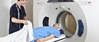
Carrying out a diagnostic study
Before administering the radiopharmaceutical, the patient needs some time to rest and drink half a liter of water. The radioform drug is administered in the supine position, after which the patient is placed in a dark room for 20 minutes in the same position.
During this time, a uniform distribution of the concentration of the administered radioactive drug in the blood and tissues is achieved, as required by the pharmacokinetic model that underlies the image processing program.
In this case, the patient should not make active movements and talk to prevent the accumulation of radioactive drug in the activated muscles, because this leads to difficulties in analyzing images and to diagnostic errors. Before starting a diagnostic scan, you must empty your bladder.
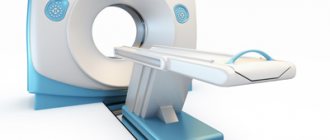
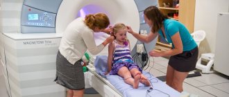
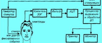
To perform the procedure, the patient lies down on a couch, which is then placed in a special PET chamber. A scanner in the form of a ring rotates inside the camera around the head during the examination.
During the diagnostic procedure, the doctor may ask you to tell or remember something; this is necessary to activate memory, speech and thinking. The duration of the study depends on the volume of the procedure and ranges from 30 to 75 minutes.
Precautionary measures
Positron emission tomography is a safe diagnostic method; only the administered radiopharmaceutical or contrast, the basis of which is most often a radioactive saccharide under the marker 18FDG, can have a negative effect on the body.
Its use makes it possible to obtain clear images and assess metabolic disorders in cells in the tissues under study.
Contraindications:
- pregnancy;
- women during lactation;
- early postoperative period;
- severe diabetes mellitus, with a sugar level more than 6.5 mmol/l;
- condition after biopsy;
- renal, hepatic and cardiovascular failure in the stage of decompensation;
- recent chemotherapy or radiation therapy;
- acute inflammatory processes in the body;
- emphysema;
- persons with an allergic reaction to the components of the radiopharmaceutical.
Possible risks
The risks associated with the administration of radiopharmaceuticals and radiation are identified. The radiopharmaceutical may provoke the development of an allergic reaction (for example, Quincke's edema) or anaphylaxis.
24 hours after the administration of the radiopharmaceutical, the patient is a source of weak ionizing radiation. During this period, it is necessary to reduce contact with other people as much as possible. The risk of radiation exposure for the patient is extremely small and is not comparable to the threat to life caused by a brain tumor.
Radiation exposure
With PET, the radiation exposure is 1-1.5 mSv. With PET/CT with methionine, the radiation exposure is higher (up to 3-5 mSv). The dose received is considered unsafe, but does not lead to health problems during or after the procedure.
Decoding the results
The results of the survey are deciphered using visual and semi-quantitative methods.
The visual assessment method uses black-and-white and color scales, which make it possible to detect the severity of the drug accumulated in the tissues and foci of impaired metabolism, their location, boundaries and sizes.
A semi-quantitative assessment involves calculating the ratio of accumulated radioactive substance in completely identical areas. In this case, pathological activity is determined in one part, reflecting a violation of metabolic processes, and the second is an identical area with the absence of pathological changes.
Indications
PET with amino acids is indicated for patients with brain tumors. The information content of studies with glucose in neuro-oncology is reduced due to the high energy functionality of the brain.
The brain parenchyma intensively accumulates glucose, against the background of which tumors become indistinguishable or are not clearly visualized. The amino acid methionine accumulates much more intensively in the neoplasm tissue. The accumulation of methionine in the brain parenchyma is weakly expressed. As a result, the tumor becomes clearly visible.
A dynamic study makes it possible to evaluate changes in the size of a space-occupying lesion over time and to identify residual tumor and relapse. Methionine PET is well suited for tumor staging.
Cost of the study
Due to insufficient equipment and high cost, this method is still limited and is available only in large positron emission tomography diagnostic centers, but its potential is high, the price of PET varies from 8,000 to 44,000 rubles throughout Moscow and throughout Russia.
The most promising method is one that involves simultaneous MRI and PET scanning with further comparison of the resulting images. This allows us to obtain a larger amount of information about structural and functional disorders of the area under study. Accordingly, the cost of the procedure increases.
Application of PET CT in malignant tumors
When oncology is detected, doctors are not faced with the question of what is better to use for diagnosing and tracking the dynamics of the tumor process - conventional CT or PET CT. The choice always falls on the second option, since only it fixes processes at the molecular level. The doctor can decide on the choice of therapeutic measures as soon as possible.
The combination of PET diagnostics with CT determines:
- exact location of the tumor;
- stage of development of the neoplasm;
- degree of aggressiveness of cancer cells;
- which organs are affected by metastases;
- result of chemotherapy.
Unlike standard scanning methods, which also detect tumors, the PET CT scan contains data on the metabolic rate in the area under study. With oncological formations, increased cell division and active absorption of glucose and other sugars occur in the places where they are localized. The sensors analyze and record metabolic processes, creating a clear picture that clearly shows areas of abnormal metabolism.
The ability to record such processes is not the only difference between the PET CT procedure and CT or MRI. Thus, after therapy, tumors often look the same as before (maintain their size and density), but lose their aggressive properties, that is, they become residual. On MRI and CT, they look the same as before treatment, and a PET scanner can differentiate residual tumor tissue from a “living” cancer tumor with 100% accuracy.