This article is about the pathology of the contents of the cranium; for pathology of the contents of pants, see Phimosis
| Halt! The page is fenced off from FGMnuttyh. Do you want to speak up? Welcome to the discussion. |
| « | If a person suddenly starts writing in his diary that he gets up in the morning on his left foot, and those who get up on his right are cattle and don’t understand anything, then he will rightly be told to go to hell. | » |
| — From the article “Vegans” . | ||
| « | Every person has a certain horizon of views. When it narrows and becomes infinitesimal, it turns into a point. Then the person says: “This is my point of view.” | » |
| — David Gilbert | ||
Phimosis
(Greek φίμωσις -
narrowing
)
of the brain
(often also in Kaschenite spelling:
“phimosis of the goil mosk”,
etc. [1].) is the main mental illness for which it is treated in Kaschenka.
A special case of FGM is the Orthodoxy of the brain (otherwise known as Christosis of the brain
).
Origin and treatment
Progressive FGM
Incidence in Africa. More details:
The picture tells us about female circumcision. Which, however, does not change the essence of the matter, since it is slightly less than in all cases due to the fault of FGM and its particular manifestations (such as Islam of the brain). It is logical to assume that in this way the map displays the minimum number of FGM patients per unit area. It is characteristic that the largest number of patients are in Somalia and Sudan, where anarchic fuckery has reigned for many decades
During the heyday of Kashchenka, its population was traditionally divided into doctors and pots-i-ents. Since the latter, in order to justify their status, had to suffer some kind of sorrow, FGM was quickly invented.
The origin of this term comes from a real disease called phimosis - a situation in which, due to an excessively rigid and inextensible foreskin, it is impossible to expose the head of the male penis, and which is treated by circumcision.
Phimosis of the brain, by analogy, began to be considered synonymous with extreme dullness and narrow-mindedness of the patient. FGM is characterized by damage to the cortex and wood of the brain (hereinafter referred to as the “phimbrain” or “interauricular ganglion”) with progressive blockage of thought pathways, an increase in the patient’s aggressiveness, as well as a catastrophic decrease in the ability to absorb information and general mental abilities. In other words, FGM is a chronic inability to use brain resources for their intended purpose. From this, in particular, it follows that high knowledge and the presence of great knowledge in various fields do not in any way guarantee the absence of FGM. As an analogy: even the most modern and sophisticated computer with a large set of software is useless while it is turned off.
The symptom complex also includes: complete rejection of criticism of one’s activities, erudition on any issue, ignoring facts that do not fit into one’s own picture of the world, attaching labels that often do not correspond to reality. The prognosis for life is favorable, but for recovery it is doubtful.
It is generally accepted that FGM is an incurable disease, therefore the only method of treating it is considered to be the use of 5 to 10 cubes of life-giving euthanasia intravenously or intravenously. An alternative to such treatment is to incense the patient, after which he stops suffering from his illness and begins to enjoy it.
Alternatively, traditional medicine suggests cutting your hands as a symptomatic treatment. True, this will not help FGM, but the patient will definitely stop rambling on forums, chats and blogs. Well then...
In advanced cases, FGM turns into boclanopotstitis.
Life forecast
The life prognosis depends on the degree of malignancy of the brain tumor. Anaplastic meningioma in more than half of cases leads to relapse 5 years after surgical treatment. A neurological syndrome may occur.
It is important to pay attention to even minor clinical manifestations. It is recommended to consult a specialist if you experience frequent headaches, nausea, vomiting, or episodes of loss of consciousness. With the help of CT and MRI, even minor pathological lesions that require surgery can be identified.
The Burdenko Research Institute provides professional surgical treatment of extracerebral neoplasms using modern technologies and safe techniques. You can make an appointment with the best neurosurgeons, conduct a comprehensive examination, and begin effective treatment that will fully correspond to the clinical situation.
Brain liquefaction
Close to FGM, but nevertheless a completely separate concept. The symptoms are similar: a complete cessation of the patient’s independent mental activity, but, in contrast to the characteristic phimosis hardening, siltation and complete impenetrability of the psyche, everything happens the other way around - the patient’s point of view blurs and turns into a shapeless blot containing any freshly collected in the skull (usually from the zombie box) substance. The contents of the skull can change rapidly and dramatically, without encountering any resistance, without even a minimal delay, and without causing any cognitive dissonance. Usually caused by an overdose of brainwash.
Also, the disease "brain liquefaction" is present in Morrowind, reducing intelligence and willpower.
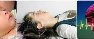
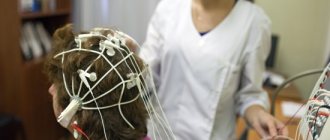
What does an electroencephalogram show?
The device for conducting an EEG is called an electroencephalograph.
The curve that appears on the monitor screen during an EEG makes it possible to diagnose various changes in the functioning of nerve cells in the brain. A specialist, assessing the activity of neurons reflected in the encephalogram, can determine the following points:
- find out the presence or absence of disorders in the cerebral cortex;
- assess the severity of brain damage if it is detected;
- accurately determine the location of the injury;
- identify those areas of the brain that are the source of epileptic seizures;
- study the features of the periodization of sleep and wakefulness;
- detect a neoplasm;
- determine how effective drug therapy was;
- find out how the brain works in the periods between attacks;
- discover the cause of fainting and other moments of crisis and much more.
Electroencephalography is a method for examining the functional state in the structures of the brain. The most pronounced readings of the device relate to deviations in sleep patterns, wakefulness, activity of the brain center or in relation to physical work.
An electroencephalogram is a painless procedure that cannot cause any harm to a person’s health or well-being. No intervention in the body or complex preparation is required to perform the procedure.
Today, the electroencephalogram of the brain is widespread and used by specialists in the field of neurology. The technique is considered extremely informative and helps to diagnose epileptic abnormalities in the brain, detect vascular pathologies, the presence of inflammatory processes or degenerative disorders of the brain. Among other things, EEG helps to identify the exact location of a tumor, cyst or hematoma in structures.
Electroencephalography is used in intensive care wards, where long-term follow-up of a patient who has fallen into a coma is indicated. The disappearance of the electrical activity of the brain center indicates the death of the patient.
Copy-paste on the topic
This phenomenon was first reliably described by the Orthodox psychiatrist P. B. Gannushkin. Already in 1933!
| GROUP OF CONSTITUTIONALLY STUPID (...) People of this kind sometimes study well (they often have a good memory) not only in secondary, but even in higher school: when they enter into life, when they have to apply their knowledge to reality, show a certain initiative, they turn out to be completely sterile. They know how to “keep themselves in society”, talk about the weather, say stereotyped, banal things, but do not show any originality (hence the expression “Salon blodsinn” - salon dementia) ... They cope well with life only within certain, narrow, long-established boundaries household and material well-being. On the other hand, this also includes elementary simple, primitive people, devoid of spiritual needs, but who cope well with the simple requirements of some craft, sometimes even working in trade, even in administration, without major misunderstandings. One of the distinguishing features of the constitutionally limited is their great suggestibility, their constant readiness to obey the voice of the majority, “public opinion” (“what will Princess Maria Alekseevna say!”); these are people of the pattern, banality, fashion, these are also people of the environment, but not quite in the same sense as unstable psychopaths: there people follow a shining example of this environment, for “vice,” but here, on the contrary, for good morals. Constitutionally limited psychopaths are always conservatives; out of a natural sense of self-defense, they cling to the old, to which they are accustomed and to which they have adapted, and are afraid of everything new. These are the “normal” people about whom Cuillier said that on the very day when there will be no more half-normal people (demi-fous), the civilized world will perish, perish not from an excess of wisdom, but from an excess of mediocrity. These are the "normal" people whom Ferry compares to a ready-made dress from big box stores; Only the law of imitation applies here. (...) Those peculiar subjects who are distinguished by great self-conceit and who, with a pompous, solemn air, utter commonplaces or florid phrases that have no meaning, which are a set of pompous words without content (a good example, however, in a caricatured form) should also be classified as constitutionally stupid , in caricature form - the sayings of Kozma Prutkov). Perhaps here it is necessary to mention some reasoners, whose desire to have their own judgment about everything leads to gross errors, to the expression as truths of absurd maxims that are based on ignorance of elementary logical requirements. It is not superfluous to emphasize that in relation to many types of constitutional stupidity, the saying of the famous German psychiatrist is confirmed that they can, they can do more than they know..., as a result of which in roughly elementary life they often turn out to be even more adapted than the so-called smart people |
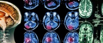
Electroencephalography for brain tumors
Electroencephalography has gained a strong place in the comprehensive diagnosis of brain tumors. With its help, it is possible to approach the resolution of a number of complex pathophysiological and local diagnostic issues that arise when recognizing brain tumors.
Issues of electroencephalography for brain tumors began to be developed in the thirties, and to date there is significant literature on this issue, both domestic and foreign.
Electroencephalographic studies in brain tumors were carried out by S. A. Sarkisov, M. N. Livanov, V. S. Rusinov, V. E. Mayorchik, A. A. Sokolova, N. P. Bekhtereva and others. Foreign authors include Walter and Dovey, Cobb, Thiry, Payat and Bonnal, Silverman, Wilke and Steinman.
Research in this area has shown that the tumor process produces certain changes in the electroencephalogram. The main characteristic feature in the electroencephalogram of patients with tumors is the appearance of delta waves and slow pathological waves (2-5 per second), i.e., waves with a much slower rhythm than normal alpha waves (8-12 per second). The tumor itself is electrically indifferent, and pathological waves arise at the border of healthy and pathologically altered brain tissue. At the same time, in addition to the appearance of slow waves, the alpha rhythm changes, until it completely disappears, and rapid oscillations of the beta wave type are observed.
Changes in the electrical activity of the cortex are of a certain nature during various processes in the brain. Processes such as trauma, epilepsy, inflammatory processes, tumors leave a certain imprint on changes in electrical activity. With brain tumors, there may be general, diffuse changes in electrical potentials that are produced by the entire pathologically altered cortical tissue, and focal changes caused by the immediate proximity of the pathologically altered brain tissue to the tumor.
Malignant tumors, spongioblastoma multiforme, sarcomas, cancer metastases, causing general intoxication and major cerebral edema, produce mainly general changes in the biocurrents of the cortex in the form of diffuse slow waves in both hemispheres.
With very high hypertension, general changes in the biocurrents of the cortex prevail over focal ones or completely obscure them.
General changes in the biocurrents of the cortex, obscuring the focus, can also occur with a benign tumor, but one that exists for a long time without any particular hypertension. According to the research of T. O. Faller, a long-existing tumor focus is a source of dynamic changes in the functional state of the entire cortex.
At a certain stage of tumor growth, the lability of nerve cells first increases, as indicated by the transition of the alpha rhythm to rapid oscillations (beta waves). Then, under the influence of a long-existing lesion, the lability of nerve cells decreases, which determines the transition to a slow rhythm throughout the entire cortex, more on the side of the lesion. Sometimes the focus of pathological electrical activity can be even better identified with increasing hypertension.
There are a number of tumors that are sometimes asymptomatic (arachnoidendotheliomas, astrocytomas, ventricular tumors). Therefore, clinically in such cases it is impossible to establish not only the location, but even the side of the lesion. Among all the existing additional research methods, electroencephalography can be of great help in this regard.
The works of V. S. Rusinov and V. E. Mayorchik show the basic principles on the basis of which one can judge the superficial or deep location of the tumor.
Benign superficial neoplasms cause changes in electrical activity in the form of delta waves only in areas of the cortex directly adjacent to the tumor. In other areas of the cortex, the normal alpha rhythm is maintained. Delta waves, as well as fast asynchronous oscillations, are not detected far from the source. In other words, attention is drawn to the delimitation of the pathological focus from other areas of the cerebral cortex.
With intracerebral tumors, there are generalized slow pathological waves of different periods in both hemispheres, most pronounced in the diseased hemisphere and especially in the tumor area. The alpha rhythm is reduced everywhere or completely disappears. Along with slow pathological waves, the intracerebral lesion causes the appearance of fast asynchronous potentials on the electroencephalogram, often in both hemispheres, running at different frequencies per unit time (18-35 per second) and resembling fast oscillations recorded from the axon. Typically, these axon-like oscillations are preserved where the cortex is intact and the focus is located deep in the brain, and are strung together into slow waves. If a tumor grows into both the cortex and subcortex, then these fast oscillations are absent in the tumor area and only slow waves remain, and fast oscillations are recorded only in the opposite hemisphere.
However, basically this is a diagram, since the focus of pathological electrical activity at the same location can manifest itself in quite a variety of forms.
For example, in arachnoidendotheliomas, the focus can be expressed in the form of an exalted alpha rhythm. This exaltation is expressed in the fact that the amplitude of the alpha rhythm increases sharply.
The focus of pathological electrical activity can also be expressed in the form of some reduction of the alpha rhythm and some of its unevenness compared to the alpha rhythm of the opposite healthy side.
Delta waves in a pathological focus can be of a wide variety of durations and amplitudes.
Thus, functional disorders of the entire cerebral cortex and in the area of the focus of pathological electrical activity are reflected in the electroencephalogram in the form of various forms of its changes. This depends on the duration of the disease, the nature of the tumor, the degree of its effect on the cortex, toxicity, and the magnitude of hypertension. The nature of the pathological focus changes as the tumor grows.
The auxiliary diagnostic value of the electroencephalogram is very great. According to the N. N. Burdenko Institute of Neurosurgery, in 80% there is a coincidence of the focus of pathological electrical activity with the location of the tumor. In 18% there are general changes in the biocurrents of the cortex without indicating pathological foci; in only 4% there are discrepancies. According to French authors, the coincidence of the focus of pathological electrical activity with the location of the tumor is observed in 70% of cases. They find very deep-lying tumors most difficult to diagnose. Spongioblastoma multiforme is best identified. In patients with brain tumors, they obtained for extracerebral tumors (meningiomas) a 68.5% coincidence of the focus of pathological electrical activity with the tumor location, 23% - general changes in biocurrents, and 8% - discrepancies. With intracerebral tumors, there were matches in 77%, mismatches in 13%, and general changes without a focus in 10%. For benign tumors, there are 73% matches, and for malignant tumors, 69.5%. In addition, the authors concluded that in tumors that are well supplied with blood vessels, such as meningeal and malignant tumors, the focus of pathological electrical activity is better identified than in tumors that are poor in blood vessels.
For neurosurgical practice, an indication of the superficial or deep location of the focus of pathological electrical activity is of great importance.
An analysis of observations from the N. N. Burdenko Institute of Neurosurgery shows that with intracerebral tumors in the vast majority of cases there are indications of a deep location of the pathological focus, and with arachnoidendotheliomas in a very small percentage of cases there are indications of a superficial location of the focus of pathological electrical activity, more often there are also indications of the deep location of the lesion.
This circumstance apparently has its own pathophysiological justification. Arachnoidendotheliomas are usually deeply embedded in the brain tissue, especially basal or located in the interhemispheric fissure, or in the Sylvian fissure. Obviously, the medulla reacts in the same way in terms of changes in bioelectric potentials if the tumor grows into the brain or strongly compresses it, and if the tumor is located only in the white matter or grows into the cortex, the electroencephalographic picture is different.
An electroencephalogram is of great importance in cases where it is neurologically very difficult, and sometimes impossible, to localize the tumor. In such cases, an electroencephalogram either complements neurological diagnosis or is the only method indicating the location of the tumor. For example, in cases where secondary brainstem symptoms suggested a diagnosis of a tumor of the posterior cranial fossa, an electroencephalogram clarified the diagnosis, indicating the location of the pathological focus.
General changes in biocurrents without indicating a pathological focus are observed mainly in cases where there is very high hypertension, obscuring the manifestations of the pathological focus, or with slight hypertension in patients with diffusely growing benign intracerebral tumors. The same picture is observed in arachnoidendotheliomas, where cerebral symptoms predominate, and local symptoms are weakly expressed. Such general changes in cortical biocurrents are observed more often in arachnoidendotheliomas than in intracerebral tumors.
For all basally located arachnoidendotheliomas, the electroencephalogram usually gives indications of the basal location of the focus of pathological electrical activity, as, for example, with tumors of the olfactory fossa. It should be noted that quite often, with a parasagittal location of the tumor, the electroencephalogram gives indications of the basal location of the pathological focus. With parasagittal tumors, the basal formations are very severely affected, which is also detected neurologically and sometimes leads to erroneous diagnosis. From this it is quite clear that the focus of pathological electrical activity can also be detected in the basal regions of the brain.
In no more than 4% there is a discrepancy between the focus of pathological activity and the location of the tumor. Since a brain tumor sometimes causes large changes in the entire brain, and sometimes distant parts of the brain suffer more than those directly adjacent to the tumor, pathophysiological changes in the cerebral cortex can also appear on electroencephalograms in parts not directly adjacent to the tumor. The neurodynamic changes present in brain tumors can be very diverse, and this also affects the nature of electroencephalograms. When analyzing these discrepancies, it is revealed that the local neurological symptoms in such patients often do not coincide with the location of the tumor, but reflect the state of the most affected parts of the brain, far located from the tumor. For example, in some cases, with tumors of the posterior cranial fossa, symptoms from the cerebral hemispheres were neurologically detected and the electroencephalogram gave indications of a lesion in the same area. However, these cases were isolated.
To better identify the focus of pathological electrical activity, various methods can be used. So, for example, with very high hypertension, obscuring the pathological focus, it can be detected by relieving this hypertension with dehydrating agents (mercusal and hypertonic solutions).
The use of afferent stimulation can also reveal a focus of pathological electrical activity in cases where the focus cannot be detected in the so-called resting electroencephalogram.
There are a number of works in the literature indicating that the epileptoid focus in patients with epilepsy can be identified by the method of activation by exposure to flickering light.
A. A. Sokolova showed that in some cases, in response to light, sound and voluntary muscle contraction, an increase in delta activity is observed near the site of the lesion.
In the case when the electroencephalogram fails to identify the focus of pathological activity and clinically there is a very slight hint of the possibility of a tumor in the motor area, it is advisable to apply adequate stimulation in the form of voluntary muscle contraction of the arm opposite to the focus, for example, clenching the hand into a fist. In this case, a focus of pathological electrical activity is revealed, which is expressed in an increase in those changes of a general nature that are observed in the electroencephalogram before irritation. So, if there is exaltation of the alpha rhythm, then it increases even more; if there is delta activity, then the latter increases. These changes are expressed in the deepening of the pathological activity that was recorded in the background recording of these electroencephalograms. In the absence of any data for the localization of the pathological focus, an inadequate stimulus is used, such as intermittent light, which can also reveal the pathological focus.
Thus, sometimes an electroencephalogram is the only method to detect the presence and clarify the location of a tumor.
Where does it occur and what else does it mean?
Article written personally by Tatyanych, notarized screenshot
- For some time, in the abbreviation database sokr.ru, invented by Artemy Tatyanovich himself, the abbreviation FGM with the meaning phimosis of the brain med.
, which delivered quite a bit. - From the point of view of Pediwiki, FGM is some kind of abstruse functional-gradient materials, a boring thing and, apparently, widely known only in narrow circles of academics. To be fair, there is an article of the same name that is a redirect to Kaschenism, for which thanks to Wikipedia.
- But in Absurdopedia it is not clear why there has been a full-fledged article since 2006.
FGM may mean:
- FGM
- physical geography of continents (and oceans) - a subject required for study at natural science faculties of various universities - FGM
- ironically, it is also Finnish Gothic Metal. - FGM-148 “Dart” is a Pindos man-portable anti-tank missile system. Knows how to change the trajectory and sting the adversary from above.
Decoding encephalogram indicators
Now about how the EEG of the brain is deciphered. A neurologist (neurophysiologist) analyzes the encephalogram and issues a conclusion, taking into account the patient’s age, his complaints, the clinical picture of existing disorders and other factors.
- The main, predominant rhythm of the encephalogram is revealed (in most healthy adults and adolescents this is the alpha rhythm).
- The symmetry of electrical potentials of nerve cells recorded from the left and right hemispheres of the brain is studied.
- Pathological rhythms present on the EEG are analyzed, for example, delta and theta rhythms in adults in a state of wakefulness.
- The regularity of bioelectrical activity and the amplitude of rhythms are checked
- Paroxysmal activity is detected on the electroencephalogram, the presence of sharp waves, peaks, spike waves
- In the absence of pathological changes in the background encephalogram, functional tests (photostimulation, hyperventilation, etc.), repeated registration of electrical potentials of the brain and EEG interpretation are performed.
Contraindications
It is important not only to know the essence of the definition of “electroencephalogram of the brain”, what this study shows and what characteristics it has, but also to understand for whom this type of diagnosis is relevant.
Initially, it is necessary to clarify the fact that no one will do an EEG without a referral. And although this procedure is not capable of causing harm to patients in any condition, doctors, before using this diagnostic resource, collect a picture of the disease of a particular person. And only if classical methods do not allow one to accurately determine the essence of the disease, an EEG is prescribed.
- presence of epileptic seizures;
- if there is a suspicion of a tumor;
- when the patient is not able to objectively assess his own feelings or is too young for this (children);
- if the patient has had a disturbed sleep pattern for a long time or suffered from insomnia;
- in case of psychopathy, nervous breakdowns and psychoses;
- if there was a brain lesion that developed from a nosological form;
- when the patient has vascular diseases;
— development of necrosis during surgery;
- if the patient is in serious condition resulting from poisoning or injury;
- in a comatose state of the patient.
With such difficulties, an electroencephalogram of the brain is extremely important, which shows the relevance of this technique in working with patients of various groups.
Among the restrictions are:
- Electrodes cannot be installed on wounds or other violations of the integrity of the epidermis;
- Particular care is taken with EEG in people with mental illness;
- Having a runny nose;
- Persistent cough.
After an EEG, it is more likely to exclude or confirm the presence of such pathologies in a person as:
- vegetative-vascular dystonia;
- brain inflammation;
- neoplasms;
- epilepsy;
- hypertonic disease;
- nervous disorders;
- cervical osteochondrosis;
- vascular disorders;
- traumatic brain injury.
An electroencephalogram of the brain reflects the state of the organ in the postoperative period, after a stroke, and the dynamics of changes after treatment. It is required when passing a medical examination to obtain a driving category C and D.
Electroencephalography has no contraindications, but in young children it is best performed during sleep. In case of exacerbation of mental illness, you need to wait for the condition to normalize. The study should be carried out with caution in people with injuries and wounds to the head.
Electroencephalography is a safe, informative research method that allows you to reliably diagnose some brain diseases. An EEG of the brain shows what underlies loss of consciousness, fatigue, insomnia, and headaches. It is used in children and adults in any condition.
Algorithm
Understanding what the electroencephalogram of the brain shows, it makes sense to pay attention to the procedure itself.
Brain research begins with an EEG procedure that is commonly called routine. At this stage, the paroxysmal state of the brain is analyzed. For 10-15 minutes, the biological potentials of the brain are recorded using a graphic recording and standard functional tests are performed.
If the use of a routine EEG does not give the desired results, doctors may prescribe an electroencephalogram with sleep deprivation. We are talking about the following procedure: the patient is woken up several hours earlier than usual or deprived of sleep all night, after which they begin to study the electronic impulses of the brain.
Within the framework of the topic “EEG of the brain - price, preparation and description,” it is worth noting that if paroxysm is suspected, a long procedure may be prescribed, during which sleep registration is performed. This approach makes it possible to obtain more accurate data.
If we talk about the most complete EEG, then this is a study that is carried out during sleep, before it and immediately after waking up. During these periods, diagnosing the state of the brain is much easier. As for the cost of the procedure, it may vary depending on the type of medical institution, as well as the region. But on average, the price of an EEG ranges from 1,500 to 2,000 rubles.
What does an EEG look like?
As for the visual component, the electroencephalogram has the form of a simple curve, which is formed in the process of recording fluctuations in the electrical activity of the brain. It is this curve that allows the doctor to get a clear picture of how brain activity manifests itself. To determine the nature of a particular disease and its degree, a special card is used.
What a brain electroencephalogram shows is extremely important information when assessing and treating problems related to the central nervous system. We are talking about the property of rhythm, with the help of which it becomes possible to accurately display the activity of all structures located in the brain. Another indicator recorded using EEG is the way the brain uses its own reserves.